
Click image to see more details
-

-
-
-
-
+1
Product Info Summary
| SKU: | A00311-2 |
|---|---|
| Size: | 100 μg/vial |
| Reactive Species: | Human |
| Host: | Rabbit |
| Application: | ELISA, Flow Cytometry, IHC, WB |
Customers Who Bought This Also Bought

Product info
Product Name
Anti-DDIT3 Antibody Picoband®
View all GADD153/CHOP Antibodies
SKU/Catalog Number
A00311-2
Size
100 μg/vial
Form
Lyophilized
Description
Boster Bio Anti-DDIT3 Antibody Picoband® catalog # A00311-2. Tested in ELISA, Flow Cytometry, IHC, WB applications. This antibody reacts with Human. The brand Picoband indicates this is a premium antibody that guarantees superior quality, high affinity, and strong signals with minimal background in Western blot applications. Only our best-performing antibodies are designated as Picoband, ensuring unmatched performance.
Storage & Handling
Store at -20˚C for one year from date of receipt. After reconstitution, at 4˚C for one month. It can also be aliquotted and stored frozen at -20˚C for six months. Avoid repeated freeze-thaw cycles.
Cite This Product
Anti-DDIT3 Antibody Picoband® (Boster Biological Technology, Pleasanton CA, USA, Catalog # A00311-2)
Host
Rabbit
Contents
Each vial contains 4mg Trehalose, 0.9mg NaCl, 0.2mg Na2HPO4, 0.05mg NaN3.
Clonality
Polyclonal
Isotype
Rabbit IgG
Immunogen
E.coli-derived human DDIT3 recombinant protein (Position: M1-A169).
*Blocking peptide can be purchased. Costs vary based on immunogen length. Contact us for pricing.
Cross-reactivity
No cross-reactivity with other proteins.
Reactive Species
A00311-2 is reactive to DDIT3 in Human
Reconstitution
Add 0.2ml of distilled water will yield a concentration of 500ug/ml.
Observed Molecular Weight
29 kDa
Calculated molecular weight
74699 MW
Background of GADD153/CHOP
DNA damage-inducible transcript 3, also known as C/EBP homologous protein (CHOP), is a pro-apoptotictranscription factor that is encoded by the DDIT3 gene. It is mapped to 12q13.3. This gene encodes a member of the CCAAT/enhancer-binding protein (C/EBP) family of transcription factors. The protein functions as a dominant-negative inhibitor by forming heterodimers with other C/EBP members, such as C/EBP and LAP (liver activator protein), and preventing their DNA binding activity. The protein is implicated in adipogenesis and erythropoiesis, is activated by endoplasmic reticulum stress, and promotes apoptosis. Fusion of this gene and FUS on chromosome 16 or EWSR1 on chromosome 22 induced by translocation generates chimeric proteins in myxoid liposarcomas or Ewing sarcoma. Multiple alternatively spliced transcript variants encoding two isoforms with different length have been identified.
Antibody Validation
Boster validates all antibodies on WB, IHC, ICC, Immunofluorescence, and ELISA with known positive control and negative samples to ensure specificity and high affinity, including thorough antibody incubations.
Application & Images
Applications
A00311-2 is guaranteed for ELISA, Flow Cytometry, IHC, WB Boster Guarantee
Assay Dilutions Recommendation
The recommendations below provide a starting point for assay optimization. The actual working concentration varies and should be decided by the user.
Western blot, 0.25-0.5μg/ml, Human
Immunohistochemistry (Paraffin-embedded Section), 0.5-1μg/ml, Human
Flow Cytometry (Fixed), 1-3μg/1x106 cells, Human
ELISA, 0.1-0.5μg/ml, -
Positive Control
WB: human U-87MG whole cell, human K562 whole cell, human THP-1 whole cell
IHC: human B lymphocytic tumor tissue, human renal cancer tissue, human lung cancer tissue
FCM: K562 cell, THP-1 cell
Validation Images & Assay Conditions
Click image to see more details
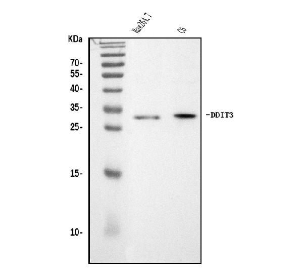
Figure 1. Western blot analysis of DDIT3 using anti-DDIT3 antibody (A00311-2).
Electrophoresis was performed on a 5-20% SDS-PAGE gel at 70V (Stacking gel) / 90V (Resolving gel) for 2-3 hours. The sample well of each lane was loaded with 50ug of sample under reducing conditions.
Lane 1: human U-87MG whole cell lysates,
Lane 2: human K562 whole cell lysates,
Lane 3: human THP-1 whole cell lysates.
After Electrophoresis, proteins were transferred to a Nitrocellulose membrane at 150mA for 50-90 minutes. Blocked the membrane with 5% Non-fat Milk/ TBS for 1.5 hour at RT. The membrane was incubated with rabbit anti-DDIT3 antigen affinity purified polyclonal antibody (Catalog # A00311-2) at 0.5 μg/mL overnight at 4°C, then washed with TBS-0.1%Tween 3 times with 5 minutes each and probed with a goat anti-rabbit IgG-HRP secondary antibody at a dilution of 1:5000 for 1.5 hour at RT. The signal is developed using an Enhanced Chemiluminescent detection (ECL) kit (Catalog # EK1002) with Tanon 5200 system. A specific band was detected for DDIT3 at approximately 29KD. The expected band size for DDIT3 is at 29KD.

Click image to see more details
Figure 2. IHC analysis of DDIT3 using anti-DDIT3 antibody (A00311-2).
DDIT3 was detected in paraffin-embedded section of human B lymphocytic tumor tissue. Heat mediated antigen retrieval was performed in EDTA buffer (pH8.0, epitope retrieval solution). The tissue section was blocked with 10% goat serum. The tissue section was then incubated with 1μg/ml rabbit anti-DDIT3 Antibody (A00311-2) overnight at 4°C. Biotinylated goat anti-rabbit IgG was used as secondary antibody and incubated for 30 minutes at 37°C. The tissue section was developed using Strepavidin-Biotin-Complex (SABC) (Catalog # SA1022) with DAB as the chromogen.

Click image to see more details
Figure 3. IHC analysis of DDIT3 using anti-DDIT3 antibody (A00311-2).
DDIT3 was detected in paraffin-embedded section of human renal cancer tissue. Heat mediated antigen retrieval was performed in EDTA buffer (pH8.0, epitope retrieval solution). The tissue section was blocked with 10% goat serum. The tissue section was then incubated with 1μg/ml rabbit anti-DDIT3 Antibody (A00311-2) overnight at 4°C. Biotinylated goat anti-rabbit IgG was used as secondary antibody and incubated for 30 minutes at 37°C. The tissue section was developed using Strepavidin-Biotin-Complex (SABC) (Catalog # SA1022) with DAB as the chromogen.

Click image to see more details
Figure 4. IHC analysis of DDIT3 using anti-DDIT3 antibody (A00311-2).
DDIT3 was detected in paraffin-embedded section of human lung cancer tissue. Heat mediated antigen retrieval was performed in EDTA buffer (pH8.0, epitope retrieval solution). The tissue section was blocked with 10% goat serum. The tissue section was then incubated with 1μg/ml rabbit anti-DDIT3 Antibody (A00311-2) overnight at 4°C. Biotinylated goat anti-rabbit IgG was used as secondary antibody and incubated for 30 minutes at 37°C. The tissue section was developed using Strepavidin-Biotin-Complex (SABC) (Catalog # SA1022) with DAB as the chromogen.

Click image to see more details
Figure 5. Flow Cytometry analysis of THP-1 cells using anti-DDIT3 antibody (A00311-2).
Overlay histogram showing THP-1 cells stained with A00311-2 (Blue line). To facilitate intracellular staining, cells were fixed with 4% paraformaldehyde and permeabilized with permeabilization buffer. The cells were blocked with 10% normal goat serum. And then incubated with rabbit anti-DDIT3 Antibody (A00311-2, 1μg/1x106 cells) for 30 min at 20°C. DyLight®488 conjugated goat anti-rabbit IgG (BA1127, 5-10μg/1x106 cells) was used as secondary antibody for 30 minutes at 20°C. Isotype control antibody (Green line) was rabbit IgG (1μg/1x106) used under the same conditions. Unlabelled sample without incubation with primary antibody and secondary antibody (Red line) was used as a blank control.
Protein Target Info & Infographic
Gene/Protein Information For DDIT3 (Source: Uniprot.org, NCBI)
Gene Name
DDIT3
Full Name
DDIT3 upstream open reading frame protein
Weight
74699 MW
Alternative Names
DNA damage-inducible transcript 3 protein; DDIT-3; C/EBP zeta; C/EBP-homologous protein; CHOP; C/EBP-homologous protein 10; CHOP-10; CCAAT/enhancer-binding protein homologous protein; Growth arrest and DNA damage-inducible protein GADD153; DDIT3; CHOP; CHOP10; GADD153 DDIT3 AltDDIT3, C/EBPzeta, CEBPZ, CHOP, CHOP-10, CHOP10, GADD153 DNA damage inducible transcript 3 DNA damage-inducible transcript 3 protein|C/EBP zeta|CCAAT/enhancer-binding protein homologous protein|alternative DDIT3 protein|c/EBP-homologous protein 10|growth arrest and DNA damage-inducible protein GADD153
*If product is indicated to react with multiple species, protein info is based on the gene entry specified above in "Species".For more info on DDIT3, check out the DDIT3 Infographic

We have 30,000+ of these available, one for each gene! Check them out.
In this infographic, you will see the following information for DDIT3: database IDs, superfamily, protein function, synonyms, molecular weight, chromosomal locations, tissues of expression, subcellular locations, post-translational modifications, and related diseases, research areas & pathways. If you want to see more information included, or would like to contribute to it and be acknowledged, please contact [email protected].
Specific Publications For Anti-DDIT3 Antibody Picoband® (A00311-2)
Hello CJ!
A00311-2 has been cited in 1 publications:
*The publications in this section are manually curated by our staff scientists. They may differ from Bioz's machine gathered results. Both are accurate. If you find a publication citing this product but is missing from this list, please let us know we will issue you a thank-you coupon.
Tension induces intervertebral disc degeneration via endoplasmic reticulum stress-mediated autophagy
Recommended Resources
Here are featured tools and databases that you might find useful.
- Boster's Pathways Library
- Protein Databases
- Bioscience Research Protocol Resources
- Data Processing & Analysis Software
- Photo Editing Software
- Scientific Literature Resources
- Research Paper Management Tools
- Molecular Biology Software
- Primer Design Tools
- Bioinformatics Tools
- Phylogenetic Tree Analysis
Customer Reviews
Have you used Anti-DDIT3 Antibody Picoband®?
Submit a review and receive an Amazon gift card.
- $30 for a review with an image
0 Reviews For Anti-DDIT3 Antibody Picoband®
Customer Q&As
Have a question?
Find answers in Q&As, reviews.
Can't find your answer?
Submit your question
