
Click image to see more details
Product Info Summary
| SKU: | M00297-2 |
|---|---|
| Size: | 100µg |
| Reactive Species: | Dog, Human, Mouse, Rat |
| Host: | Mouse |
| Application: | ELISA, IHC, WB |
Customers Who Bought This Also Bought

Product info
Product Name
Anti-Fos Monoclonal Antibody (8B5)
SKU/Catalog Number
M00297-2
Size
100µg
Form
Lyophilized
Description
Boster Bio Anti-Fos Monoclonal Antibody (8B5) (Catalog# M00297-2). Tested in ELISA, IHC, WB application(s). This antibody reacts with Human, Mouse, Rat, Dog.
Storage & Handling
Store at -20°C for one year. For short-term storage and frequent use, store at 4°C for up to one month. Avoid repeated freeze-thaw cycles.
Cite This Product
Anti-Fos Monoclonal Antibody (8B5) (Boster Biological Technology, Pleasanton CA, USA, Catalog # M00297-2)
Host
Mouse
Contents
Lyophilized from 1ml of 2x PBS containing 0.09% sodium azide, PEG, and sucrose.
Clonality
Monoclonal
Clone Number
8B5
Isotype
IgG1
Immunogen
Synthetic peptide corresponding to a portion of human c-Fos
*Blocking peptide can be purchased. Costs vary based on immunogen length. Contact us for pricing.
Reactive Species
M00297-2 is reactive to FOS in Dog, Human, Mouse, Rat
Reconstitution
Reconstitute with 1ml water (15 minutes at room temperature).
Observed Molecular Weight
Calculated molecular weight
73882 MW
Background of c-Fos
This monoclonal antibody recognizes a protein of 72kDa, which is identified as MMP2. The matrix metalloproteinases (MMP) are a family of peptidase enzymes responsible for the degradation of extracellular matrix components, including collagen, gelatin, Fibronectin, Laminin and proteoglycan. Transcription of MMP genes is differentially activated by phorbol ester, lipopolysaccharide (LPS) or staphylococcal enterotoxin B (SEB). MMP catalysis requires both calcium and zinc. MMP-2 (also designated type IV collagenase) cleaves collagen types IV,V, VII and X and gelatin type I. Activation of MMP-2 secretion requires the Ras signaling pathway.
Antibody Validation
Boster validates all antibodies on WB, IHC, ICC, Immunofluorescence, and ELISA with known positive control and negative samples to ensure specificity and high affinity, including thorough antibody incubations.
Application & Images
Applications
M00297-2 is guaranteed for ELISA, IHC, WB Boster Guarantee
Assay Dilutions Recommendation
The recommendations below provide a starting point for assay optimization. The actual working concentration varies and should be decided by the user.
ELISA (0.1µg/ml)
Immunohistochemistry (1:50)
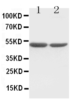
Western Blot (0.5µg/ml, ECL). Detects a band of ~50kDa.
Suggested dilutions/conditions may not be available for all applications. Optimal conditions must be determined individually for each application.
Positive Control
Validation Images & Assay Conditions
Click image to see more details
Figure 1. Western blot analysis of FOS using anti-FOS antibody (M00297-2).
Electrophoresis was performed on a 5-20% SDS-PAGE gel at 70V (Stacking gel) / 90V (Resolving gel) for 2-3 hours. The sample well of each lane was loaded with 50ug of sample under reducing conditions.
After Electrophoresis, proteins were transferred to a Nitrocellulose membrane at 150mA for 50-90 minutes. Blocked the membrane with 5% Non-fat Milk/ TBS for 1.5 hour at RT. The membrane was incubated with rabbit anti-FOS antigen affinity purified polyclonal antibody (Catalog # M00297-2) at 0.5 ug/mL overnight at 4°C, then washed with TBS-0.1%Tween 3 times with 5 minutes each and probed with a goat anti-Mouse IgG-HRP secondary antibody at a dilution of 1:10000 for 1.5 hour at RT. The signal is developed using an Enhanced Chemiluminescent detection (ECL) kit (Catalog # SA1021) with Tanon 5200 system. A specific band was detected for FOS.

Click image to see more details
Figure 2. IHC analysis of FOS using anti-FOS antibody (M00297-2).
FOS was detected in paraffin-embedded section. Heat mediated antigen retrieval was performed in citrate buffer (pH6, epitope retrieval solution) for 20 mins. The tissue section was blocked with 10% goat serum. The tissue section was then incubated with 1ug/ml rabbit anti-FOS Antibody (M00297-2) overnight at 4°C. Biotinylated goat anti Mouse IgG antibody was used as secondary antibody and incubated for 30 minutes at 37°C. The tissue section was developed using Strepavidin-Biotin-Complex (SABC)(Catalog # SA1021) with DAB as the chromogen.
Protein Target Info & Infographic
Gene/Protein Information For FOS (Source: Uniprot.org, NCBI)
Gene Name
FOS
Full Name
Proto-oncogene c-Fos
Weight
73882 MW
Superfamily
bZIP family
Alternative Names
activator protein 1; AP-1; cellular oncogene c-fos; Cellular oncogene fos; cFos; c-Fos; FBJ murine osteosarcoma viral (v-fos) oncogene homolog (oncogene FOS); FBJ murine osteosarcoma viral oncogene homolog; FOS; G0/G1 switch regulatory protein 7; G0S7; proto-oncogene c-Fos; v-fos FBJ murine osteosarcoma viral oncogene homolog FOS AP-1, C-FOS, p55 Fos proto-oncogene, AP-1 transcription factor subunit proto-oncogene c-Fos|FBJ murine osteosarcoma viral (v-fos) oncogene homolog (oncogene FOS)|FBJ murine osteosarcoma viral oncogene homolog|Fos proto-oncogene, AP-1 trancription factor subunit|G0/G1 switch regulatory protein 7|activator protein 1|cellular oncogene c-fos
*If product is indicated to react with multiple species, protein info is based on the gene entry specified above in "Species".For more info on FOS, check out the FOS Infographic

We have 30,000+ of these available, one for each gene! Check them out.
In this infographic, you will see the following information for FOS: database IDs, superfamily, protein function, synonyms, molecular weight, chromosomal locations, tissues of expression, subcellular locations, post-translational modifications, and related diseases, research areas & pathways. If you want to see more information included, or would like to contribute to it and be acknowledged, please contact [email protected].
Specific Publications For Anti-Fos Monoclonal Antibody (8B5) (M00297-2)
Hello CJ!
M00297-2 has been cited in 6 publications:
*The publications in this section are manually curated by our staff scientists. They may differ from Bioz's machine gathered results. Both are accurate. If you find a publication citing this product but is missing from this list, please let us know we will issue you a thank-you coupon.
Overexpression of c-fos in Helicobacter pylori-induced gastric precancerosis of Mongolian gerbil
Changes in the expression of c-fos & heat shock protein genes & blood flow velocity in the brain of rats undergoing myocardial ischaemia/reperfusion
Positional and expressive alteration of prohibitin during the induced differentiation of human hepatocarcinoma SMMC-7721 cells
Analgesic Neural Circuits Are Activated by Electroacupuncture at Two Sets of Acupoints
miR-144 and targets, c-fos and cyclooxygenase-2 (COX2), modulate synthesis of PGE2 in the amnion during pregnancy and labor
The Expression Patterns of c-Fos and c-Jun Induced by Different Frequencies of Electroacupuncture in the Brain
Recommended Resources
Here are featured tools and databases that you might find useful.
- Boster's Pathways Library
- Protein Databases
- Bioscience Research Protocol Resources
- Data Processing & Analysis Software
- Photo Editing Software
- Scientific Literature Resources
- Research Paper Management Tools
- Molecular Biology Software
- Primer Design Tools
- Bioinformatics Tools
- Phylogenetic Tree Analysis
Customer Reviews
Have you used Anti-Fos Monoclonal Antibody (8B5)?
Submit a review and receive an Amazon gift card.
- $30 for a review with an image
0 Reviews For Anti-Fos Monoclonal Antibody (8B5)
Customer Q&As
Have a question?
Find answers in Q&As, reviews.
Can't find your answer?
Submit your question
15 Customer Q&As for Anti-Fos Monoclonal Antibody (8B5)
Question
We are currently using anti-Fos Monoclonal antibody (8B5) M00297-2 for human tissue, and we are happy with the WB results. The species of reactivity given in the datasheet says human, mouse, rat. Is it possible that the antibody can work on feline tissues as well?
Verified Customer
Verified customer
Asked: 2020-04-30
Answer
The anti-Fos Monoclonal antibody (8B5) (M00297-2) has not been tested for cross reactivity specifically with feline tissues, but there is a good chance of cross reactivity. We have an innovator award program that if you test this antibody and show it works in feline you can get your next antibody for free. Please contact me if I can help you with anything.
Boster Scientific Support
Answered: 2020-04-30
Question
Will anti-Fos Monoclonal antibody (8B5) M00297-2 work for IHC with mucosa of stomach?
Verified Customer
Verified customer
Asked: 2020-02-27
Answer
According to the expression profile of mucosa of stomach, FOS is highly expressed in mucosa of stomach. So, it is likely that anti-Fos Monoclonal antibody (8B5) M00297-2 will work for IHC with mucosa of stomach.
Boster Scientific Support
Answered: 2020-02-27
Question
Can you help my question with product M00297-2, anti-Fos Monoclonal antibody (8B5) . I was wondering if it would be possible to conjugate this antibody with biotin. I would need it to be without BSA or sodium azide. I am planning on using a buffer exchange of sodium azide with PBS only. Would there be problems for me to conjugate the antibody and store it in -20 degrees in small aliquots?
Verified Customer
Verified customer
Asked: 2019-12-24
Answer
It is not recommended storing this antibody with PBS buffer only in -20 degrees. If you want to store it in -20 degrees it is best to add some cryoprotectant like glycerol. If you want carrier free M00297-2 anti-Fos Monoclonal antibody (8B5) , we can provide it to you in a special formula with trehalose and/or glycerol. These molecules will not interfere with conjugation chemistry and provide a good level of protection for the antibody from degradation. Please be sure to specify this in your purchase order.
Boster Scientific Support
Answered: 2019-12-24
Question
Will M00297-2 anti-Fos Monoclonal antibody (8B5) work on parafin embedded sections? If so, which fixation method do you recommend we use (PFA, paraformaldehyde, other)?
Verified Customer
Verified customer
Asked: 2019-11-20
Answer
It shows on the product datasheet, M00297-2 anti-Fos Monoclonal antibody (8B5) as been tested on IHC. It is best to use PFA for fixation because it has better tissue penetration ability. PFA needs to be prepared fresh before use. Long term stored PFA turns into formalin, as the PFA molecules congregate and become formalin.
Boster Scientific Support
Answered: 2019-11-20
Question
I was wanting to use your anti-Fos Monoclonal antibody (8B5) for IHC for rat mucosa of stomach on frozen tissues, but I want to know if it has been validated for this particular application. Has this antibody been validated and is this antibody a good choice for rat mucosa of stomach identification?
Verified Customer
Verified customer
Asked: 2019-09-27
Answer
It shows on the product datasheet, M00297-2 anti-Fos Monoclonal antibody (8B5) has been tested for ELISA, IHC, WB on human, mouse, rat tissues. We have an innovator award program that if you test this antibody and show it works in rat mucosa of stomach in IHC-frozen, you can get your next antibody for free.
Boster Scientific Support
Answered: 2019-09-27
Question
We have observed staining in mouse colon. Any tips? Is anti-Fos Monoclonal antibody (8B5) supposed to stain colon positively?
Verified Customer
Verified customer
Asked: 2019-09-18
Answer
From literature colon does express FOS. From Uniprot.org, FOS is expressed in mucosa of stomach, colon, pancreas, leukemic t-cell, among other tissues. Regarding which tissues have FOS expression, here are a few articles citing expression in various tissues:
Colon, Pubmed ID: 14702039
Leukemic T-cell, Pubmed ID: 19690332
Pancreas, Pubmed ID: 15489334
Boster Scientific Support
Answered: 2019-09-18
Question
Is this M00297-2 anti-Fos Monoclonal antibody (8B5) reactive to the isotypes of FOS?
Verified Customer
Verified customer
Asked: 2019-07-05
Answer
The immunogen of M00297-2 anti-Fos Monoclonal antibody (8B5) is Synthetic peptide corresponding to a portion of human c-Fos . Could you tell me which isotype you are interested in so I can help see if the immunogen is part of this isotype?
Boster Scientific Support
Answered: 2019-07-05
Question
I appreciate helping with my inquiry over the phone. Here are the WB image, lot number and protocol we used for mucosa of stomach using anti-Fos Monoclonal antibody (8B5) M00297-2. Let me know if you need anything else.
Verified Customer
Verified customer
Asked: 2019-01-24
Answer
We appreciate the data. You have provided everything we needed. Our lab team are working to resolve your inquiry as quickly as possible, and we appreciate your patience and understanding! Please let me know if there is anything you need in the meantime.
Boster Scientific Support
Answered: 2019-01-24
Question
I would like to test anti-Fos Monoclonal antibody (8B5) M00297-2 on rat mucosa of stomach for research purposes, then I may be interested in using anti-Fos Monoclonal antibody (8B5) M00297-2 for diagnostic purposes as well. Is the antibody suitable for diagnostic purposes?
Verified Customer
Verified customer
Asked: 2018-12-31
Answer
The products we sell, including anti-Fos Monoclonal antibody (8B5) M00297-2, are only intended for research use. They would not be suitable for use in diagnostic work. If you have the means to develop a product into diagnostic use, and are interested in collaborating with us and develop our product into an IVD product, please contact us for more discussions.
Boster Scientific Support
Answered: 2018-12-31
Question
Here is the WB image, lot number and protocol we used for mucosa of stomach using anti-Fos Monoclonal antibody (8B5) M00297-2. Please let me know if you require anything else.
O. Williams
Verified customer
Asked: 2018-06-27
Answer
Thank you very much for the data. Our lab team are working to resolve this as quickly as possible, and we appreciate your patience and understanding! You have provided everything we needed. Please let me know if there is anything you need in the meantime.
Boster Scientific Support
Answered: 2018-06-27
Question
We purchased anti-Fos Monoclonal antibody (8B5) for IHC on colon in the past. I am using rat, and We are going to use the antibody for WB next. I was wanting to use examining colon as well as leukemic t-cell in our next experiment. Could give a recommendation on which antibody would work the best for WB?
P. Patel
Verified customer
Asked: 2018-03-21
Answer
I have checked the website and datasheets of our anti-Fos Monoclonal antibody (8B5) and it seems that M00297-2 has been validated on rat in both IHC and WB. Thus M00297-2 should work for your application. Our Boster satisfaction guarantee will cover this product for WB in rat even if the specific tissue type has not been validated. We do have a comprehensive range of products for WB detection and you can check out our website bosterbio.com to find out more information about them.
Boster Scientific Support
Answered: 2018-03-21
Question
Is a blocking peptide available for product anti-Fos Monoclonal antibody (8B5) (M00297-2)?
Verified Customer
Verified customer
Asked: 2018-03-14
Answer
We do provide the blocking peptide for product anti-Fos Monoclonal antibody (8B5) (M00297-2). If you would like to place an order for it please contact [email protected] and make a special request.
Boster Scientific Support
Answered: 2018-03-14
Question
Do you have a BSA free version of anti-Fos Monoclonal antibody (8B5) M00297-2 available?
Verified Customer
Verified customer
Asked: 2018-02-01
Answer
We appreciate your recent telephone inquiry. I can confirm that some lots of this anti-Fos Monoclonal antibody (8B5) M00297-2 are BSA free. For now, these lots are available and we can make a BSA free formula for you free of charge. It will take 3 extra days to prepare. If you require this antibody BSA free again in future, please do not hesitate to contact me and I will be pleased to check which lots we have in stock that are BSA free.
Boster Scientific Support
Answered: 2018-02-01
Question
We were content with the WB result of your anti-Fos Monoclonal antibody (8B5) . However we have observed positive staining in colon nucleus. endoplasmic reticulum. cytoplasm, using this antibody. Is that expected? Could you tell me where is FOS supposed to be expressed?
N. Evans
Verified customer
Asked: 2017-03-13
Answer
Based on literature, colon does express FOS. Generally FOS expresses in nucleus. endoplasmic reticulum. cytoplasm,. Regarding which tissues have FOS expression, here are a few articles citing expression in various tissues:
Colon, Pubmed ID: 14702039
Leukemic T-cell, Pubmed ID: 19690332
Pancreas, Pubmed ID: 15489334
Boster Scientific Support
Answered: 2017-03-13
Question
I see that the anti-Fos Monoclonal antibody (8B5) M00297-2 works with IHC, what is the protocol used to produce the result images on the product page?
J. Lewis
Verified customer
Asked: 2016-09-06
Answer
You can find protocols for IHC on the "support/technical resources" section of our navigation menu. If you have any further questions, please send an email to [email protected]
Boster Scientific Support
Answered: 2016-09-06
