
Click image to see more details
Product Info Summary
| SKU: | PB9079 |
|---|---|
| Size: | 100 μg/vial |
| Reactive Species: | Human, Rat |
| Host: | Rabbit |
| Application: | IHC, WB |
Customers Who Bought This Also Bought

Product info
Product Name
Anti-CXCR3 Antibody Picoband™
SKU/Catalog Number
PB9079
Size
100 μg/vial
Form
Lyophilized
Description
Boster Bio Anti-CXCR3 Antibody Picoband™ catalog # PB9079. Tested in IHC, WB applications. This antibody reacts with Human, Rat.
Storage & Handling
Store at -20˚C for one year from date of receipt. After reconstitution, at 4˚C for one month. It can also be aliquotted and stored frozen at -20˚C for six months. Avoid repeated freeze-thaw cycles.
Cite This Product
Anti-CXCR3 Antibody Picoband™ (Boster Biological Technology, Pleasanton CA, USA, Catalog # PB9079)
Host
Rabbit
Contents
Each vial contains 5mg BSA, 0.9mg NaCl, 0.2mg Na2HPO4, 0.05mg NaN3.
Clonality
Polyclonal
Isotype
Rabbit IgG
Immunogen
E.coli-derived human CXCR3 recombinant protein (Position: M1-L368). Human CXCR3 shares 86% amino acid (aa) sequence identity with both mouse and rat CXCR3.
*Blocking peptide can be purchased. Costs vary based on immunogen length. Contact us for pricing.
Cross-reactivity
No cross-reactivity with other proteins
Reactive Species
PB9079 is reactive to CXCR3 in Human, Rat
Applications
PB9079 is guaranteed for IHC, WB Boster Guarantee
Observed Molecular Weight
41 kDa
Calculated molecular weight
40660 MW
Background of CXCR3
Chemokine receptor CXCR3 is a Galphai protein-coupled receptor in the CXC chemokine receptor family. Other names for CXCR3 are G protein-coupled receptor 9 (GPR9) and CD183. It is mapped to Xq13.1. CXCR3 is expressed on malignant B cells from chronic lymphoproliferative disorders, particularly in patients with CLL, and represents a fully functional receptor involved in chemotaxis of malignant B lymphocytes. It is found that in the absence of known etiologic agents, CXCR3 represents a novel target for therapeutic interference early in type 1 diabetes. CXCR3 signaling is associated with MG pathogenesis and proposed that and CXCR3 may serve as novel drug targets to treat MG. CXCR3A and CXCR3B are involved in the chemotactic and vascular effects of CXCL4L1.
Antibody Validation
Boster validates all antibodies on WB, IHC, ICC, Immunofluorescence, and ELISA with known positive control and negative samples to ensure specificity and high affinity, including thorough antibody incubations.
Assay dilution & Images
Reconstitution
Add 0.2ml of distilled water will yield a concentration of 500ug/ml.
Assay Dilutions Recommendation
The recommendations below provide a starting point for assay optimization. The actual working concentration varies and should be decided by the user.
Immunohistochemistry (Paraffin-embedded Section), 0.5-1μg/ml, Human, Rat, By Heat
Western blot, 0.1-0.5μg/ml, Human
Validation Images & Assay Conditions
Click image to see more details
Figure 1. IHC analysis of CXCR3 using anti-CXCR3 antibody (PB9079).
CXCR3 was detected in a paraffin-embedded section of rat kidney tissue. Heat mediated antigen retrieval was performed in EDTA buffer (pH 8.0, epitope retrieval solution). The tissue section was blocked with 10% goat serum. The tissue section was then incubated with 1 μg/ml rabbit anti-CXCR3 Antibody (PB9079) overnight at 4°C. Biotinylated goat anti-rabbit IgG was used as secondary antibody and incubated for 30 minutes at 37°C. The tissue section was developed using Strepavidin-Biotin-Complex (SABC) (Catalog # SA1022) with DAB as the chromogen.

Click image to see more details
Figure 2. IHC analysis of CXCR3 using anti-CXCR3 antibody (PB9079).
CXCR3 was detected in a paraffin-embedded section of human tonsil tissue. Heat mediated antigen retrieval was performed in EDTA buffer (pH 8.0, epitope retrieval solution). The tissue section was blocked with 10% goat serum. The tissue section was then incubated with 1 μg/ml rabbit anti-CXCR3 Antibody (PB9079) overnight at 4°C. Biotinylated goat anti-rabbit IgG was used as secondary antibody and incubated for 30 minutes at 37°C. The tissue section was developed using Strepavidin-Biotin-Complex (SABC) (Catalog # SA1022) with DAB as the chromogen.

Click image to see more details
Figure 3. IHC analysis of CXCR3 using anti-CXCR3 antibody (PB9079).
CXCR3 was detected in a paraffin-embedded section of human lung cancer tissue. Heat mediated antigen retrieval was performed in EDTA buffer (pH 8.0, epitope retrieval solution). The tissue section was blocked with 10% goat serum. The tissue section was then incubated with 1 μg/ml rabbit anti-CXCR3 Antibody (PB9079) overnight at 4°C. Biotinylated goat anti-rabbit IgG was used as secondary antibody and incubated for 30 minutes at 37°C. The tissue section was developed using Strepavidin-Biotin-Complex (SABC) (Catalog # SA1022) with DAB as the chromogen.

Click image to see more details
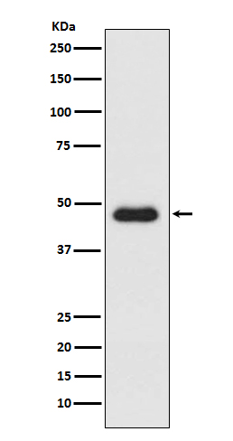
Figure 4. Western blot analysis of CXCR3 using anti-CXCR3 antibody (PB9079).
Electrophoresis was performed on a 5-20% SDS-PAGE gel at 70V (Stacking gel) / 90V (Resolving gel) for 2-3 hours. The sample well of each lane was loaded with 30 ug of sample under reducing conditions.
Lane 1: human Colo320 whole cell lysates,
Lane 2: human SGC whole cell lysates.
After electrophoresis, proteins were transferred to a nitrocellulose membrane at 150 mA for 50-90 minutes. Blocked the membrane with 5% non-fat milk/TBS for 1.5 hour at RT. The membrane was incubated with rabbit anti-CXCR3 antigen affinity purified polyclonal antibody (Catalog # PB9079) at 0.5 μg/mL overnight at 4°C, then washed with TBS-0.1%Tween 3 times with 5 minutes each and probed with a goat anti-rabbit IgG-HRP secondary antibody at a dilution of 1:5000 for 1.5 hour at RT. The signal is developed using an Enhanced Chemiluminescent detection (ECL) kit (Catalog # EK1002) with Tanon 5200 system. A specific band was detected for CXCR3 at approximately 41 kDa. The expected band size for CXCR3 is at 41 kDa.
Protein Target Info & Infographic
Gene/Protein Information For CXCR3 (Source: Uniprot.org, NCBI)
Gene Name
CXCR3
Full Name
C-X-C chemokine receptor type 3
Weight
40660 MW
Superfamily
G-protein coupled receptor 1 family
Alternative Names
CD183 antigen; CD183; chemokine (C-X-C motif) receptor 3; chemokine (C-X-C) receptor 3; CKR-L2IP-10 receptor; CMKAR3; C-X-C chemokine receptor type 3; CXCR3; CXC-R3; CXCR-3; G protein-coupled receptor 9CD182; GPR9; GPR9Mig-R; Interferon-inducible protein 10 receptor; IP10 receptor; IP10-R; Mig receptor; MigR CXCR3 CD182, CD183, CKR-L2, CMKAR3, GPR9, IP10-R, Mig-R, MigR C-X-C motif chemokine receptor 3 C-X-C chemokine receptor type 3|G protein-coupled receptor 9|IP-10 receptor|Mig receptor|chemokine (C-X-C motif) receptor 3|chemokine receptor 3|interferon-inducible protein 10 receptor
*If product is indicated to react with multiple species, protein info is based on the gene entry specified above in "Species".For more info on CXCR3, check out the CXCR3 Infographic

We have 30,000+ of these available, one for each gene! Check them out.
In this infographic, you will see the following information for CXCR3: database IDs, superfamily, protein function, synonyms, molecular weight, chromosomal locations, tissues of expression, subcellular locations, post-translational modifications, and related diseases, research areas & pathways. If you want to see more information included, or would like to contribute to it and be acknowledged, please contact [email protected].
Specific Publications For Anti-CXCR3 Antibody Picoband™ (PB9079)
Hello CJ!
PB9079 has been cited in 5 publications:
*The publications in this section are manually curated by our staff scientists. They may differ from Bioz's machine gathered results. Both are accurate. If you find a publication citing this product but is missing from this list, please let us know we will issue you a thank-you coupon.
CXCL10 Produced by HPV-Positive Cervical Cancer Cells Stimulates Exosomal PDL1 Expression by Fibroblasts via CXCR3 and JAK-STAT Pathways
Bu-Shen-Fang-Chuan formula attenuates T-lymphocytes recruitment in the lung of rats with COPD through suppressing CXCL9/CXCL10/CXCL11-CXCR3 axis
Li Q,Sun J,Cao Y,Liu B,Li L,Mohammadtursun N,Zhang H,Dong J,Wu J.Bu-Shen-Fang-Chuan formula attenuates T-lymphocytes recruitment in the lung of rats with COPD through suppressing CXCL9/CXCL10/CXCL11-CXCR3 axis.Biomed Pharmacother.2020 Mar;123:109735.doi:1
Species: Rat
PB9079 usage in article: APP:IHC, SAMPLE:LUNG TISSUE, DILUTION:NA
Impact of chemokine receptor CXCR3 on tumor-infiltrating lymphocyte recruitment associated with favorable prognosis in advanced gastric cancer
An alternatively spliced variant of CXCR3 mediates the metastasis of CD133+ liver cancer cells induced by CXCL9
Recommended Resources
Here are featured tools and databases that you might find useful.
- Boster's Pathways Library
- Protein Databases
- Bioscience Research Protocol Resources
- Data Processing & Analysis Software
- Photo Editing Software
- Scientific Literature Resources
- Research Paper Management Tools
- Molecular Biology Software
- Primer Design Tools
- Bioinformatics Tools
- Phylogenetic Tree Analysis
Customer Reviews
Have you used Anti-CXCR3 Antibody Picoband™?
Submit a review and receive an Amazon gift card.
- $30 for a review with an image
0 Reviews For Anti-CXCR3 Antibody Picoband™
Customer Q&As
Have a question?
Find answers in Q&As, reviews.
Can't find your answer?
Submit your question
16 Customer Q&As for Anti-CXCR3 Antibody Picoband™
Question
Please see the WB image, lot number and protocol we used for fetal astrocyte using anti-CXCR3 antibody PB9079. Please let me know if you require anything else.
R. Jackson
Verified customer
Asked: 2020-01-24
Answer
Thank you very much for the data. Our lab team are working to resolve this as quickly as possible, and we appreciate your patience and understanding! You have provided everything we needed. Please let me know if there is anything you need in the meantime.
Boster Scientific Support
Answered: 2020-01-24
Question
I am interested in using your anti-CXCR3 antibody for positive regulation of cell population proliferation studies. Has this antibody been tested with western blotting on lung cancer tissue? We would like to see some validation images before ordering.
Verified Customer
Verified customer
Asked: 2020-01-24
Answer
We appreciate your inquiry. This PB9079 anti-CXCR3 antibody is tested on rat kidney tissue, human tonsil tissue, lung cancer tissue, colo320 whole cell lysate, sgc whole cell lysate. It is guaranteed to work for IHC, WB in human, rat. Our Boster guarantee will cover your intended experiment even if the sample type has not been be directly tested.
Boster Scientific Support
Answered: 2020-01-24
Question
Will PB9079 anti-CXCR3 antibody work on parafin embedded sections? If so, which fixation method do you recommend we use (PFA, paraformaldehyde, other)?
Verified Customer
Verified customer
Asked: 2019-08-15
Answer
It shows on the product datasheet, PB9079 anti-CXCR3 antibody as been validated on IHC. It is best to use PFA for fixation because it has better tissue penetration ability. PFA needs to be prepared fresh before use. Long term stored PFA turns into formalin, as the PFA molecules congregate and become formalin.
Boster Scientific Support
Answered: 2019-08-15
Question
We are currently using anti-CXCR3 antibody PB9079 for human tissue, and we are satisfied with the WB results. The species of reactivity given in the datasheet says human, rat. Is it true that the antibody can work on monkey tissues as well?
Verified Customer
Verified customer
Asked: 2019-06-26
Answer
The anti-CXCR3 antibody (PB9079) has not been validated for cross reactivity specifically with monkey tissues, though there is a good chance of cross reactivity. We have an innovator award program that if you test this antibody and show it works in monkey you can get your next antibody for free. Please contact me if I can help you with anything.
Boster Scientific Support
Answered: 2019-06-26
Question
I see that the anti-CXCR3 antibody PB9079 works with IHC, what is the protocol used to produce the result images on the product page?
Verified Customer
Verified customer
Asked: 2019-06-17
Answer
You can find protocols for IHC on the "support/technical resources" section of our navigation menu. If you have any further questions, please send an email to [email protected]
Boster Scientific Support
Answered: 2019-06-17
Question
Is a blocking peptide available for product anti-CXCR3 antibody (PB9079)?
Verified Customer
Verified customer
Asked: 2019-06-14
Answer
We do provide the blocking peptide for product anti-CXCR3 antibody (PB9079). If you would like to place an order for it please contact [email protected] and make a special request.
Boster Scientific Support
Answered: 2019-06-14
Question
Our team were happy with the WB result of your anti-CXCR3 antibody. However we have been able to see positive staining in lung testis isoform 2: cell membrane using this antibody. Is that expected? Could you tell me where is CXCR3 supposed to be expressed?
Verified Customer
Verified customer
Asked: 2019-01-03
Answer
From literature, lung testis does express CXCR3. Generally CXCR3 expresses in isoform 1: cell membrane, isoform 2: cell membrane. Regarding which tissues have CXCR3 expression, here are a few articles citing expression in various tissues:
Blood, Pubmed ID: 9064356
Brain, Lung, and Testis, Pubmed ID: 15489334
Colon, Pubmed ID: 14702039
Endothelial cell, and Thymus, Pubmed ID: 18291705
Fetal astrocyte, Pubmed ID: 9625760
Boster Scientific Support
Answered: 2019-01-03
Question
My question regarding product PB9079, anti-CXCR3 antibody. I was wondering if it would be possible to conjugate this antibody with biotin. I would need it to be without BSA or sodium azide. I am planning on using a buffer exchange of sodium azide with PBS only. Would there be problems for me to conjugate the antibody and store it in -20 degrees in small aliquots?
Verified Customer
Verified customer
Asked: 2017-12-15
Answer
We suggest not storing this antibody with PBS buffer only in -20 degrees. If you want to store it in -20 degrees it is best to add some cryoprotectant like glycerol. If you want carrier free PB9079 anti-CXCR3 antibody, we can provide it to you in a special formula with trehalose and/or glycerol. These molecules will not interfere with conjugation chemistry and provide a good level of protection for the antibody from degradation. Please be sure to specify this in your purchase order.
Boster Scientific Support
Answered: 2017-12-15
Question
We bought anti-CXCR3 antibody for IHC on brain last year. I am using human, and We are going to use the antibody for WB next. I am looking for examining brain as well as blood in our next experiment. Do you have any suggestion on which antibody would work the best for WB?
Verified Customer
Verified customer
Asked: 2017-10-16
Answer
I took a look at the website and datasheets of our anti-CXCR3 antibody and it appears that PB9079 has been validated on human in both IHC and WB. Thus PB9079 should work for your application. Our Boster satisfaction guarantee will cover this product for WB in human even if the specific tissue type has not been validated. We do have a comprehensive range of products for WB detection and you can check out our website bosterbio.com to find out more information about them.
Boster Scientific Support
Answered: 2017-10-16
Question
Is this PB9079 anti-CXCR3 antibody reactive to the isotypes of CXCR3?
Verified Customer
Verified customer
Asked: 2017-10-03
Answer
The immunogen of PB9079 anti-CXCR3 antibody is E.coli-derived human CXCR3 recombinant protein (Position: M1-L368). Human CXCR3 shares 86% amino acid (aa) sequence identity with both mouse and rat CXCR3. Could you tell me which isotype you are interested in so I can help see if the immunogen is part of this isotype?
Boster Scientific Support
Answered: 2017-10-03
Question
Would anti-CXCR3 antibody PB9079 work for IHC with fetal astrocyte?
Verified Customer
Verified customer
Asked: 2017-06-29
Answer
According to the expression profile of fetal astrocyte, CXCR3 is highly expressed in fetal astrocyte. So, it is likely that anti-CXCR3 antibody PB9079 will work for IHC with fetal astrocyte.
Boster Scientific Support
Answered: 2017-06-29
Question
We have seen staining in rat endothelial cell thymus. Are there any suggestions? Is anti-CXCR3 antibody supposed to stain endothelial cell thymus positively?
O. Wu
Verified customer
Asked: 2017-04-17
Answer
From what I have seen in literature endothelial cell thymus does express CXCR3. From what I have seen in Uniprot.org, CXCR3 is expressed in leukocyte, blood, endothelial cell thymus, colon, brain, lung testis, fetal astrocyte, among other tissues. Regarding which tissues have CXCR3 expression, here are a few articles citing expression in various tissues:
Blood, Pubmed ID: 9064356
Brain, Lung, and Testis, Pubmed ID: 15489334
Colon, Pubmed ID: 14702039
Endothelial cell, and Thymus, Pubmed ID: 18291705
Fetal astrocyte, Pubmed ID: 9625760
Boster Scientific Support
Answered: 2017-04-17
Question
Is there a BSA free version of anti-CXCR3 antibody PB9079 available?
R. Bhatt
Verified customer
Asked: 2017-03-20
Answer
Thanks for your recent telephone inquiry. I can confirm that some lots of this anti-CXCR3 antibody PB9079 are BSA free. For now, these lots are available and we can make a BSA free formula for you free of charge. It will take 3 extra days to prepare. If you require this antibody BSA free again in future, please do not hesitate to contact me and I will be pleased to check which lots we have in stock that are BSA free.
Boster Scientific Support
Answered: 2017-03-20
Question
I appreciate helping with my inquiry over the phone. Here are the WB image, lot number and protocol we used for fetal astrocyte using anti-CXCR3 antibody PB9079. Let me know if you need anything else.
B. Edwards
Verified customer
Asked: 2016-12-14
Answer
Thanks for the data. You have provided everything we needed. Our lab team are working to resolve your inquiry as quickly as possible, and we appreciate your patience and understanding! Please let me know if there is anything you need in the meantime.
Boster Scientific Support
Answered: 2016-12-14
Question
I was wanting to use your anti-CXCR3 antibody for IHC for human fetal astrocyte on frozen tissues, but I want to know if it has been validated for this particular application. Has this antibody been validated and is this antibody a good choice for human fetal astrocyte identification?
R. Parker
Verified customer
Asked: 2016-08-15
Answer
As indicated on the product datasheet, PB9079 anti-CXCR3 antibody has been tested for IHC, WB on human, rat tissues. We have an innovator award program that if you test this antibody and show it works in human fetal astrocyte in IHC-frozen, you can get your next antibody for free.
Boster Scientific Support
Answered: 2016-08-15
Question
We want to test anti-CXCR3 antibody PB9079 on human fetal astrocyte for research purposes, then I may be interested in using anti-CXCR3 antibody PB9079 for diagnostic purposes as well. Is the antibody suitable for diagnostic purposes?
K. Johnson
Verified customer
Asked: 2013-02-11
Answer
The products we sell, including anti-CXCR3 antibody PB9079, are only intended for research use. They would not be suitable for use in diagnostic work. If you have the means to develop a product into diagnostic use, and are interested in collaborating with us and develop our product into an IVD product, please contact us for more discussions.
Boster Scientific Support
Answered: 2013-02-11
