This website uses cookies to ensure you get the best experience on our website.
- Table of Contents
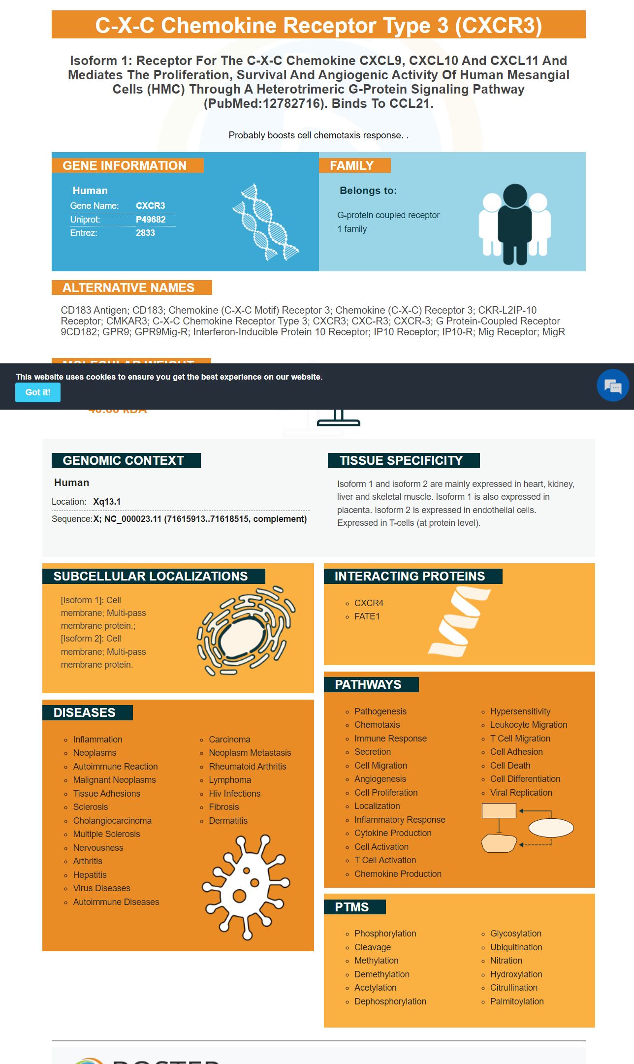
Facts about C-X-C chemokine receptor type 3.
Probably boosts cell chemotaxis response. .
Human | |
---|---|
Gene Name: | CXCR3 |
Uniprot: | P49682 |
Entrez: | 2833 |
Belongs to: |
---|
G-protein coupled receptor 1 family |
CD183 antigen; CD183; chemokine (C-X-C motif) receptor 3; chemokine (C-X-C) receptor 3; CKR-L2IP-10 receptor; CMKAR3; C-X-C chemokine receptor type 3; CXCR3; CXC-R3; CXCR-3; G protein-coupled receptor 9CD182; GPR9; GPR9Mig-R; Interferon-inducible protein 10 receptor; IP10 receptor; IP10-R; Mig receptor; MigR
Mass (kDA):
40.66 kDA
Human | |
---|---|
Location: | Xq13.1 |
Sequence: | X; NC_000023.11 (71615913..71618515, complement) |
Isoform 1 and isoform 2 are mainly expressed in heart, kidney, liver and skeletal muscle. Isoform 1 is also expressed in placenta. Isoform 2 is expressed in endothelial cells. Expressed in T-cells (at protein level).
[Isoform 1]: Cell membrane; Multi-pass membrane protein.; [Isoform 2]: Cell membrane; Multi-pass membrane protein.
BD Biosciences and Boster Bio are two companies that are making anti-CXCR3 antibodies. These antibodies react with Human and Mouse proteins. They can be stored at -20degC or 4degC for one month. Boster Bio validates all of their antibodies with known positive and negative samples, and the Boster Bio Anti-CXCR3 Antibody is no exception.
The boster bio Anti-CXCR3 antibody is a member of the Picoband(tm) catalog. This antibody reacts with Human and Rat CXCR3 proteins. The antibody can be stored at -20°C for up to one year. The antibody contains 5 mg of BSA. The boster bio Anti-CXCR3 antibody is suitable for various applications.
The CXCR3 receptor is a G-protein-coupled 7-transmembrane protein. It is expressed in high concentrations on plasmacytoid monocytes, natural killer cells, and activated T cells. It also plays an important role in the regulation of immune responses, inflammation, and infection. The CXCR3 ligands are essential for regulating leukocyte migration.
The BD Biosciences CXCR3 marker has many applications in immunohistochemistry, including assays for the identification of immune cells. This marker is expressed on T cells in the immune system. It has recently been discovered that CXCL10 is also expressed on B cells. Hence, the CXCR3 and CXCL10 marker are involved in the inflammatory microenvironment.
The role of the chemokine CXCR3 in the formation of granulomas after intracellular bacterial infection is well established. Infection with Brucella causes increased expression of CXCR3 and its ligands in PBMCs and tissues of infected patients. This study reveals that the chemokine receptor CXCR3 and its ligands play a critical role in the regulation of leukocyte migration.
A comprehensive clinical examination and detailed history will help determine whether brucellosis is the cause of your symptoms. Molecular tests for Brucella infection will reveal elevated C-reactive protein, elevated erythrocyte sedimentation rate (ESR), and positive cultures. Further, your doctor may perform imaging tests to determine if the infection has affected your gastrointestinal tract. A CAT scan or MRI of your brain and spinal cord may be necessary to confirm the diagnosis.
Recent genetic studies indicate that the IL-8/IL-8 gene is associated with a higher risk of brucellosis in patients with high CXCR3 gene expression. This genetic variation in IL-8 has been linked with a high risk of developing brucellosis in developing countries. Further, a recent study has found that increased levels of IL-8-251AA in blood are associated with a higher risk of developing brucellosis in humans.
In the Saudi Arabia study, 73 episodes of brucellosis were found in 55 patients. Nearly half of these patients suffered from rheumatic symptoms. Moreover, patients with hemodynamic stability and no complications could be treated with a combination of antimicrobials. If the disease is detected early and treated promptly, it is rare to require surgical intervention. This is especially true when the disease is endemic in an area.
The CXCR3 marker is expressed on T cells in peripheral blood and has been associated with inflammation. Exogenous IL-2 and anti-T cell receptor antibodies have induced CXCR3 surface expression. In humans, CXCR3 is found on T cells that have a Th2 bias. A recent study by Hancock and colleagues showed that mice deficient in CXCR3 exhibited resistance to acute allograft rejection.
The CXCR3 marker is an effective biomarker of inflammation. This receptor is expressed on T cells that can promote anti-tumor immune responses. This article will discuss the biology of CXCR3 and how it is expressed in inflammation and carcinogenesis. Let's begin by discussing the potential uses of CXCR3 in inflammation. Here are some examples. You'll notice that CXCR3-expressing cells have fewer naive cells and more EM cells than their counterparts.
It is widely known that CXCR3 contributes to preferential recruitment of Th2 cells to inflammatory sites. However, this process requires many other molecules such as chemokine receptors, adhesion molecules, and members of the selectin family. CXCR3 alone may not be sufficient for the process of Th2 migration, but it is necessary for it. But, the best uses of the CXCR3 marker for inflammation are still being discovered.
Using a TCR transgenic model of inflammatory disease, researchers were able to analyze the role of CXCR3 on T cells. Inflammatory-infiltrating T cells express CXCR3 ligands, which promote the trafficking of T cells into peripheral sites. As a result, CXCR3 inhibition can inhibit this response. This has implications for the design of future vaccines.
A new study reveals that acute respiratory virus (ARV) infections trigger changes in the peripheral blood that could help in the development of new treatment options. These changes, which occur in coronavirus infections, include an increase in chemokines and a recruitment of leukocytes to the lungs. Chemokines are molecules that attract cells with receptors for which CXCR3 is a key player.
The authors used various techniques to assess the role of CXCR3 in acute bacterial infections. These methods included flow cytometry, transcript analysis, and single cell RNA sequencing datasets. They also evaluated the functions of CD8 and NK cells. They observed that activated NK cells accumulate in the BALF in patients with moderate COVID-19 infection. They also found that activated T cells, including memory T cells, were enriched in CXCR3 expression in patients with COVID-19 infection.
While CXCR3 is involved in many aspects of immune cell function, it is particularly important in the skin. It is associated with inflammation caused by infection, including autoimmunity. CXCR3 expression can be important for re-epithelisation during the healing process. While CXCR3 has a dual role in inflammation, it is not yet fully understood how it contributes to the immune response.
The best uses of the CXCR3 marker in immunoregulation are still largely unknown. While the role of CD4+ Tregs has been thoroughly investigated, CXCR3 expression was also found in many CD8+ T cells. The CXCR3 marker is a reliable marker of T cell memory, indicating that they are able to recall a viral infection. However, further studies are needed to investigate the function of CXCR3-/ T cells and their interactions with a variety of immune cell populations.
This antibody recognizes the CXCR3 receptor on human cells. It is highly expressed in PBMCs and tissues from patients with brucellosis. Its ligands CXCL9 and CXCL10 are important mediators of the immune system. Hence, this protein has been used as an immunotherapy target. For this purpose, many studies have been conducted.
Besides its roles in T cell migration, the CXCR3 chemokine receptor is also expressed on innate lymphocytes. These lymphocytes are thought to participate in the localization of first-line defenders during inflammation and infection. The protein also is expressed on plasmacytoid dendritic cells and a subset of B cells. The receptor also appears to play a role in migration in inflamed lymph nodes.
NK cells that express CXCR3 showed increased levels of protective activity against L. monocytogenes in utero. This protective effect was observed in fetal resorption triggered by partial ablation of maternal Tregs, which expand during pregnancy to sustain fetal tolerance. In autoimmune rheumatoid arthritis, deficiency of CXCR3 reduces autoimmune infiltration into the kidneys and diabetes in systemic lupus erythematosus.
PMID: 9064356 by Loetscher M., et al. Chemokine receptor specific for IP10 and mig: structure, function, and expression in activated T-lymphocytes.
PMID: 12782716 by Lasagni L., et al. An alternatively spliced variant of CXCR3 mediates the inhibition of endothelial cell growth induced by IP-10, Mig, and I-TAC, and acts as functional receptor for platelet factor 4.
*More publications can be found for each product on its corresponding product page