Click image to see more details
Product Info Summary
| SKU: | PA2232 |
|---|---|
| Size: | 100 μg/vial |
| Reactive Species: | Human, Mouse, Rat |
| Host: | Rabbit |
| Application: | IHC, ICC, WB |
Customers Who Bought This Also Bought


Product info
Product Name
Anti-AIF/AIFM1 Antibody
SKU/Catalog Number
PA2232
BA3715 is an alternative SKU for this antibody, used in previous lots.
Size
100 μg/vial
Form
Lyophilized
Description
Boster Bio Anti-AIF/AIFM1 Antibody catalog # PA2232. Tested in IHC, ICC, WB applications. This antibody reacts with Human, Mouse, Rat.
Storage & Handling
Store at -20˚C for one year from date of receipt. After reconstitution, at 4˚C for one month. It can also be aliquotted and stored frozen at -20˚C for six months. Avoid repeated freeze-thaw cycles.
Cite This Product
Anti-AIF/AIFM1 Antibody (Boster Biological Technology, Pleasanton CA, USA, Catalog # PA2232)
Host
Rabbit
Contents
Each vial contains 5mg BSA, 0.9mg NaCl, 0.2mg Na2HPO4, 0.05mg Thimerosal, 0.05mg NaN3.
Clonality
Polyclonal
Isotype
Rabbit IgG
Immunogen
A synthetic peptide corresponding to a sequence at the C-terminus of human AIF, identical to the related rat and mouse sequences.
*Blocking peptide can be purchased. Costs vary based on immunogen length. Contact us for pricing.
Cross-reactivity
No cross-reactivity with other proteins
Reactive Species
PA2232 is reactive to AIFM1 in Human, Mouse, Rat
Reconstitution
Add 0.2ml of distilled water will yield a concentration of 500ug/ml.
Observed Molecular Weight
67 kDa
Calculated molecular weight
66901 MW
Background of AIF
Apoptosis-inducing factor 1, mitochondrial, also known as AIF or PDCD8 is a protein that in humans is encoded by the AIFM1 gene. AIFM1 gene is mapped to Xq26.1 based on an alignment of the AIFM1 sequence with the genomic sequence. This gene encodes a flavoprotein essential for nuclear disassembly in apoptotic cells, and it is found in the mitochondrial intermembrane space in healthy cells. Induction of apoptosis results in the translocation of this protein to the nucleus where it affects chromosome condensation and fragmentation. In addition, this gene product induces mitochondria to release the apoptogenic proteins cytochrome c and caspase-9. Mutations in this gene cause combined oxidative phosphorylation deficiency 6, which results in a severe mitochondrial encephalomyopathy. A related pseudogene has been identified on chromosome 10.
Antibody Validation
Boster validates all antibodies on WB, IHC, ICC, Immunofluorescence, and ELISA with known positive control and negative samples to ensure specificity and high affinity, including thorough antibody incubations.
Application & Images
Applications
PA2232 is guaranteed for IHC, ICC, WB Boster Guarantee
Assay Dilutions Recommendation
The recommendations below provide a starting point for assay optimization. The actual working concentration varies and should be decided by the user.
Immunocytochemistry , 0.5-1μg/ml, Human, Mouse, Rat
Immunohistochemistry (Frozen Section), 0.5-1μg/ml, Rat, Human, Mouse
Immunohistochemistry (Paraffin-embedded Section), 0.5-1μg/ml, Human, Rat, Mouse, By Heat
Western blot, 0.1-0.5μg/ml, Human, Mouse, Rat
Positive Control
WB: human MCF-7 whole cell, human K562 whole cell, human A549 whole cell, human HepG2 whole cell, rat kidney tissue, rat spleen tissue, mouse kidney tissue, mouse spleen tissue
IHC: Human Lung Cancer Tissue, Rat Cardiac Muscle Tissue
ICC/IF: MCF-7 cells
Validation Images & Assay Conditions
Click image to see more details
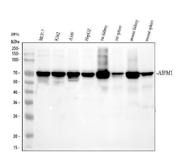
Figure 1. Western blot analysis of AIFM1 using anti-AIFM1 antibody (PA2232).
Electrophoresis was performed on a 5-20% SDS-PAGE gel at 70V (Stacking gel) / 90V (Resolving gel) for 2-3 hours. The sample well of each lane was loaded with 30 ug of sample under reducing conditions.
Lane 1: human MCF-7 whole cell lysates,
Lane 2: human K562 whole cell lysates,
Lane 3: human A549 whole cell lysates,
Lane 4: human HepG2 whole cell lysates,
Lane 5: rat kidney tissue lysates,
Lane 6: rat spleen tissue lysates,
Lane 7: mouse kidney tissue lysates,
Lane 8: mouse spleen tissue lysates.
After electrophoresis, proteins were transferred to a nitrocellulose membrane at 150 mA for 50-90 minutes. Blocked the membrane with 5% non-fat milk/TBS for 1.5 hour at RT. The membrane was incubated with rabbit anti-AIFM1 antigen affinity purified polyclonal antibody (Catalog # PA2232) at 0.5 μg/mL overnight at 4°C, then washed with TBS-0.1%Tween 3 times with 5 minutes each and probed with a goat anti-rabbit IgG-HRP secondary antibody at a dilution of 1:5000 for 1.5 hour at RT. The signal is developed using an Enhanced Chemiluminescent detection (ECL) kit (Catalog # EK1002) with Tanon 5200 system. A specific band was detected for AIFM1 at approximately 67 kDa. The expected band size for AIFM1 is at 67 kDa.

Click image to see more details
Anti-AIF antibody, PA2232, IHC(P)
IHC(P): Human Lung Cancer Tissue

Click image to see more details
Anti-AIF antibody, PA2232, IHC(P)
IHC(P): Rat Cardiac Muscle Tissue

Click image to see more details
Figure 4. IF analysis of AIFM1 using anti-AIFM1 antibody (PA2232).
AIFM1 was detected in an immunocytochemical section of MCF-7 cells. Enzyme antigen retrieval was performed using IHC enzyme antigen retrieval reagent (AR0022) for 15 mins. The cells were blocked with 10% goat serum. And then incubated with 5 μg/mL rabbit anti-AIFM1 Antibody (PA2232) overnight at 4°C. DyLight®488 Conjugated Goat Anti-Rabbit IgG (BA1127) was used as secondary antibody at 1:500 dilution and incubated for 30 minutes at 37°C. The section was counterstained with DAPI. Visualize using a fluorescence microscope and filter sets appropriate for the label used.
Protein Target Info & Infographic
Gene/Protein Information For AIFM1 (Source: Uniprot.org, NCBI)
Gene Name
AIFM1
Full Name
Apoptosis-inducing factor 1, mitochondrial
Weight
66901 MW
Superfamily
FAD-dependent oxidoreductase family
Alternative Names
Apoptosis-inducing factor 1, mitochondrial;1.1.1.-;Programmed cell death protein 8;AIFM1;AIF, PDCD8; AIFM1 AIF, AUNX1, CMT2D, CMTX4, COWCK, COXPD6, DFNX5, NADMR, NAMSD, PDCD8, SEMDHL apoptosis inducing factor mitochondria associated 1 apoptosis-inducing factor 1, mitochondrial|apoptosis-inducing factor, mitochondrion-associated, 1|auditory neuropathy, X-linked recessive 1|programmed cell death 8 (apoptosis-inducing factor)|striatal apoptosis-inducing factor|testicular secretory protein Li 4
*If product is indicated to react with multiple species, protein info is based on the gene entry specified above in "Species".For more info on AIFM1, check out the AIFM1 Infographic

We have 30,000+ of these available, one for each gene! Check them out.
In this infographic, you will see the following information for AIFM1: database IDs, superfamily, protein function, synonyms, molecular weight, chromosomal locations, tissues of expression, subcellular locations, post-translational modifications, and related diseases, research areas & pathways. If you want to see more information included, or would like to contribute to it and be acknowledged, please contact [email protected].
Specific Publications For Anti-AIF/AIFM1 Antibody (PA2232)
Hello CJ!
PA2232 has been cited in 3 publications:
*The publications in this section are manually curated by our staff scientists. They may differ from Bioz's machine gathered results. Both are accurate. If you find a publication citing this product but is missing from this list, please let us know we will issue you a thank-you coupon.
Wnt5a Increases Properties of Lung Cancer Stem Cells and Resistance to Cisplatin through Activation of Wnt5a/PKC Signaling Pathway
Mechanism of apoptosis induction in human hepatocellular carcinoma cells following treatment with a gecko peptides mixture
Feng T, Liu Y, Li C, Li Z. Cell Biochem Biophys. 2015 Jan;71(1):345-51. Doi: 10.1007/S12013-014-0204-1. Protective Effects Of Nigranoic Acid On Cerebral Ischemia-Reperfusion Injury And Its Mechanism Involving Apoptotic Signaling Pathway.
Recommended Resources
Here are featured tools and databases that you might find useful.
- Boster's Pathways Library
- Protein Databases
- Bioscience Research Protocol Resources
- Data Processing & Analysis Software
- Photo Editing Software
- Scientific Literature Resources
- Research Paper Management Tools
- Molecular Biology Software
- Primer Design Tools
- Bioinformatics Tools
- Phylogenetic Tree Analysis
Customer Reviews
Have you used Anti-AIF/AIFM1 Antibody?
Submit a review and receive an Amazon gift card.
- $30 for a review with an image
0 Reviews For Anti-AIF/AIFM1 Antibody
Customer Q&As
Have a question?
Find answers in Q&As, reviews.
Can't find your answer?
Submit your question
5 Customer Q&As for Anti-AIF/AIFM1 Antibody
Question
We have seen staining in mouse cervix carcinoma. Are there any suggestions? Is anti-AIF/AIFM1 antibody supposed to stain cervix carcinoma positively?
Verified Customer
Verified customer
Asked: 2019-12-27
Answer
From what I have seen in literature cervix carcinoma does express AIFM1. From what I have seen in Uniprot.org, AIFM1 is expressed in apex of heart, kidney, brain, cervix carcinoma, cervix carcinoma erythroleukemia, liver, among other tissues. Regarding which tissues have AIFM1 expression, here are a few articles citing expression in various tissues:
Brain, Pubmed ID: 10913597, 15489334
Cervix carcinoma, Pubmed ID: 18669648, 18691976
Cervix carcinoma, and Erythroleukemia, Pubmed ID: 23186163
Kidney, Pubmed ID: 14702039
Liver, Pubmed ID: 24275569
Boster Scientific Support
Answered: 2019-12-27
Question
We purchased anti-AIF/AIFM1 antibody for IHC on kidney a few years ago. I am using mouse, and We are going to use the antibody for WB next. I am looking for examining kidney as well as cervix carcinoma in our next experiment. Do you have any suggestion on which antibody would work the best for WB?
Verified Customer
Verified customer
Asked: 2019-12-11
Answer
I looked at the website and datasheets of our anti-AIF/AIFM1 antibody and I see that PA2232 has been validated on mouse in both IHC and WB. Thus PA2232 should work for your application. Our Boster satisfaction guarantee will cover this product for WB in mouse even if the specific tissue type has not been validated. We do have a comprehensive range of products for WB detection and you can check out our website bosterbio.com to find out more information about them.
Boster Scientific Support
Answered: 2019-12-11
Question
My boss were happy with the WB result of your anti-AIF/AIFM1 antibody. However we have seen positive staining in cervix carcinoma erythroleukemia isoform 3: mitochondrion intermembrane space using this antibody. Is that expected? Could you tell me where is AIFM1 supposed to be expressed?
M. Huang
Verified customer
Asked: 2019-09-17
Answer
From literature, cervix carcinoma erythroleukemia does express AIFM1. Generally AIFM1 expresses in mitochondrion intermembrane space., isoform 3: mitochondrion intermembrane space, isoform 5: cytoplasm. Regarding which tissues have AIFM1 expression, here are a few articles citing expression in various tissues:
Brain, Pubmed ID: 10913597, 15489334
Cervix carcinoma, Pubmed ID: 18669648, 18691976
Cervix carcinoma, and Erythroleukemia, Pubmed ID: 23186163
Kidney, Pubmed ID: 14702039
Liver, Pubmed ID: 24275569
Boster Scientific Support
Answered: 2019-09-17
Question
Our lab want to know about using your anti-AIF/AIFM1 antibody for cellular response to oxygen-glucose deprivation studies. Has this antibody been tested with western blotting on tissue lysate? We would like to see some validation images before ordering.
W. Rodriguez
Verified customer
Asked: 2019-04-29
Answer
We appreciate your inquiry. This PA2232 anti-AIF/AIFM1 antibody is validated on lung cancer tissue, tissue lysate, rat heart tissue, cardiac muscle tissue, brain tissue, k562 cell lysate, hepg2 cell lysate, a431 cell lysate, nih3t3 cell lysate. It is guaranteed to work for IHC, ICC, WB in human, mouse, rat. Our Boster guarantee will cover your intended experiment even if the sample type has not been be directly tested.
Boster Scientific Support
Answered: 2019-04-29
Question
We are currently using anti-AIF/AIFM1 antibody PA2232 for mouse tissue, and we are happy with the IHC results. The species of reactivity given in the datasheet says human, mouse, rat. Is it true that the antibody can work on feline tissues as well?
C. Miller
Verified customer
Asked: 2019-04-09
Answer
The anti-AIF/AIFM1 antibody (PA2232) has not been validated for cross reactivity specifically with feline tissues, though there is a good chance of cross reactivity. We have an innovator award program that if you test this antibody and show it works in feline you can get your next antibody for free. Please contact me if I can help you with anything.
Boster Scientific Support
Answered: 2019-04-09
