Click image to see more details
Product Info Summary
| SKU: | M00251 |
|---|---|
| Size: | 100 μl |
| Reactive Species: | Human, Mouse |
| Host: | Rabbit |
| Application: | Flow Cytometry, IHC, WB |
Customers Who Bought This Also Bought

Product info
Product Name
Anti-ACE1 Monoclonal Antibody
SKU/Catalog Number
M00251
Size
100 μl
Form
Liquid
Description
Boster Bio Anti-ACE1 Monoclonal Antibody catalog # M00251. Tested in WB, IHC, Flow Cytometry applications. This antibody reacts with Human, Mouse.
Storage & Handling
Store at -20°C for one year. For short term storage and frequent use, store at 4°C for up to one month. Avoid repeated freeze-thaw cycles.
Cite This Product
Anti-ACE1 Monoclonal Antibody (Boster Biological Technology, Pleasanton CA, USA, Catalog # M00251)
Host
Rabbit
Contents
Rabbit IgG in phosphate buffered saline, pH 7.4, 150mM NaCl, 0.02% sodium azide and 50% glycerol, 0.4-0.5mg/ml BSA.
Clonality
Monoclonal
Clone Number
ADCO-1
Isotype
Rabbit IgG
Immunogen
A synthesized peptide derived from human ACE1
*Blocking peptide can be purchased. Costs vary based on immunogen length. Contact us for pricing.
Reactive Species
M00251 is reactive to ACE in Human, Mouse
Reconstitution
Restore with deionized water (or equivalent) for reconstitution volume of 1.0 mL
Observed Molecular Weight
180 kDa
Calculated molecular weight
50 kDa
Antibody Validation
Boster validates all antibodies on WB, IHC, ICC, Immunofluorescence, and ELISA with known positive control and negative samples to ensure specificity and high affinity, including thorough antibody incubations.
Application & Images
Applications
M00251 is guaranteed for Flow Cytometry, IHC, WB Boster Guarantee
Assay Dilutions Recommendation
The recommendations below provide a starting point for assay optimization. The actual working concentration varies and should be decided by the user.
WB 1:500-1:1000
IHC 1:50-1:200
FC 1:30
Positive Control
WB: human fetal kidney, rat lung tissue, mouse lung tissue, mouse kidney tissue
Validation Images & Assay Conditions
Click image to see more details
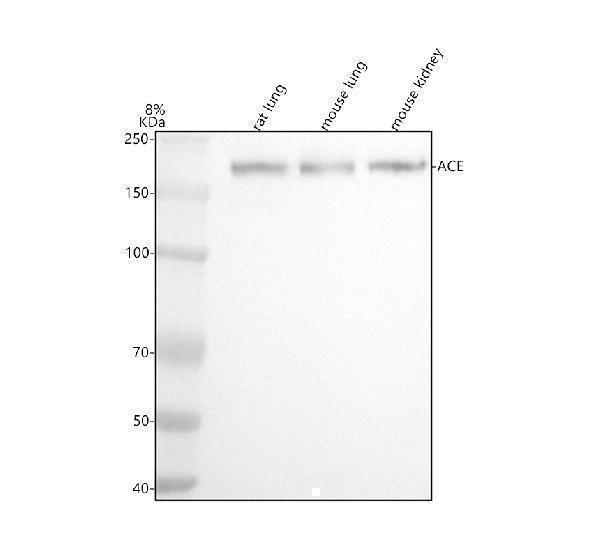
Figure 1. Western blot analysis of ACE1 using anti-ACE1 antibody (M00251).
Electrophoresis was performed on a 5-20% SDS-PAGE gel at 70V (Stacking gel) / 90V (Resolving gel) for 2-3 hours. The sample well of each lane was loaded with 30 ug of sample under reducing conditions.
Lane 1: rat lung tissue lysates,
Lane 2: mouse lung tissue lysates,
Lane 3: mouse kidney tissue lysates.
After electrophoresis, proteins were transferred to a nitrocellulose membrane at 150 mA for 50-90 minutes. Blocked the membrane with 5% non-fat milk/TBS for 1.5 hour at RT. The membrane was incubated with rabbit anti-ACE1 antigen affinity purified monoclonal antibody (Catalog # M00251) at 1:500 overnight at 4°C, then washed with TBS-0.1%Tween 3 times with 5 minutes each and probed with a goat anti-rabbit IgG-HRP secondary antibody at a dilution of 1:500 for 1.5 hour at RT. The signal is developed using an Enhanced Chemiluminescent detection (ECL) kit (Catalog # EK1002) with Tanon 5200 system. A specific band was detected for ACE1 at approximately 180 kDa. The expected band size for ACE1 is at 54 kDa.

Click image to see more details
Western blot analysis of ACE1 expression in human fetal kidney lysate.
Protein Target Info & Infographic
Gene/Protein Information For ACE (Source: Uniprot.org, NCBI)
Gene Name
ACE
Full Name
Angiotensin-converting enzyme
Weight
50 kDa
Superfamily
peptidase M2 family
Alternative Names
C-reactive protein;C-reactive protein (1-205);CRP;PTX1; ACE ACE1, CD143, DCP, DCP1 angiotensin I converting enzyme angiotensin-converting enzyme|CD143 |angiotensin I converting enzyme (peptidyl-dipeptidase A) 1|carboxycathepsin|dipeptidyl carboxypeptidase 1|dipeptidyl carboxypeptidase I|kininase II|peptidase P
*If product is indicated to react with multiple species, protein info is based on the gene entry specified above in "Species".For more info on ACE, check out the ACE Infographic

We have 30,000+ of these available, one for each gene! Check them out.
In this infographic, you will see the following information for ACE: database IDs, superfamily, protein function, synonyms, molecular weight, chromosomal locations, tissues of expression, subcellular locations, post-translational modifications, and related diseases, research areas & pathways. If you want to see more information included, or would like to contribute to it and be acknowledged, please contact [email protected].
Specific Publications For Anti-ACE1 Monoclonal Antibody (M00251)
Hello CJ!
M00251 has been cited in 1 publications:
*The publications in this section are manually curated by our staff scientists. They may differ from Bioz's machine gathered results. Both are accurate. If you find a publication citing this product but is missing from this list, please let us know we will issue you a thank-you coupon.
Telmisartan protects chronic intermittent hypoxic mice via modulating cardiac renin-angiotensin system activity
Recommended Resources
Here are featured tools and databases that you might find useful.
- Boster's Pathways Library
- Protein Databases
- Bioscience Research Protocol Resources
- Data Processing & Analysis Software
- Photo Editing Software
- Scientific Literature Resources
- Research Paper Management Tools
- Molecular Biology Software
- Primer Design Tools
- Bioinformatics Tools
- Phylogenetic Tree Analysis
Customer Reviews
Have you used Anti-ACE1 Monoclonal Antibody?
Submit a review and receive an Amazon gift card.
- $30 for a review with an image
0 Reviews For Anti-ACE1 Monoclonal Antibody
Customer Q&As
Have a question?
Find answers in Q&As, reviews.
Can't find your answer?
Submit your question
3 Customer Q&As for Anti-ACE1 Monoclonal Antibody
Question
Our team were well pleased with the WB result of your anti-ACE1 Monoclonal antibody. However we have seen positive staining in umbilical vein endothelial cell soluble form: using this antibody. Is that expected? Could you tell me where is ACE supposed to be expressed?
Verified Customer
Verified customer
Asked: 2019-12-05
Answer
Based on literature, umbilical vein endothelial cell does express ACE. Generally ACE expresses in angiotensin-converting enzyme, soluble form:, cell membrane. Regarding which tissues have ACE expression, here are a few articles citing expression in various tissues:
Brain, Pubmed ID: 16625196
Liver, Pubmed ID: 19159218
Lung, Pubmed ID: 2558109
Plasma, Pubmed ID: 16335952
Testis, Pubmed ID: 14702039
Umbilical vein endothelial cell, Pubmed ID: 9642152
Boster Scientific Support
Answered: 2019-12-05
Question
We are currently using anti-ACE1 Monoclonal antibody M00251 for mouse tissue, and we are happy with the WB results. The species of reactivity given in the datasheet says human, mouse. Is it true that the antibody can work on dog tissues as well?
Verified Customer
Verified customer
Asked: 2019-04-17
Answer
The anti-ACE1 Monoclonal antibody (M00251) has not been validated for cross reactivity specifically with dog tissues, though there is a good chance of cross reactivity. We have an innovator award program that if you test this antibody and show it works in dog you can get your next antibody for free. Please contact me if I can help you with anything.
Boster Scientific Support
Answered: 2019-04-17
Question
We have been able to see staining in mouse plasma. Any tips? Is anti-ACE1 Monoclonal antibody supposed to stain plasma positively?
V. Jackson
Verified customer
Asked: 2014-09-22
Answer
Based on literature plasma does express ACE. Based on Uniprot.org, ACE is expressed in testis, brain, lung, umbilical vein endothelial cell, plasma, liver, among other tissues. Regarding which tissues have ACE expression, here are a few articles citing expression in various tissues:
Brain, Pubmed ID: 16625196
Liver, Pubmed ID: 19159218
Lung, Pubmed ID: 2558109
Plasma, Pubmed ID: 16335952
Testis, Pubmed ID: 14702039
Umbilical vein endothelial cell, Pubmed ID: 9642152
Boster Scientific Support
Answered: 2014-09-22
