Click image to see more details
Product Info Summary
| SKU: | M00210 |
|---|---|
| Size: | 100 μl |
| Reactive Species: | Human, Mouse, Rat |
| Host: | Rabbit |
| Application: | IP, WB |
Customers Who Bought This Also Bought


Product info
Product Name
Anti-Vitamin D Receptor VDR Rabbit Monoclonal Antibody
View all VDR/NR1I1/Vitamin D Receptor Antibodies
SKU/Catalog Number
M00210
Size
100 μl
Form
Liquid
Description
Boster Bio Anti-Vitamin D Receptor VDR Rabbit Monoclonal Antibody catalog # M00210. Tested in WB, IP applications. This antibody reacts with Human, Mouse, Rat.
Storage & Handling
Store at -20°C for one year. For short term storage and frequent use, store at 4°C for up to one month. Avoid repeated freeze-thaw cycles.
Cite This Product
Anti-Vitamin D Receptor VDR Rabbit Monoclonal Antibody (Boster Biological Technology, Pleasanton CA, USA, Catalog # M00210)
Host
Rabbit
Contents
Rabbit IgG in phosphate buffered saline, pH 7.4, 150mM NaCl, 0.02% sodium azide and 50% glycerol, 0.4-0.5mg/ml BSA.
Clonality
Monoclonal
Clone Number
AAGE-22
Isotype
Rabbit IgG
Immunogen
A synthesized peptide derived from human Vitamin D Receptor
*Blocking peptide can be purchased. Costs vary based on immunogen length. Contact us for pricing.
Reactive Species
M00210 is reactive to VDR in Human, Mouse, Rat
Applications
M00210 is guaranteed for IP, WB Boster Guarantee
Observed Molecular Weight
48 kDa
Calculated molecular weight
48289 MW
Antibody Validation
Boster validates all antibodies on WB, IHC, ICC, Immunofluorescence, and ELISA with known positive control and negative samples to ensure specificity and high affinity, including thorough antibody incubations.
Assay dilution & Images
Reconstitution
Restore with deionized water (or equivalent) for reconstitution volume of 1.0 mL
Assay Dilutions Recommendation
The recommendations below provide a starting point for assay optimization. The actual working concentration varies and should be decided by the user.
WB 1:500-1:2000
IP 1:50
Validation Images & Assay Conditions
Click image to see more details
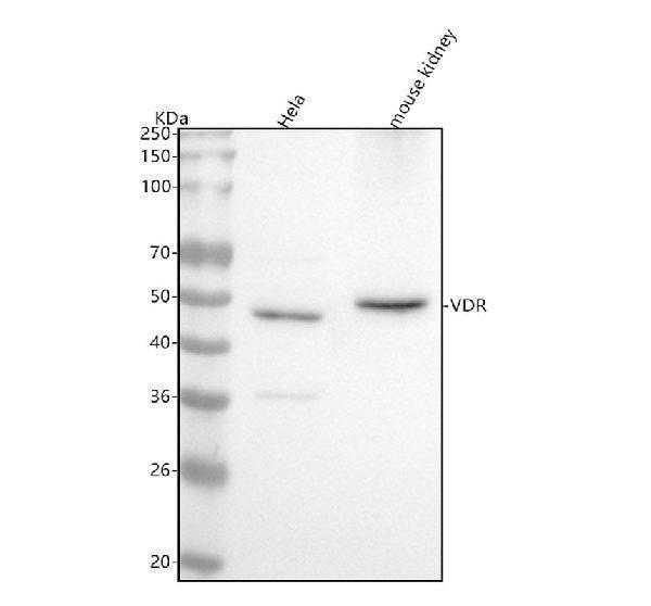
Figure 1. Western blot analysis of VDR using anti-VDR antibody (M00210).
Electrophoresis was performed on a 5-20% SDS-PAGE gel at 70V (Stacking gel) / 90V (Resolving gel) for 2-3 hours. The sample well of each lane was loaded with 30 ug of sample under reducing conditions.
Lane 1: human Hela whole cell lysates,
Lane 2: mouse kidney tissue lysates.
After electrophoresis, proteins were transferred to a nitrocellulose membrane at 150 mA for 50-90 minutes. Blocked the membrane with 5% non-fat milk/TBS for 1.5 hour at RT. The membrane was incubated with rabbit anti-VDR antigen affinity purified monoclonal antibody (Catalog # M00210) at 1:500 overnight at 4°C, then washed with TBS-0.1%Tween 3 times with 5 minutes each and probed with a goat anti-rabbit IgG-HRP secondary antibody at a dilution of 1:5000 for 1.5 hour at RT. The signal is developed using an Enhanced Chemiluminescent detection (ECL) kit (Catalog # EK1002) with Tanon 5200 system. A specific band was detected for VDR at approximately 48 kDa. The expected band size for VDR is at 48 kDa.

Click image to see more details
All lanes use the Antibody at 1:1K dilution for 1 hour at room temperature.
Protein Target Info & Infographic
Gene/Protein Information For VDR (Source: Uniprot.org, NCBI)
Gene Name
VDR
Full Name
Vitamin D3 receptor
Weight
48289 MW
Superfamily
nuclear hormone receptor family
Alternative Names
NR1I1; NR1I1Nuclear receptor subfamily 1 group I member 11,25-dihydroxyvitamin D3 receptor; VDR; vitamin D1,25- dihydroxyvitamin D3 receptor; vitamin D3 receptor VDR NR1I1, PPP1R163 vitamin D receptor vitamin D3 receptor|1,25-dihydroxyvitamin D3 receptor|nuclear receptor subfamily 1 group I member 1|protein phosphatase 1, regulatory subunit 163|vitamin D (1,25- dihydroxyvitamin D3) receptor|vitamin D nuclear receptor variant 1
*If product is indicated to react with multiple species, protein info is based on the gene entry specified above in "Species".For more info on VDR, check out the VDR Infographic

We have 30,000+ of these available, one for each gene! Check them out.
In this infographic, you will see the following information for VDR: database IDs, superfamily, protein function, synonyms, molecular weight, chromosomal locations, tissues of expression, subcellular locations, post-translational modifications, and related diseases, research areas & pathways. If you want to see more information included, or would like to contribute to it and be acknowledged, please contact [email protected].
Specific Publications For Anti-Vitamin D Receptor VDR Rabbit Monoclonal Antibody (M00210)
Hello CJ!
M00210 has been cited in 1 publications:
*The publications in this section are manually curated by our staff scientists. They may differ from Bioz's machine gathered results. Both are accurate. If you find a publication citing this product but is missing from this list, please let us know we will issue you a thank-you coupon.
Comparative Transcriptome Analysis of Fetal Skin Reveals Key Genes Related to Hair Follicle Morphogenesis in Cashmere Goats
Recommended Resources
Here are featured tools and databases that you might find useful.
- Boster's Pathways Library
- Protein Databases
- Bioscience Research Protocol Resources
- Data Processing & Analysis Software
- Photo Editing Software
- Scientific Literature Resources
- Research Paper Management Tools
- Molecular Biology Software
- Primer Design Tools
- Bioinformatics Tools
- Phylogenetic Tree Analysis
Customer Reviews
Have you used Anti-Vitamin D Receptor VDR Rabbit Monoclonal Antibody?
Submit a review and receive an Amazon gift card.
- $30 for a review with an image
0 Reviews For Anti-Vitamin D Receptor VDR Rabbit Monoclonal Antibody
Customer Q&As
Have a question?
Find answers in Q&As, reviews.
Can't find your answer?
Submit your question
4 Customer Q&As for Anti-Vitamin D Receptor VDR Rabbit Monoclonal Antibody
Question
I would like using your anti-Vitamin D Receptor Rabbit Monoclonal antibody for skeletal system development studies. Has this antibody been tested with western blotting on hela cell lysate? We would like to see some validation images before ordering.
Verified Customer
Verified customer
Asked: 2020-01-17
Answer
We appreciate your inquiry. This M00210 anti-Vitamin D Receptor Rabbit Monoclonal antibody is tested on hela cell lysate, mouse kidney. It is guaranteed to work for WB in human, mouse, rat. Our Boster guarantee will cover your intended experiment even if the sample type has not been be directly tested.
Boster Scientific Support
Answered: 2020-01-17
Question
We have observed staining in rat placenta. Are there any suggestions? Is anti-Vitamin D Receptor Rabbit Monoclonal antibody supposed to stain placenta positively?
Verified Customer
Verified customer
Asked: 2019-12-31
Answer
From what I have seen in literature placenta does express VDR. From what I have seen in Uniprot.org, VDR is expressed in tibia, lens epithelium, placenta, peripheral blood, prostate, among other tissues. Regarding which tissues have VDR expression, here are a few articles citing expression in various tissues:
Lens epithelium, Pubmed ID: 9212063
Peripheral blood, Pubmed ID: 1850412, 16252240
Placenta, Pubmed ID: 15489334
Prostate, Pubmed ID: 22323358
Boster Scientific Support
Answered: 2019-12-31
Question
My colleagues were well pleased with the WB result of your anti-Vitamin D Receptor Rabbit Monoclonal antibody. However we have observed positive staining in tibia nucleus using this antibody. Is that expected? Could you tell me where is VDR supposed to be expressed?
Verified Customer
Verified customer
Asked: 2018-04-16
Answer
From literature, tibia does express VDR. Generally VDR expresses in nucleus. Regarding which tissues have VDR expression, here are a few articles citing expression in various tissues:
Lens epithelium, Pubmed ID: 9212063
Peripheral blood, Pubmed ID: 1850412, 16252240
Placenta, Pubmed ID: 15489334
Prostate, Pubmed ID: 22323358
Boster Scientific Support
Answered: 2018-04-16
Question
We are currently using anti-Vitamin D Receptor Rabbit Monoclonal antibody M00210 for mouse tissue, and we are content with the WB results. The species of reactivity given in the datasheet says human, mouse, rat. Is it true that the antibody can work on zebrafish tissues as well?
F. Collins
Verified customer
Asked: 2015-01-23
Answer
The anti-Vitamin D Receptor Rabbit Monoclonal antibody (M00210) has not been tested for cross reactivity specifically with zebrafish tissues, but there is a good chance of cross reactivity. We have an innovator award program that if you test this antibody and show it works in zebrafish you can get your next antibody for free. Please contact me if I can help you with anything.
Boster Scientific Support
Answered: 2015-01-23
