Click image to see more details
-

-
-
-
-
+1
Product Info Summary
| SKU: | PA1957 |
|---|---|
| Size: | 100 μg/vial |
| Reactive Species: | Human, Mouse, Rat |
| Host: | Rabbit |
| Application: | Flow Cytometry, IF, IHC, IHC-F, ICC, WB |
Customers Who Bought This Also Bought


Product info
Product Name
Anti-Tight junction protein ZO-2 TJP2 Antibody
View all Tight Junction Protein 2 Antibodies
SKU/Catalog Number
PA1957
Size
100 μg/vial
Form
Lyophilized
Description
Boster Bio Anti-Tight junction protein ZO-2 TJP2 Antibody catalog # PA1957. Tested in Flow Cytometry, IF, IHC, IHC-F, ICC, WB applications. This antibody reacts with Human, Mouse, Rat.
Storage & Handling
Store at -20˚C for one year from date of receipt. After reconstitution, at 4˚C for one month. It can also be aliquotted and stored frozen at -20˚C for six months. Avoid repeated freeze-thaw cycles.
Cite This Product
Anti-Tight junction protein ZO-2 TJP2 Antibody (Boster Biological Technology, Pleasanton CA, USA, Catalog # PA1957)
Host
Rabbit
Contents
Each vial contains 5mg BSA, 0.9mg NaCl, 0.2mg Na2HPO4, 0.05mg Thimerosal, 0.01mg NaN3.
Clonality
Polyclonal
Isotype
Rabbit IgG
Immunogen
A synthetic peptide corresponding to a sequence at the C-terminus of human TJP2.
*Blocking peptide can be purchased. Costs vary based on immunogen length. Contact us for pricing.
Cross-reactivity
No cross-reactivity with other proteins
Reactive Species
PA1957 is reactive to TJP2 in Human, Mouse, Rat
Applications
PA1957 is guaranteed for Flow Cytometry, IF, IHC, IHC-F, ICC, WB Boster Guarantee
Observed Molecular Weight
60 kDa
Calculated molecular weight
133958 MW
Background of Tight Junction Protein 2
TJP2 (Tight Junction Protein 2), also known as Zona Occludens 2 or ZO2 is a protein that in humans is encoded by the TJP2 gene. Tight junction proteins (TJPs) belong to a family of membrane-associated guanylate kinase (MAGUK) homologs that are involved in the organization of epithelial and endothelial intercellular junctions. Duclos et al. (1994) mapped the TJP2 gene telomeric to the Friedreich ataxia critical region on chromosome 9q13-q21. TJP2 lies about 70 kb centromeric to the X123 gene and is transcribed in the centromere-to-telomere direction. Using in vitro assays and immunoprecipitation studies, Itoh et al. (1999) showed that the mouse Tjp1, Tjp2, and Tjp3 PDZ1 domains interacted with the C-terminal cytoplasmic domains of Cldn1 through Cldn8. In the mouse inner ear, Walsh et al. (2010) found that Tjp2 expression decreased rapidly between E16.5 and age 1 week to a level in adult mice that was approximately 50% of the level at birth (P0).
Antibody Validation
Boster validates all antibodies on WB, IHC, ICC, Immunofluorescence, and ELISA with known positive control and negative samples to ensure specificity and high affinity, including thorough antibody incubations.
Assay dilution & Images
Reconstitution
Add 0.2ml of distilled water will yield a concentration of 500ug/ml.
Assay Dilutions Recommendation
The recommendations below provide a starting point for assay optimization. The actual working concentration varies and should be decided by the user.
Western blot, 0.1-0.5μg/ml, Human, Mouse, Rat
Immunohistochemistry (Paraffin-embedded Section), 0.5-1μg/ml, Human, By Heat
Immunohistochemistry (Frozen Section), 0.5-1μg/ml, Human, -
Immunocytochemistry/Immunofluorescence, 5μg/ml, Human
Flow Cytometry (Fixed), 1-3μg/1x106 cells, Human
Validation Images & Assay Conditions
Click image to see more details
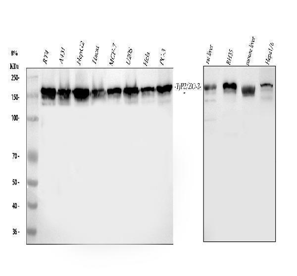
Figure 1. Western blot analysis of TJP2 using anti-TJP2 antibody (PA1957).
Electrophoresis was performed on a 5-20% SDS-PAGE gel at 70V (Stacking gel) / 90V (Resolving gel) for 2-3 hours. The sample well of each lane was loaded with 30 ug of sample under reducing conditions.
Lane 1: human RT4 whole cell lysates,
Lane 2: human A431 whole cell lysates,
Lane 3: human HepG2 whole cell lysates,
Lane 4: human Hacat whole cell lysates,
Lane 5: human MCF-7 whole cell lysates,
Lane 6: human U20S whole cell lysates,
Lane 7: human Hela whole cell lysates,
Lane 8: human PC-3 whole cell lysates,
Lane 9: rat liver tissue lysates,
Lane 10: rat RH35 whole cell lysates,
Lane 11: mouse liver tissue lysates,
Lane 12: mouse HEPA1-6 whole cell lysates.
After electrophoresis, proteins were transferred to a nitrocellulose membrane at 150 mA for 50-90 minutes. Blocked the membrane with 5% non-fat milk/TBS for 1.5 hour at RT. The membrane was incubated with rabbit anti-TJP2 antigen affinity purified polyclonal antibody (Catalog # PA1957) at 0.5 μg/mL overnight at 4°C, then washed with TBS-0.1%Tween 3 times with 5 minutes each and probed with a goat anti-rabbit IgG-HRP secondary antibody at a dilution of 1:5000 for 1.5 hour at RT. The signal is developed using an Enhanced Chemiluminescent detection (ECL) kit (Catalog # EK1002) with Tanon 5200 system. A specific band was detected for TJP2 at approximately 150 kDa. The expected band size for TJP2 is at 134 kDa.

Click image to see more details
Figure 2. IHC analysis of TJP2 using anti-TJP2 antibody (PA1957).
TJP2 was detected in a paraffin-embedded section of Human Intestinal Cancer tissue. Heat mediated antigen retrieval was performed in EDTA buffer (pH 8.0, epitope retrieval solution). The tissue section was blocked with 10% goat serum. The tissue section was then incubated with 1 μg/ml rabbit anti-TJP2 Antibody (PA1957) overnight at 4°C. Peroxidase Conjugated Goat Anti-rabbit IgG was used as secondary antibody and incubated for 30 minutes at 37°C. The tissue section was developed using HRP Conjugated Rabbit IgG Super Vision Assay Kit (Catalog # SV0002) with DAB as the chromogen.

Click image to see more details
Figure 3. IF analysis of TJP2 using anti-TJP2 antibody (PA1957).
TJP2 was detected in immunocytochemical section of A431 cells. Enzyme antigen retrieval was performed using IHC enzyme antigen retrieval reagent (AR0022) for 15 mins. The cells were blocked with 10% goat serum. And then incubated with 5μg/mL rabbit anti-TJP2 Antibody (PA1957) overnight at 4°C. DyLight®488 Conjugated Goat Anti-Rabbit IgG (BA1127) was used as secondary antibody at 1:100 dilution and incubated for 30 minutes at 37°C. The section was counterstained with DAPI. Visualize using a fluorescence microscope and filter sets appropriate for the label used.

Click image to see more details
Figure 4. IF analysis of TJP2 using anti-TJP2 antibody (PA1957).
TJP2 was detected in immunocytochemical section of U2OS cells. Enzyme antigen retrieval was performed using IHC enzyme antigen retrieval reagent (AR0022) for 15 mins. The cells were blocked with 10% goat serum. And then incubated with 5μg/mL rabbit anti-TJP2 Antibody (PA1957) overnight at 4°C. DyLight®488 Conjugated Goat Anti-Rabbit IgG (BA1127) was used as secondary antibody at 1:500 dilution and incubated for 30 minutes at 37°C. The section was counterstained with DAPI. Visualize using a fluorescence microscope and filter sets appropriate for the label used.

Click image to see more details
Figure 5. Flow Cytometry analysis of MCF-7 cells using anti-TJP2 antibody (PA1957).
Overlay histogram showing MCF-7 cells stained with PA1957 (Blue line). To facilitate intracellular staining, cells were fixed with 4% paraformaldehyde and permeabilized with permeabilization buffer. The cells were blocked with 10% normal goat serum. And then incubated with rabbit anti-TJP2 Antibody (PA1957, 1μg/1x106 cells) for 30 min at 20°C. DyLight®488 conjugated goat anti-rabbit IgG (BA1127, 5-10μg/1x106 cells) was used as secondary antibody for 30 minutes at 20°C. Isotype control antibody (Green line) was rabbit IgG (1μg/1x106) used under the same conditions. Unlabelled sample without incubation with primary antibody and secondary antibody (Red line) was used as a blank control.
Protein Target Info & Infographic
Gene/Protein Information For TJP2 (Source: Uniprot.org, NCBI)
Gene Name
TJP2
Full Name
Tight junction protein ZO-2
Weight
133958 MW
Superfamily
MAGUK family
Alternative Names
C9DUPq21.11; DFNA51; DUP9q21.11; MGC26306; tight junction protein 2 (zona occludens 2); Tight junction protein 2; TJP2; X104; X104tight junction protein ZO-2; ZO2; ZO-2; ZO2Friedreich ataxia region gene X104 (tight junction protein ZO-2); Zona occludens protein 2; Zonula occludens protein 2 TJP2 C9DUPq21.11, DFNA51, DUP9q21.11, PFIC4, X104, ZO2 tight junction protein 2 tight junction protein ZO-2|Friedreich ataxia region gene X104 (tight junction protein ZO-2)|zona occludens 2|zonula occludens protein 2
*If product is indicated to react with multiple species, protein info is based on the gene entry specified above in "Species".For more info on TJP2, check out the TJP2 Infographic

We have 30,000+ of these available, one for each gene! Check them out.
In this infographic, you will see the following information for TJP2: database IDs, superfamily, protein function, synonyms, molecular weight, chromosomal locations, tissues of expression, subcellular locations, post-translational modifications, and related diseases, research areas & pathways. If you want to see more information included, or would like to contribute to it and be acknowledged, please contact [email protected].
Specific Publications For Anti-Tight junction protein ZO-2 TJP2 Antibody (PA1957)
Hello CJ!
No publications found for PA1957
*Do you have publications using this product? Share with us and receive a reward. Ask us for more details.
Recommended Resources
Here are featured tools and databases that you might find useful.
- Boster's Pathways Library
- Protein Databases
- Bioscience Research Protocol Resources
- Data Processing & Analysis Software
- Photo Editing Software
- Scientific Literature Resources
- Research Paper Management Tools
- Molecular Biology Software
- Primer Design Tools
- Bioinformatics Tools
- Phylogenetic Tree Analysis
Customer Reviews
Have you used Anti-Tight junction protein ZO-2 TJP2 Antibody?
Submit a review and receive an Amazon gift card.
- $30 for a review with an image
0 Reviews For Anti-Tight junction protein ZO-2 TJP2 Antibody
Customer Q&As
Have a question?
Find answers in Q&As, reviews.
Can't find your answer?
Submit your question
3 Customer Q&As for Anti-Tight junction protein ZO-2 TJP2 Antibody
Question
Is there a BSA free version of anti-TJP2 antibody PA1957 available?
Verified Customer
Verified customer
Asked: 2019-11-25
Answer
I appreciate your recent telephone inquiry. I can confirm that some lots of this anti-TJP2 antibody PA1957 are BSA free. For now, these lots are available and we can make a BSA free formula for you free of charge. It will take 3 extra days to prepare. If you require this antibody BSA free again in future, please do not hesitate to contact me and I will be pleased to check which lots we have in stock that are BSA free.
Boster Scientific Support
Answered: 2019-11-25
Question
you antibody to test anti-TJP2 antibody PA1957 on human cervix carcinoma erythroleukemia for research purposes, then I may be interested in using anti-TJP2 antibody PA1957 for diagnostic purposes as well. Is the antibody suitable for diagnostic purposes?
Verified Customer
Verified customer
Asked: 2019-10-03
Answer
The products we sell, including anti-TJP2 antibody PA1957, are only intended for research use. They would not be suitable for use in diagnostic work. If you have the means to develop a product into diagnostic use, and are interested in collaborating with us and develop our product into an IVD product, please contact us for more discussions.
Boster Scientific Support
Answered: 2019-10-03
Question
I was wanting to use your anti-TJP2 antibody for IHC for human cervix carcinoma erythroleukemia on frozen tissues, but I want to know if it has been validated for this particular application. Has this antibody been validated and is this antibody a good choice for human cervix carcinoma erythroleukemia identification?
Verified Customer
Verified customer
Asked: 2017-10-27
Answer
You can see on the product datasheet, PA1957 anti-TJP2 antibody has been tested for IHC, WB on human tissues. We have an innovator award program that if you test this antibody and show it works in human cervix carcinoma erythroleukemia in IHC-frozen, you can get your next antibody for free.
Boster Scientific Support
Answered: 2017-10-27
