Click image to see more details
-

-
-
-
-
+1
Product Info Summary
| SKU: | M03133-2 |
|---|---|
| Size: | 100 μg/vial |
| Reactive Species: | Human |
| Host: | Mouse |
| Application: | Flow Cytometry, IF, IHC, ICC, WB |
Customers Who Bought This Also Bought


Product info
Product Name
Anti-TIF1 gamma/TRIM33 Antibody Picoband® (monoclonal, 8I8)
View all TIF1 gamma Antibodies
SKU/Catalog Number
M03133-2
Size
100 μg/vial
Form
Lyophilized
Description
Boster Bio Anti-TIF1 gamma/TRIM33 Antibody Picoband® (monoclonal, 8I8) catalog # M03133-2. Tested in Flow Cytometry, IF, IHC, ICC, WB applications. This antibody reacts with Human. The brand Picoband indicates this is a premium antibody that guarantees superior quality, high affinity, and strong signals with minimal background in Western blot applications. Only our best-performing antibodies are designated as Picoband, ensuring unmatched performance.
Storage & Handling
Store at -20˚C for one year from date of receipt. After reconstitution, at 4˚C for one month. It can also be aliquotted and stored frozen at -20˚C for six months. Avoid repeated freeze-thaw cycles.
Cite This Product
Anti-TIF1 gamma/TRIM33 Antibody Picoband® (monoclonal, 8I8) (Boster Biological Technology, Pleasanton CA, USA, Catalog # M03133-2)
Host
Mouse
Contents
Each vial contains 4mg Trehalose, 0.9mg NaCl, 0.2mg Na2HPO4, 0.01mg NaN3.
Clonality
Monoclonal
Clone Number
8I8
Isotype
Mouse IgG2b
Immunogen
E.coli-derived human TIF1 gamma recombinant protein (Position: M1001-K1127). Human TIF1 gamma shares 96.1% amino acid (aa) sequence identity with mouse TIF1 gamma.
*Blocking peptide can be purchased. Costs vary based on immunogen length. Contact us for pricing.
Cross-reactivity
No cross-reactivity with other proteins.
Reactive Species
M03133-2 is reactive to TRIM33 in Human
Reconstitution
Add 0.2ml of distilled water will yield a concentration of 500ug/ml.
Observed Molecular Weight
150 kDa
Calculated molecular weight
122.533kDa
Background of TIF1 gamma
Tripartite motif-containing 33 (TRIM33), also known as transcriptional intermediary factor 1 gamma (TIF1-γ), is a human gene. The TRIM33 gene is mapped to chromosome 1p13 by FISH. The protein encoded by this gene is thought to be a transcriptional corepressor. However, molecules that interact with this protein have not yet been identified. The protein is a member of the tripartite motif family. This motif includes three zinc-binding domains, a RING, a B-box type 1 and a B-box type 2, and a coiled-coil region. Three alternatively spliced transcript variants for this gene have been described; however, the full-length nature of one variant has not been determined.
Antibody Validation
Boster validates all antibodies on WB, IHC, ICC, Immunofluorescence, and ELISA with known positive control and negative samples to ensure specificity and high affinity, including thorough antibody incubations.
Application & Images
Applications
M03133-2 is guaranteed for Flow Cytometry, IF, IHC, ICC, WB Boster Guarantee
Assay Dilutions Recommendation
The recommendations below provide a starting point for assay optimization. The actual working concentration varies and should be decided by the user.
Western blot, 0.1-0.5μg/ml, Human
Immunohistochemistry (Paraffin-embedded Section), 0.5-1μg/ml, Human
Immunocytochemistry/Immunofluorescence, 4μg/ml, Human
Flow Cytometry (Fixed), 1-3μg/1x106 cells, Human
Positive Control
WB: human MCF-7 whole cell, human SW620 whole cell, human K562 whole cell
IHC: human mammary cancer tissue, human rectal cancer tissue
ICC/IF: Hela cell
FCM: HL-60 cell
Validation Images & Assay Conditions
Click image to see more details
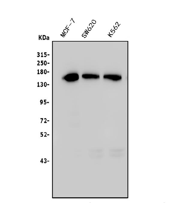
Figure 1. Western blot analysis of TIF1 gamma/TRIM33 using anti-TIF1 gamma/TRIM33 antibody (M03133-2).
Electrophoresis was performed on a 5-20% SDS-PAGE gel at 70V (Stacking gel) / 90V (Resolving gel) for 2-3 hours. The sample well of each lane was loaded with 50ug of sample under reducing conditions.
Lane 1: human MCF-7 whole cell lysates,
Lane 2: human SW620 whole cell lysates,
Lane 3: human K562 whole cell lysates.
After Electrophoresis, proteins were transferred to a Nitrocellulose membrane at 150mA for 50-90 minutes. Blocked the membrane with 5% Non-fat Milk/ TBS for 1.5 hour at RT. The membrane was incubated with mouse anti-TIF1 gamma/TRIM33 antigen affinity purified monoclonal antibody (Catalog # M03133-2) at 0.5 μg/mL overnight at 4°C, then washed with TBS-0.1%Tween 3 times with 5 minutes each and probed with a goat anti-mouse IgG-HRP secondary antibody at a dilution of 1:10000 for 1.5 hour at RT. The signal is developed using an Enhanced Chemiluminescent detection (ECL) kit (Catalog # EK1001) with Tanon 5200 system. A specific band was detected for TIF1 gamma/TRIM33 at approximately 150KD. The expected band size for TIF1 gamma/TRIM33 is at 150KD.

Click image to see more details
Figure 2. IHC analysis of TIF1 gamma/TRIM33 using anti TIF1 gamma/TRIM33 antibody (M03133-2).
TIF1 gamma/TRIM33 was detected in paraffin-embedded section of human mammary cancer tissue. Heat mediated antigen retrieval was performed in EDTA buffer (pH8.0, epitope retrieval solution). The tissue section was blocked with 10% goat serum. The tissue section was then incubated with 1μg/ml mouse anti-TIF1 gamma/TRIM33 Antibody (M03133-2) overnight at 4°C. Biotinylated goat anti-mouse IgG was used as secondary antibody and incubated for 30 minutes at 37°C. The tissue section was developed using Strepavidin-Biotin-Complex (SABC) (Catalog # SA1021) with DAB as the chromogen.

Click image to see more details
Figure 3. IHC analysis of TIF1 gamma/TRIM33 using anti TIF1 gamma/TRIM33 antibody (M03133-2).
TIF1 gamma/TRIM33 was detected in paraffin-embedded section of human rectal cancer tissue. Heat mediated antigen retrieval was performed in EDTA buffer (pH8.0, epitope retrieval solution). The tissue section was blocked with 10% goat serum. The tissue section was then incubated with 1μg/ml mouse anti-TIF1 gamma/TRIM33 Antibody (M03133-2) overnight at 4°C. Biotinylated goat anti-mouse IgG was used as secondary antibody and incubated for 30 minutes at 37°C. The tissue section was developed using Strepavidin-Biotin-Complex (SABC) (Catalog # SA1021) with DAB as the chromogen.

Click image to see more details
Figure 4. IF analysis of TIF1 gamma/TRIM33 using anti-TIF1 gamma/TRIM33 antibody (M03133-2).
TIF1 gamma/TRIM33 was detected in immunocytochemical section of Hela cells. Enzyme antigen retrieval was performed using IHC enzyme antigen retrieval reagent (AR0022) for 15 mins. The cells were blocked with 10% goat serum. And then incubated with 4μg/mL mouse anti-TIF1 gamma/TRIM33 Antibody (M03133-2) overnight at 4°C. DyLight®488 Conjugated Goat Anti-Mouse IgG (BA1126) was used as secondary antibody at 1:100 dilution and incubated for 30 minutes at 37°C. The section was counterstained with DAPI. Visualize using a fluorescence microscope and filter sets appropriate for the label used.

Click image to see more details
Figure 5. Flow Cytometry analysis of HL-60 cells using anti- TIF1 gamma/TRIM33 antibody (M03133-2).
Overlay histogram showing HL-60 cells stained with M03133-2 (Blue line). To facilitate intracellular staining, cells were fixed with 4% paraformaldehyde and permeabilized with permeabilization buffer. The cells were blocked with 10% normal goat serum. And then incubated with mouse anti-TIF1 gamma/TRIM33 Antibody (M03133-2, 1μg/1x106 cells) for 30 min at 20°C. DyLight®488 conjugated goat anti-mouse IgG (BA1126, 5-10μg/1x106 cells) was used as secondary antibody for 30 minutes at 20°C. Isotype control antibody (Green line) was mouse IgG (1μg/1x106) used under the same conditions. Unlabelled sample without incubation with primary antibody and secondary antibody (Red line) was used as a blank control.
Protein Target Info & Infographic
Gene/Protein Information For TRIM33 (Source: Uniprot.org, NCBI)
Gene Name
TRIM33
Full Name
E3 ubiquitin-protein ligase TRIM33
Weight
122.533kDa
Superfamily
TRIM/RBCC family
Alternative Names
"chromosomal protein, Nonhistone, HMG4 antibody|High mobility group (nonhistone chromosomal) protein 4 antibody|High mobility group box 3 antibody|High mobility group protein 2a antibody|High mobility group protein 4 antibody|High mobility group protein B3 antibody| High mobility group protein HMG4 antibody|HMG 4 antibody|HMG-2a antibody|HMG-4 antibody|HMG2A antibody|HMGB 3 antibody|HMGB3 antibody| HMGB3_HUMAN antibody|MGC90319 antibody|Non histone chromosomal protein antibody|Nonhistone chromosomal protein HMG4 antibody" TRIM33 ECTO, PTC7, RFG7, TF1G, TIF1G, TIF1GAMMA, TIFGAMMA tripartite motif containing 33 E3 ubiquitin-protein ligase TRIM33|RET-fused gene 7 protein|RING-type E3 ubiquitin transferase TRIM33|TIF1-gamma|ectodermin homolog|protein Rfg7|transcriptional intermediary factor 1 gamma
*If product is indicated to react with multiple species, protein info is based on the gene entry specified above in "Species".For more info on TRIM33, check out the TRIM33 Infographic

We have 30,000+ of these available, one for each gene! Check them out.
In this infographic, you will see the following information for TRIM33: database IDs, superfamily, protein function, synonyms, molecular weight, chromosomal locations, tissues of expression, subcellular locations, post-translational modifications, and related diseases, research areas & pathways. If you want to see more information included, or would like to contribute to it and be acknowledged, please contact [email protected].
Specific Publications For Anti-TIF1 gamma/TRIM33 Antibody Picoband® (monoclonal, 8I8) (M03133-2)
Hello CJ!
No publications found for M03133-2
*Do you have publications using this product? Share with us and receive a reward. Ask us for more details.
Recommended Resources
Here are featured tools and databases that you might find useful.
- Boster's Pathways Library
- Protein Databases
- Bioscience Research Protocol Resources
- Data Processing & Analysis Software
- Photo Editing Software
- Scientific Literature Resources
- Research Paper Management Tools
- Molecular Biology Software
- Primer Design Tools
- Bioinformatics Tools
- Phylogenetic Tree Analysis
Customer Reviews
Have you used Anti-TIF1 gamma/TRIM33 Antibody Picoband® (monoclonal, 8I8)?
Submit a review and receive an Amazon gift card.
- $30 for a review with an image
0 Reviews For Anti-TIF1 gamma/TRIM33 Antibody Picoband® (monoclonal, 8I8)
Customer Q&As
Have a question?
Find answers in Q&As, reviews.
Can't find your answer?
Submit your question
