Click image to see more details
-

-
-
-
-
+3
Product Info Summary
| SKU: | A03223-2 |
|---|---|
| Size: | 100 µg/vial |
| Reactive Species: | Human, Mouse, Rat |
| Host: | Rabbit |
| Application: | ELISA, Flow Cytometry, IF, IHC, ICC, WB |
Customers Who Bought This Also Bought

Product info
Product Name
Anti-PRDM2 Antibody Picoband®
SKU/Catalog Number
A03223-2
Size
100 µg/vial
Form
Lyophilized
Description
Boster Bio Anti-PRDM2 Antibody Picoband® catalog # A03223-2. Tested in ELISA, IF, IHC, ICC, WB, Flow Cytometry applications. This antibody reacts with Human, Mouse, Rat. The brand Picoband indicates this is a premium antibody that guarantees superior quality, high affinity, and strong signals with minimal background in Western blot applications. Only our best-performing antibodies are designated as Picoband, ensuring unmatched performance.
Storage & Handling
At -20°C for one year from date of receipt. After reconstitution, at 4°C for one month. It can also be aliquotted and stored frozen at -20°C for six months. Avoid repeated freezing and thawing.
Cite This Product
Anti-PRDM2 Antibody Picoband® (Boster Biological Technology, Pleasanton CA, USA, Catalog # A03223-2)
Host
Rabbit
Contents
Each vial contains 4 mg Trehalose, 0.9 mg NaCl, 0.2 mg Na2HPO4.
Clonality
Polyclonal
Isotype
Rabbit IgG
Immunogen
E.coli-derived human PRDM2 recombinant protein (Position: M1-Q1378).
*Blocking peptide can be purchased. Costs vary based on immunogen length. Contact us for pricing.
Cross-reactivity
No cross-reactivity with other proteins.
Reactive Species
A03223-2 is reactive to PRDM2 in Human, Mouse, Rat
Reconstitution
Adding 0.2 ml of distilled water will yield a concentration of 500 µg/ml.
Observed Molecular Weight
280 kDa
Calculated molecular weight
37836 MW
Background of PRDM2
PR domain zinc finger protein 2 is a protein that in humans is encoded by the PRDM2 gene. This tumor suppressor gene is a member of a nuclear histone/protein methyltransferase superfamily. It encodes a zinc finger protein that can bind to retinoblastoma protein, estrogen receptor, and the TPA-responsive element (MTE) of the heme-oxygenase-1 gene. Although the functions of this protein have not been fully characterized, it may (1) play a role in transcriptional regulation during neuronal differentiation and pathogenesis of retinoblastoma, (2) act as a transcriptional activator of the heme-oxygenase-1 gene, and (3) be a specific effector of estrogen action. Multiple transcript variants encoding different isoforms have been found for this gene.
Antibody Validation
Boster validates all antibodies on WB, IHC, ICC, Immunofluorescence, and ELISA with known positive control and negative samples to ensure specificity and high affinity, including thorough antibody incubations.
Application & Images
Applications
A03223-2 is guaranteed for ELISA, Flow Cytometry, IF, IHC, ICC, WB Boster Guarantee
Assay Dilutions Recommendation
The recommendations below provide a starting point for assay optimization. The actual working concentration varies and should be decided by the user.
Western blot, 0.25-0.5 µg/ml, Human
Immunohistochemistry(Paraffin-embedded Section), 2-5 µg/ml, Human, Mouse, Rat
Immunocytochemistry/Immunofluorescence, 5 µg/ml, Human
Flow Cytometry (Fixed), 1-3 µg/1x106 cells, Human
ELISA, 0.1-0.5 µg/ml, Human
Positive Control
WB: human Hela whole cell, human K562 whole cell
IHC: human rectum adenocarcinoma tissue, human testicular seminoma tissue, mouse testis tissue, rat testis tissue
ICC/IF: U2OS cell
FCM: HepG2 cell
Validation Images & Assay Conditions
Click image to see more details
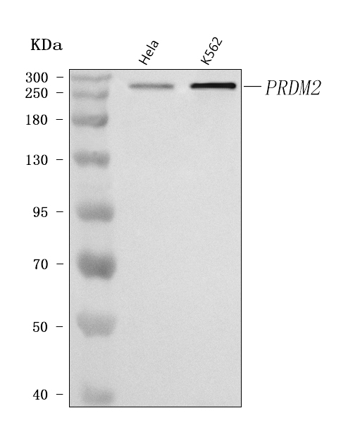
Figure 1. Western blot analysis of PRDM2 using anti-PRDM2 antibody (A03223-2).
Electrophoresis was performed on a 5-20% SDS-PAGE gel at 70V (Stacking gel) / 90V (Resolving gel) for 2-3 hours. The sample well of each lane was loaded with 30 ug of sample under reducing conditions.
Lane 1: human Hela whole cell lysates,
Lane 2: human K562 whole cell lysates.
After electrophoresis, proteins were transferred to a nitrocellulose membrane at 150 mA for 50-90 minutes. Blocked the membrane with 5% non-fat milk/TBS for 1.5 hour at RT. The membrane was incubated with rabbit anti-PRDM2 antigen affinity purified polyclonal antibody (Catalog # A03223-2) at 0.5 μg/mL overnight at 4°C, then washed with TBS-0.1%Tween 3 times with 5 minutes each and probed with a goat anti-rabbit IgG-HRP secondary antibody at a dilution of 1:5000 for 1.5 hour at RT. The signal is developed using an Enhanced Chemiluminescent detection (ECL) kit (Catalog # EK1002) with Tanon 5200 system. A specific band was detected for PRDM2 at approximately 280 kDa. The expected band size for PRDM2 is at 189,250-280 kDa.

Click image to see more details
Figure 2. IHC analysis of PRDM2 using anti-PRDM2 antibody (A03223-2).
PRDM2 was detected in a paraffin-embedded section of human rectum adenocarcinoma tissue. Heat mediated antigen retrieval was performed in EDTA buffer (pH 8.0, epitope retrieval solution). The tissue section was blocked with 10% goat serum. The tissue section was then incubated with 2 μg/ml rabbit anti-PRDM2 Antibody (A03223-2) overnight at 4°C. Peroxidase Conjugated Goat Anti-rabbit IgG was used as secondary antibody and incubated for 30 minutes at 37°C. The tissue section was developed using HRP Conjugated Rabbit IgG Super Vision Assay Kit (Catalog # SV0002) with DAB as the chromogen.

Click image to see more details
Figure 3. IHC analysis of PRDM2 using anti-PRDM2 antibody (A03223-2).
PRDM2 was detected in a paraffin-embedded section of human testicular seminoma tissue. Heat mediated antigen retrieval was performed in EDTA buffer (pH 8.0, epitope retrieval solution). The tissue section was blocked with 10% goat serum. The tissue section was then incubated with 2 μg/ml rabbit anti-PRDM2 Antibody (A03223-2) overnight at 4°C. Peroxidase Conjugated Goat Anti-rabbit IgG was used as secondary antibody and incubated for 30 minutes at 37°C. The tissue section was developed using HRP Conjugated Rabbit IgG Super Vision Assay Kit (Catalog # SV0002) with DAB as the chromogen.

Click image to see more details
Figure 4. IHC analysis of PRDM2 using anti-PRDM2 antibody (A03223-2).
PRDM2 was detected in a paraffin-embedded section of mouse testis tissue. Heat mediated antigen retrieval was performed in EDTA buffer (pH 8.0, epitope retrieval solution). The tissue section was blocked with 10% goat serum. The tissue section was then incubated with 2 μg/ml rabbit anti-PRDM2 Antibody (A03223-2) overnight at 4°C. Peroxidase Conjugated Goat Anti-rabbit IgG was used as secondary antibody and incubated for 30 minutes at 37°C. The tissue section was developed using HRP Conjugated Rabbit IgG Super Vision Assay Kit (Catalog # SV0002) with DAB as the chromogen.

Click image to see more details
Figure 5. IHC analysis of PRDM2 using anti-PRDM2 antibody (A03223-2).
PRDM2 was detected in a paraffin-embedded section of rat testis tissue. Heat mediated antigen retrieval was performed in EDTA buffer (pH 8.0, epitope retrieval solution). The tissue section was blocked with 10% goat serum. The tissue section was then incubated with 2 μg/ml rabbit anti-PRDM2 Antibody (A03223-2) overnight at 4°C. Peroxidase Conjugated Goat Anti-rabbit IgG was used as secondary antibody and incubated for 30 minutes at 37°C. The tissue section was developed using HRP Conjugated Rabbit IgG Super Vision Assay Kit (Catalog # SV0002) with DAB as the chromogen.

Click image to see more details
Figure 6. IF analysis of PRDM2 using anti-PRDM2 antibody (A03223-2) and anti-Beta Tubulin antibody (M01857-3).
PRDM2 was detected in immunocytochemical section of U2OS cell. Enzyme antigen retrieval was performed using IHC enzyme antigen retrieval reagent (AR0022) for 15 mins. The cells were blocked with 10% goat serum. And then incubated with 5 μg/mL rabbit anti-PRDM2 Antibody (A03223-2) and mouse anti-Beta Tubulin antibody (M01857-3) overnight at 4°C. Cy3 Conjugated Goat Anti-Rabbit IgG (BA1032) and DyLight®488 Conjugated Goat Anti-Mouse IgG (BA1126) were used as secondary antibody at 1:500 dilution and incubated for 30 minutes at 37°C. Visualize using a fluorescence microscope and filter sets appropriate for the label used.

Click image to see more details
Figure 7. Flow Cytometry analysis of HepG2 cells using anti-PRDM2 antibody (A03223-2).
Overlay histogram showing HepG2 cells stained with A03223-2 (Blue line). To facilitate intracellular staining, cells were fixed with 4% paraformaldehyde and permeabilized with permeabilization buffer. The cells were blocked with 10% normal goat serum. And then incubated with rabbit anti-PRDM2 Antibody (A03223-2, 1 μg/1x106 cells) for 30 min at 20°C. DyLight®488 conjugated goat anti-rabbit IgG (BA1127, 5-10 μg/1x106 cells) was used as secondary antibody for 30 minutes at 20°C. Isotype control antibody (Green line) was rabbit IgG (1 μg/1x106) used under the same conditions. Unlabelled sample (Red line) was also used as a control.
Protein Target Info & Infographic
Gene/Protein Information For PRDM2 (Source: Uniprot.org, NCBI)
Gene Name
PRDM2
Full Name
PR domain zinc finger protein 2
Weight
37836 MW
Superfamily
class V-like SAM-binding methyltransferase superfamily
Alternative Names
Thrombopoietin; C-mpl ligand; ML; Megakaryocyte colony-stimulating factor; Megakaryocyte growth and development factor; MGDF; Myeloproliferative leukemia virus oncogene ligand; THPO; MGDF PRDM2 HUMHOXY1, KMT8, KMT8A, MTB-ZF, RIZ, RIZ1, RIZ2 PR/SET domain 2 PR domain zinc finger protein 2|GATA-3 binding protein G3B|MTE-binding protein|PR domain 2|PR domain containing 2, with ZNF domain|PR domain-containing protein 2|lysine N-methyltransferase 8|retinoblastoma protein-binding zinc finger protein|retinoblastoma protein-interacting zinc finger protein|zinc finger protein RIZ|zinc-finger DNA-binding protein
*If product is indicated to react with multiple species, protein info is based on the gene entry specified above in "Species".For more info on PRDM2, check out the PRDM2 Infographic

We have 30,000+ of these available, one for each gene! Check them out.
In this infographic, you will see the following information for PRDM2: database IDs, superfamily, protein function, synonyms, molecular weight, chromosomal locations, tissues of expression, subcellular locations, post-translational modifications, and related diseases, research areas & pathways. If you want to see more information included, or would like to contribute to it and be acknowledged, please contact [email protected].
Specific Publications For Anti-PRDM2 Antibody Picoband® (A03223-2)
Hello CJ!
No publications found for A03223-2
*Do you have publications using this product? Share with us and receive a reward. Ask us for more details.
Recommended Resources
Here are featured tools and databases that you might find useful.
- Boster's Pathways Library
- Protein Databases
- Bioscience Research Protocol Resources
- Data Processing & Analysis Software
- Photo Editing Software
- Scientific Literature Resources
- Research Paper Management Tools
- Molecular Biology Software
- Primer Design Tools
- Bioinformatics Tools
- Phylogenetic Tree Analysis
Customer Reviews
Have you used Anti-PRDM2 Antibody Picoband®?
Submit a review and receive an Amazon gift card.
- $30 for a review with an image
0 Reviews For Anti-PRDM2 Antibody Picoband®
Customer Q&As
Have a question?
Find answers in Q&As, reviews.
Can't find your answer?
Submit your question
