Click image to see more details
Product Info Summary
| SKU: | A00187-4 |
|---|---|
| Size: | 100 μg/vial |
| Reactive Species: | Human |
| Host: | Rabbit |
| Application: | ELISA, IHC |
Customers Who Bought This Also Bought


Product info
Product Name
Anti-MUC1 Antibody Picoband™
SKU/Catalog Number
A00187-4
Size
100 μg/vial
Form
Lyophilized
Description
Boster Bio Anti-MUC1 Antibody Picoband™ catalog # A00187-4. Tested in ELISA, IHC applications. This antibody reacts with Human.
Storage & Handling
At -20°C for one year from date of receipt. After reconstitution, at 4°C for one month. It can also be aliquotted and stored frozen at -20°C for six months. Avoid repeated freezing and thawing.
Cite This Product
Anti-MUC1 Antibody Picoband™ (Boster Biological Technology, Pleasanton CA, USA, Catalog # A00187-4)
Host
Rabbit
Contents
Each vial contains 4 mg Trehalose, 0.9 mg NaCl, 0.2 mg Na2HPO4.
Clonality
Polyclonal
Isotype
Rabbit IgG
Immunogen
E.coli-derived human MUC1 recombinant protein (Position: A979-R1095).
*Blocking peptide can be purchased. Costs vary based on immunogen length. Contact us for pricing.
Cross-reactivity
No cross-reactivity with other proteins.
Reactive Species
A00187-4 is reactive to MUC1 in Human
Applications
A00187-4 is guaranteed for ELISA, IHC Boster Guarantee
Observed Molecular Weight
122 kDa
Calculated molecular weight
21184 MW
Background of MUC1
Mucin 1, cell surface associated (MUC1) or polymorphic epithelial mucin (PEM) is a mucin encoded by the MUC1 gene in humans. This gene encodes a membrane-bound protein that is a member of the mucin family. Mucins are O-glycosylated proteins that play an essential role in forming protective mucous barriers on epithelial surfaces. It is mapped to 1q22. Mucin 1 is a transmembrane mucin normally expressed on the apical borders of secretory epithelial cells. Overexpression of Mucin 1 is often associated with colon, breast, ovarian, lung and pancreatic cancers. The protein serves a protective function by binding to pathogens and also functions in a cell signaling capacity. Mucin 1 stimulated ESR1-mediated transcription and contributed to estradiol-mediated growth and survival of breast cancer cells. This gene also can suppress pulmonary innate immunity, and its antiinflammatory activity may play an important modulatory role during microbial infection.
Antibody Validation
Boster validates all antibodies on WB, IHC, ICC, Immunofluorescence, and ELISA with known positive control and negative samples to ensure specificity and high affinity, including thorough antibody incubations.
Assay dilution & Images
Reconstitution
Adding 0.2 ml of distilled water will yield a concentration of 500 μg/ml.
Assay Dilutions Recommendation
The recommendations below provide a starting point for assay optimization. The actual working concentration varies and should be decided by the user.
Immunohistochemistry(Paraffin-embedded Section), 2-5 μg/ml, Human
Direct ELISA, 0.1-0.5 μg/ml, Human
Validation Images & Assay Conditions
Click image to see more details
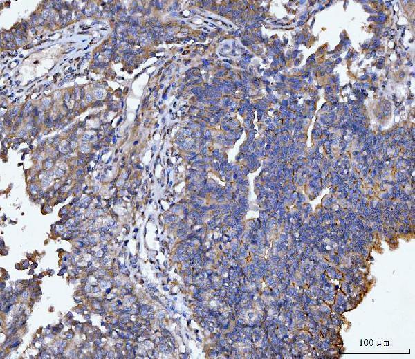
Figure 1. IHC analysis of MUC1 using anti-MUC1 antibody (A00187-4).
MUC1 was detected in a paraffin-embedded section of human bladder epithelial carcinoma tissue. Heat mediated antigen retrieval was performed in EDTA buffer (pH 8.0, epitope retrieval solution). The tissue section was blocked with 10% goat serum. The tissue section was then incubated with 2 μg/ml rabbit anti-MUC1 Antibody (A00187-4) overnight at 4°C. Peroxidase Conjugated Goat Anti-rabbit IgG was used as secondary antibody and incubated for 30 minutes at 37°C. The tissue section was developed using HRP Conjugated Rabbit IgG Super Vision Assay Kit (Catalog # SV0002) with DAB as the chromogen.

Click image to see more details
Figure 2. IHC analysis of MUC1 using anti-MUC1 antibody (A00187-4).
MUC1 was detected in a paraffin-embedded section of human endometrial cancer tissue. Heat mediated antigen retrieval was performed in EDTA buffer (pH 8.0, epitope retrieval solution). The tissue section was blocked with 10% goat serum. The tissue section was then incubated with 2 μg/ml rabbit anti-MUC1 Antibody (A00187-4) overnight at 4°C. Peroxidase Conjugated Goat Anti-rabbit IgG was used as secondary antibody and incubated for 30 minutes at 37°C. The tissue section was developed using HRP Conjugated Rabbit IgG Super Vision Assay Kit (Catalog # SV0002) with DAB as the chromogen.

Click image to see more details
Figure 3. IHC analysis of MUC1 using anti-MUC1 antibody (A00187-4).
MUC1 was detected in a paraffin-embedded section of human ovarian carcinoma tissue. Heat mediated antigen retrieval was performed in EDTA buffer (pH 8.0, epitope retrieval solution). The tissue section was blocked with 10% goat serum. The tissue section was then incubated with 2 μg/ml rabbit anti-MUC1 Antibody (A00187-4) overnight at 4°C. Peroxidase Conjugated Goat Anti-rabbit IgG was used as secondary antibody and incubated for 30 minutes at 37°C. The tissue section was developed using HRP Conjugated Rabbit IgG Super Vision Assay Kit (Catalog # SV0002) with DAB as the chromogen.
Protein Target Info & Infographic
Gene/Protein Information For MUC1 (Source: Uniprot.org, NCBI)
Gene Name
MUC1
Full Name
Mucin-1
Weight
21184 MW
Alternative Names
Breast carcinoma-associated antigen DF3; Carcinoma-associated mucin; CD227 antigen; CD227; DF3 antigen; EMA; Episialin; H23 antigen; H23AG; KL-6; MAM6; MUC-1; MUC1/ZD; mucin 1, cell surface associated; mucin 1, transmembrane; Mucin-1; Peanut-reactive urinary mucin; PEM; PEMMUC-1/SEC; PEMT; Polymorphic epithelial mucin; PUMMUC-1/X; tumor associated epithelial mucin; Tumor-associated epithelial membrane antigen; Tumor-associated mucin MUC1 ADMCKD, ADMCKD1, ADTKD2, CA 15-3, CD227, EMA, H23AG, KL-6, MAM6, MCD, MCKD, MCKD1, MUC-1, MUC-1/SEC, MUC-1/X/ZD, PEM, PEMT, PUM, MUC1 mucin 1, cell surface associated mucin-1|H23 antigen|breast carcinoma-associated antigen DF3|cancer antigen 15-3|carcinoma-associated mucin|episialin|krebs von den Lungen-6|mucin 1, transmembrane|peanut-reactive urinary mucin|polymorphic epithelial mucin|tumor associated epithelial mucin|tumor-associated epithelial membrane antigen
*If product is indicated to react with multiple species, protein info is based on the gene entry specified above in "Species".For more info on MUC1, check out the MUC1 Infographic

We have 30,000+ of these available, one for each gene! Check them out.
In this infographic, you will see the following information for MUC1: database IDs, superfamily, protein function, synonyms, molecular weight, chromosomal locations, tissues of expression, subcellular locations, post-translational modifications, and related diseases, research areas & pathways. If you want to see more information included, or would like to contribute to it and be acknowledged, please contact [email protected].
Specific Publications For Anti-MUC1 Antibody Picoband™ (A00187-4)
Hello CJ!
No publications found for A00187-4
*Do you have publications using this product? Share with us and receive a reward. Ask us for more details.
Recommended Resources
Here are featured tools and databases that you might find useful.
- Boster's Pathways Library
- Protein Databases
- Bioscience Research Protocol Resources
- Data Processing & Analysis Software
- Photo Editing Software
- Scientific Literature Resources
- Research Paper Management Tools
- Molecular Biology Software
- Primer Design Tools
- Bioinformatics Tools
- Phylogenetic Tree Analysis
Customer Reviews
Have you used Anti-MUC1 Antibody Picoband™?
Submit a review and receive an Amazon gift card.
- $30 for a review with an image
0 Reviews For Anti-MUC1 Antibody Picoband™
Customer Q&As
Have a question?
Find answers in Q&As, reviews.
Can't find your answer?
Submit your question
