Product Info Summary
| SKU: | M00082 |
|---|---|
| Size: | 80 µl |
| Reactive Species: | Human |
| Host: | Mouse |
| Application: | Flow Cytometry, IHC-P, WB |
Customers Who Bought This Also Bought
Product info
Product Name
Anti-CSF1R Antibody
View all M-CSFR/CD115 Antibodies
SKU/Catalog Number
M00082
Size
80 µl
Description
Boster Bio Anti-CSF1R Antibody (Catalog # M00082). Tested in WB, Flow Cytometry, IHC-P application(s). This antibody reacts with Human.
Storage & Handling
Maintain refrigerated at 2-8°C for up to 2 weeks. For long-term storage, store at -20°C in small aliquots to prevent freeze-thaw cycles.
Cite This Product
Anti-CSF1R Antibody (Boster Biological Technology, Pleasanton CA, USA, Catalog # M00082)
Host
Mouse
Contents
Purified monoclonal antibody supplied in PBS with 0.09% (W/V) sodium azide.
Clonality
Monoclonal
Clone Number
1486CT328.53.37
Isotype
IgG1,k
Immunogen
This CSF1R antibody is generated from a mouse immunized with a recombinant protein.
*Blocking peptide can be purchased. Costs vary based on immunogen length. Contact us for pricing.
Reactive Species
M00082 is reactive to CSF1R in Human
Reconstitution
Calculated molecular weight
107984 Da
Background of M-CSFR/CD115
Tyrosine-protein kinase that acts as cell-surface receptor for CSF1 and IL34 and plays an essential role in the regulation of survival, proliferation and differentiation of hematopoietic precursor cells, especially mononuclear phagocytes, such as macrophages and monocytes. Promotes the release of proinflammatory chemokines in response to IL34 and CSF1, and thereby plays an important role in innate immunity and in inflammatory processes. Plays an important role in the regulation of osteoclast proliferation and differentiation, the regulation of bone resorption, and is required for normal bone and tooth development. Required for normal male and female fertility, and for normal development of milk ducts and acinar structures in the mammary gland during pregnancy. Promotes reorganization of the actin cytoskeleton, regulates formation of membrane ruffles, cell adhesion and cell migration, and promotes cancer cell invasion. Activates several signaling pathways in response to ligand binding. Phosphorylates PIK3R1, PLCG2, GRB2, SLA2 and CBL. Activation of PLCG2 leads to the production of the cellular signaling molecules diacylglycerol and inositol 1,4,5- trisphosphate, that then lead to the activation of protein kinase C family members, especially PRKCD. Phosphorylation of PIK3R1, the regulatory subunit of phosphatidylinositol 3-kinase, leads to activation of the AKT1 signaling pathway. Activated CSF1R also mediates activation of the MAP kinases MAPK1/ERK2 and/or MAPK3/ERK1, and of the SRC family kinases SRC, FYN and YES1. Activated CSF1R transmits signals both via proteins that directly interact with phosphorylated tyrosine residues in its intracellular domain, or via adapter proteins, such as GRB2. Promotes activation of STAT family members STAT3, STAT5A and/or STAT5B. Promotes tyrosine phosphorylation of SHC1 and INPP5D/SHIP- 1. Receptor signaling is down-regulated by protein phosphatases, such as INPP5D/SHIP-1, that dephosphorylate the receptor and its downstream effectors, and by rapid internalization of the activated receptor.
Antibody Validation
Boster validates all antibodies on WB, IHC, ICC, Immunofluorescence, and ELISA with known positive control and negative samples to ensure specificity and high affinity, including thorough antibody incubations.
Application & Images
Applications
M00082 is guaranteed for Flow Cytometry, IHC-P, WB Boster Guarantee
Assay Dilutions Recommendation
The recommendations below provide a starting point for assay optimization. The actual working concentration varies and should be decided by the user.
WB: 1:4000
IHC-P: 1:25
FC: 1:25
Validation Images & Assay Conditions

Click image to see more details
Anti-CSF1R Antibodyat 1:2000 dilution + human placenta lysates
Lysates/proteins at 20 μg per lane.
Secondary
Goat Anti-mouse IgG, (H+L), Peroxidase conjugated at 1/10000 dilution
Predicted band size : 108 kDa
Blocking/Dilution buffer: 5% NFDM/TBST.

Click image to see more details
Anti-CSF1R Antibodyat 1:4000 dilution + U-87MG whole cell lysates
Lysates/proteins at 20 μg per lane.
Secondary
Goat Anti-mouse IgG, (H+L), Peroxidase conjugated at 1/10000 dilution
Predicted band size : 108 kDa
Blocking/Dilution buffer: 5% NFDM/TBST.

Click image to see more details
M00082 staining CSF1R in human skin sections by Immunohistochemistry (IHC-P -paraformaldehyde-fixed, paraffin-embedded sections). Tissue was fixed with formaldehyde and blocked with 3% BSA for 0. 5 hour at room temperature; antigen retrieval was by heat mediation with a citrate buffer (pH6). Samples were incubated with primary antibody (1/25) for 1 hours at 37°C. A undiluted biotinylated goat polyvalent antibody was used as the secondary antibody.

Click image to see more details
Overlay histogram showing U-87MG cells stained with M00082 (green line). The cells were fixed with 2% paraformaldehyde (10 min). The cells were then icubated in 2% bovine serum albumin to block non-specific protein-protein interactions followed by the antibody (M00082, 1:25 dilution) for 60 min at 37ºC. The secondary antibody used was Goat-Anti-mouse IgG, DyLight® 488 Conjugated Highly Cross-Adsorbed at 1/400 dilution for 40 min at 37ºC. Isotype control antibody (blue line) was rabbit IgG (1g/1x10^6 cells) used under the same conditions. Acquisition of >10, 000 events was performed.
Protein Target Info & Infographic
Gene/Protein Information For CSF1R (Source: Uniprot.org, NCBI)
Gene Name
CSF1R
Full Name
Macrophage colony-stimulating factor 1 receptor
Weight
107984 Da
Superfamily
protein kinase superfamily
Alternative Names
Macrophage colony-stimulating factor 1 receptor, CSF-1 receptor, CSF-1-R, CSF-1R, M-CSF-R, Proto-oncogene c-Fms, CD115, CSF1R, FMS CSF1R BANDDOS, C-FMS, CD115, CSF-1R, CSFR, FIM2, FMS, HDLS, M-CSF-R colony stimulating factor 1 receptor macrophage colony-stimulating factor 1 receptor|CD115 |CSF-1 receptor|FMS proto-oncogene|McDonough feline sarcoma viral (v-fms) oncogene homolog|macrophage colony stimulating factor I receptor|proto-oncogene c-Fms
*If product is indicated to react with multiple species, protein info is based on the gene entry specified above in "Species".For more info on CSF1R, check out the CSF1R Infographic

We have 30,000+ of these available, one for each gene! Check them out.
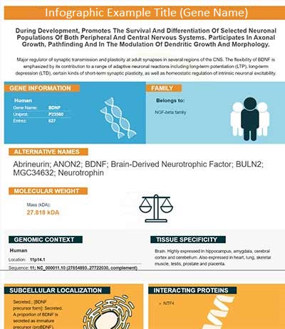
In this infographic, you will see the following information for CSF1R: database IDs, superfamily, protein function, synonyms, molecular weight, chromosomal locations, tissues of expression, subcellular locations, post-translational modifications, and related diseases, research areas & pathways. If you want to see more information included, or would like to contribute to it and be acknowledged, please contact [email protected].
Specific Publications For Anti-CSF1R Antibody (M00082)
Loading publications
Recommended Resources
Here are featured tools and databases that you might find useful.
- Boster's Pathways Library
- Protein Databases
- Bioscience Research Protocol Resources
- Data Processing & Analysis Software
- Photo Editing Software
- Scientific Literature Resources
- Research Paper Management Tools
- Molecular Biology Software
- Primer Design Tools
- Bioinformatics Tools
- Phylogenetic Tree Analysis
Customer Reviews
Have you used Anti-CSF1R Antibody?
Submit a review and receive an Amazon gift card.
- $30 for a review with an image
0 Reviews For Anti-CSF1R Antibody
Customer Q&As
Have a question?
Find answers in Q&As, reviews.
Can't find your answer?
Submit your question


