This website uses cookies to ensure you get the best experience on our website.
- Table of Contents
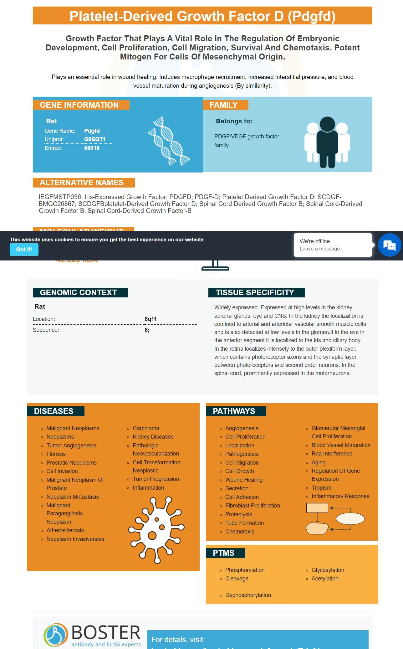


Facts about Platelet-derived growth factor D.
Plays an essential role in wound healing. Induces macrophage recruitment, increased interstitial pressure, and blood vessel maturation during angiogenesis (By similarity).
| Rat | |
|---|---|
| Gene Name: | Pdgfd |
| Uniprot: | Q9EQT1 |
| Entrez: | 66018 |

| Belongs to: |
|---|
| PDGF/VEGF growth factor family |

IEGFMSTP036; Iris-expressed growth factor; PDGFD; PDGF-D; platelet derived growth factor D; SCDGF-BMGC26867; SCDGFBplatelet-derived growth factor D; spinal cord derived growth factor B; Spinal cord-derived growth factor B; spinal cord-derived growth factor-B
Mass (kDA):
42.809 kDA

| Rat | |
|---|---|
| Location: | 8q11 |
| Sequence: | 8; |
Widely expressed. Expressed at high levels in the kidney, adrenal glands, eye and CNS. In the kidney the localization is confined to arterial and arteriolar vascular smooth muscle cells and is also detected at low levels in the glomeruli In the eye in the anterior segment it is localized to the iris and ciliary body. In the retina localizes intensely to the outer plexiform layer, which contains photoreceptor axons and the synaptic layer between photoreceptors and second order neurons. In the spinal cord, prominently expressed in the motorneurons.


If you are searching for a product that can detect PDGFD, you may want to consider purchasing Boster Bio's Anti-PDGF Receptor beta/PDGFRB Antibody. Boster validates antibodies with positive and/or negative samples to ensure that they are specific and high in affinity. Boster also offers product credits for the first reviewers of its products. This unique tool is now available to scientists all over the world.
A new antibody developed by Boster Bio identifies PDGF receptor beta (PDGFRB) in human tissue. This antibody is part of the Picoband(tm) catalog. It reacts with the Human PDGF receptor. This antibody is highly specific in detecting PDGFR. It is available for use in many biomedical applications.
The Boster Bio Anti-PDGF receptors beta/PDGFRB Antibody, part of Picoband(tm), reacts with human and mouse cells. This antibody was tested for its ability of recognizing phospho–Y751 PDGF-receptors. The PDGFD marker is used for identifying the epitopes necessary to PDGF receptor gene biogenesis.
The PDGF signaling system is a key component that promotes vascular remodelling and narrowing. The signaling cascades that result from its binding to the cognate-ligand ligand are initiated by it. The type of response depends upon the ligand attached to the receptor, and the heterodimer PDGFRB/PDGFRA. The phosphorylation PLCG1 activates the production signaling molecules PTPN11/PIK3R1 as well as mobilizes cytosolic Ca2+.
This bispecific antibody binds CD3 Epsilon and EPCAM, and is designed to prevent adverse reactions from T cells. Its PDGFD mark is used to distinguish the two groups. While the two bispecific antibodies are effective for identifying tumor-derived PDGF receptors, they are not equally efficient for treating tumors.
This antibody inhibits platelet activation, and measures the activity PDGF. It has high binding affinity. It also inhibits platelet activity in heparinized hematopoiesis blood. It can detect PDGFD in patients with anemia. To test the effectiveness, please contact your pharmacy.
Boster Bio Anti PDGF Beta/PDGFRB Antibody uses a PDGFD unique marker. The PDGFD marker is a valuable biomarker for determining PDGFRB/PDGFRB activation. The PDGFD indicator is used to determine PDGFD activity. This antibody is also useful for assessing the activity of PDGF in various cancers.
The antigenbinding molecule must at least have one bond between regions VH and VL of the first antigenbinding domain. The antigen-binding molecules that bind to both the light or heavy chain variable (VH) must be included in the corresponding antibodies.
Boster Bio Zvegf4 protein preparations can be achieved using a variety if chemical synthesis methods. These include classical solution Chemistry, partial-solid synthesis, fragment condensation and exclusive solid phase synthesis. Merrifield has described some of the methods in his J. Am. Chem. Soc. 85:2149 in 1963, Stewart et al. in Pierce Chemical Co. Rockford, Ill. 1984, Bayer and Rapp. Peptide Prot. 3, 1986, and Atherton and Rapp in Solid Phase Peptide Synthesis: A Practical Approach
Recombinant human zvegf4 PCR fragments were prepared using a conventional baculovirus expressing system and a carboxyl terminal Glu Glu affinity mark. The resulting cDNA from the amplifying process was then subcloned into the pZyTrack gene by EcoRV -AscI. After cloning, pZyTrack-transformation was performed with adenovirus in 293A cells. The resulting product of cloning was purified by CsCl and named AdZyvegf4.
Aerosolization is another method to deliver zvegf4 in the body. Aerosolization has been developed by a few companies. These polypeptides can also be called zvegf4 nue-GFD. These molecules prevent fibrotic tissue formation by inhibiting zvegf4 genes transcription.
Mitogenesis assays were used to determine whether zvegf4 polypeptides activate or inhibit cell proliferation. Various assays were performed on human cells. For example, antibodies to Zvegf4 in human cells might be considered specifically binding if the protein is bound at 10x the concentration as control proteins. Non-human antibodies to the zvegf4 protein might bind to it from other species.
Another method is to combine zvegf4 polypeptides with immunoglobulins. This combination produces immunogens which are highly effective at producing antibodies. It also allows the incorporation hapten-like components of the Zvegf4 polypeptide to immunoglobulin-binding proteins. These combinations can be combined to form a macromolecular carrier.
In addition to polypeptides, these antagonists can be delivered in various forms. Some can be exogenous, while others are delivered via viral delivery systems. The use of these compounds in combination with other agents enables a more comprehensive understanding of how zvegf4 interacts with cells. These compounds can ultimately lead to the development and application of new technologies and treatments for cancer patients.
The first component of these proteins is a ribozyme. It consists primarily of a catalytic central and a target-RNA binding portion. It regulates cell differentiation, proliferation, metabolism, growth, and migration. It is composed of two polypeptides which can selectively bind zvegf4 (mRNA) as a heterodimer.
PMID: 11162582 by Hamada T., et al. Molecular cloning of SCDGF-B, a novel growth factor homologous to SCDGF/PDGF-C/fallotein.
PMID: 11850188 by Hamada T., et al. The expression of SCDGF/PDGF-C/fallotein and SCDGF-B/PDGF-D in the rat central nervous system.


