This website uses cookies to ensure you get the best experience on our website.
- Table of Contents
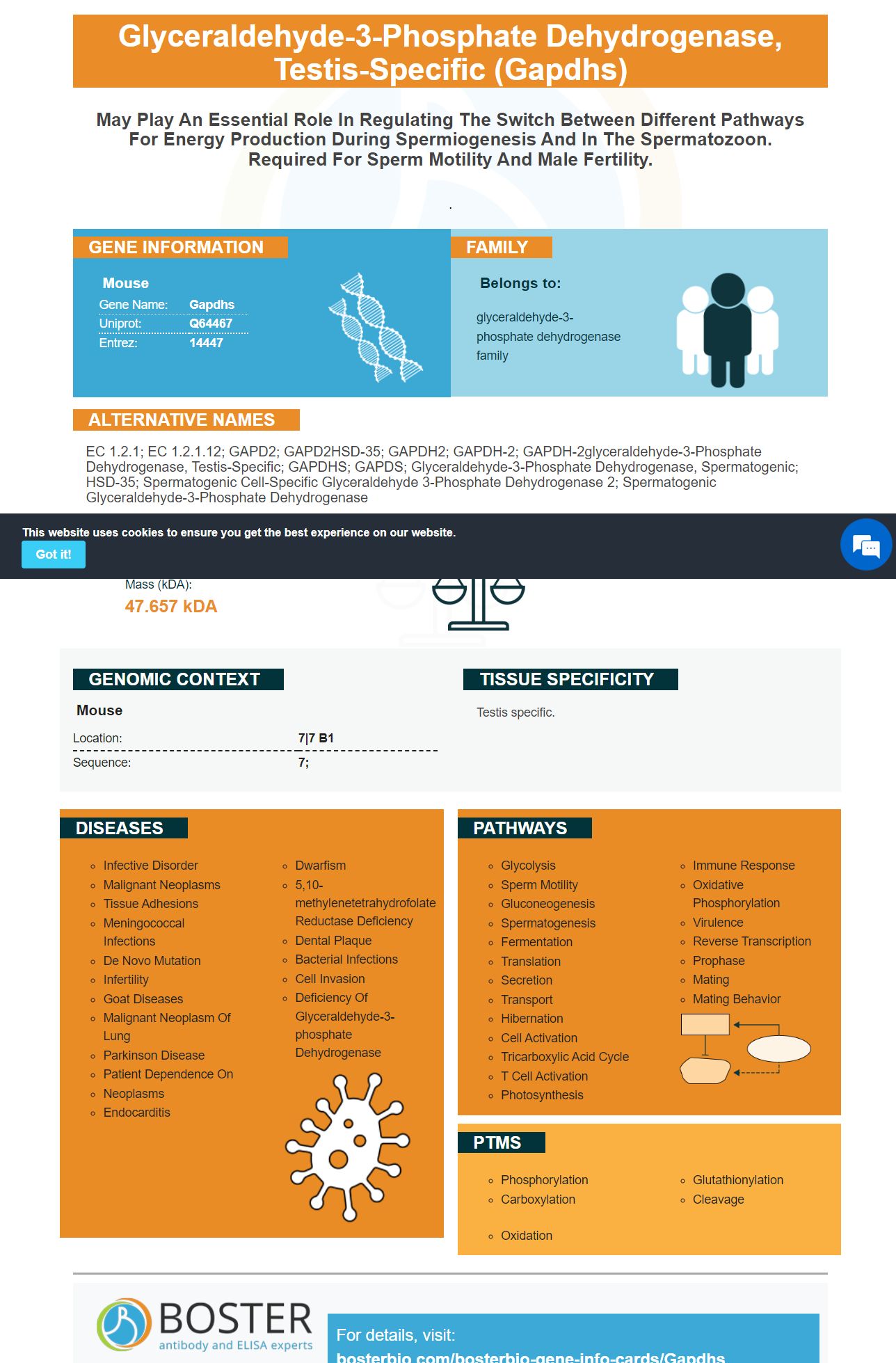
Facts about Glyceraldehyde-3-phosphate dehydrogenase, testis-specific.
.
| Mouse | |
|---|---|
| Gene Name: | Gapdhs |
| Uniprot: | Q64467 |
| Entrez: | 14447 |

| Belongs to: |
|---|
| glyceraldehyde-3-phosphate dehydrogenase family |

EC 1.2.1; EC 1.2.1.12; GAPD2; GAPD2HSD-35; GAPDH2; GAPDH-2; GAPDH-2glyceraldehyde-3-phosphate dehydrogenase, testis-specific; GAPDHS; GAPDS; glyceraldehyde-3-phosphate dehydrogenase, spermatogenic; HSD-35; Spermatogenic cell-specific glyceraldehyde 3-phosphate dehydrogenase 2; Spermatogenic glyceraldehyde-3-phosphate dehydrogenase
Mass (kDA):
47.657 kDA

| Mouse | |
|---|---|
| Location: | 7|7 B1 |
| Sequence: | 7; |
Testis specific.


Boster Bio produces the GAPDH marker. It has many applications in medicine. It is a surface marker on the cells of three Mycoplasma genera: Mycoplasma pneumoniae; Mycoplasma hoopneumoniae; and Mycoplasma sou. These bacteria also have cell-surface markers and a special relationship with host cells.
An ELISA for GAPDHS is a specific antibody that detects activity of the protein GAPDH. The protein plays a number of important roles in the body, including membrane binding, phosphotransferase activity and nuclear RNA export. Recent studies have shown that the protein may be involved in cancer progression, neuronal diseases, and apoptosis. For researchers interested in these processes, an anti-GAPDH antibodies is a great choice.
This assay shows that rGAPDH protein binds with different ECM components. Cell membrane proteins could be prepared using a commercial Membrane Cytosol Protein Exctraction Kit (Tiangen Biotech China). A microtiter plate with 96 wells was coated with 100mL cell membrane protein. The plate was incubated at 37 degC with blocking solution, followed by two hours at room temperature. PBS was used as a control.
Western blot analysis validated the GAPDHS marker. GAPDH, which regulates expression of TNFa, is found in parasite EVs. However, it is not clear if GAPDH is a direct regulator for TNF-a. Further research is needed to confirm these findings. In addition, this test may identify the presence of TNF-a-producing macrophages in a variety of conditions.
An ELISA for the GAPDHS marker measures the level of this protein in cell lysates and tissue homogenates. GAPDH is an enzyme found in all cells, in both the cytoplasmic and nucleus. GAPDH is an excellent housekeeping gene due to its widespread expression. It is also an important marker to study normal cellular biology.
The EV-associated protein GAPDH (GAPDH) is a key component of the membrane of exosomes. While some studies have found this protein within the EV lumen (some reports it only being associated with its outer surface), others have reported it to be only present in the EV lumen. This study used a protease digestion assay that distinguishes between the internal and external proteins. Although it was unable to detect GAPDH endogenous in human SCs it did show that the molecule travels into the exolysosomal.
The G58 peptide was synthesized and expressed in E. coli. It contains GAPDH's PS-binding motif and TARBP2's double-strandedRNA-binding region. The siRNA-loaded EVs G58T for 2 h at 40°C were incubated with the EVs. Gel-binding assays showed extensive binding of the G58 peptide to the EV surface.
GAPDH was seen to be co-associated in the present study with human EVs. It co-localized with a subset of CD63-positive EVs and bound to their surface via the PS-binding domain. This antibody also binds to EVs with no CD63 markers. Data also indicate that cell-derived GAPDH may have an effect on GAPDH activity in the EVs.
GAPDH is a key player in tumorigenesis in eukaryotes. GAPDH stabilizes active Akt and inhibits protease-independent cell death. It also participates in the upregulation ATG12, an autophagy protein. It also clears damaged mitochondria. GAPDH is also involved in several neurological diseases. It has been shown to be a major component in Alzheimer's disease amyloid plaques. It interacts with amyloid-b precursor proteins.
Antibodies that recognize specific protein targets are used to detect the GAPDH marker by SDS-PAGE. The amount per lane of lysate can vary depending upon the target protein. This amount is consistent regardless of the antibody or target protein used. Often, a protein will be transferred directly from the gel into the membrane without checking for consistency in the in-gel proteins loading consistency. To avoid this, stain-free gel technology was used, which allowed for the visualization of SDS-PAGE separation prior to transfer.
Since their initial experiments, stripping buffers' performance has improved significantly. Commercial preparations claim to be more gentle than the originals. They generally contain SDS and glycine as well as detergents. They also work at roomtemperature. To make sure that these preparations work well and do not affect sample integrity, it is still a good idea. The membranes can now be detected using ECL-reagents or other detection techniques after they have been removed of any antibodies.
There are many steps involved when performing Western blotting. The steps include deposition of the sample, lysing the proteins and denaturing them, incubation with the primary and second antibodies, and detection and quantification of the target protein. The Western blotting workflow can be automated and has new techniques. Learn more about Western blotting. These are some steps you can follow to perform the analysis.
Western blotting has become a standard in the field physiology. This technique is most commonly used in skeletal muscular physiology. The process is important for resolving the mechanisms underpinning the adaptation of skeletal muscles to exercise and provides mechanistic insight into regulatory processes. The procedure is inexpensive and easy to use, but poor quality data can lead to inaccurate results.
Typically, Western Blotting can be done by using an antibody which recognizes LDHB. Monoclonal antibodies that identify GAPDH or Histone H3 can be purchased separately or in a duo. The duos can be used for immunostaining studies by providing different MW targets. For example, one pair of antibodies detects GAPDH and the other a specific nucleus protein.
This technique can be used to treat myocardial ischemia in animals. The samples used for the experiment are tissue, cells, or other solutions. Gel electrophoresis is used to separate the protein molecules. The resulting macromolecules can be transferred onto a second matrix. The second matrix is usually made of nitrocellulose. You can block non-specific locations with PVDF membrane.
Detection by recombinants plasmid of GAPDHS is a method of detecting the presence of the GAPDH protein in human sera. The GAPDHS-recombinant plasmid contains six histidines fused at its N terminus. It is made from E.coli cell. After the cells had grown, they were harvested and the lysate was stained in Coomassie blue to show the presence GAPDH.
GAPDH regulates many biological functions. GAPDH was cloned with the pTZ57R/T-plasmid. The plasmid was ligated with DH5a-competent cells and linearized using TransformAid bacterial transform kit. The expression host was Invitrogen's BL21 (DE3) pLys-S cell line.
Two cis elements between the cre and PAOX1 uORF genes were integrated using a primer pairing lox71-PAOX1. The resulting recombinant proteins were named pMC (or pMCU) respectively. These two recombinant plasmids contain the same PAOX1-lacO gene and can be detected using PCR.
Researchers identified the GAPDH gene sequence within E. tarda by using recombinant GAPDHS-plasmids. This gene is more closely related to the sequences of A. hydrophila and V. cholerae than to that of the human. These differences in sequence suggest that these recombinant plasmids are useful for the study of GAPDH in a wide range of human species.
GAPDH is a 45-kDa immunomodulatory gene that was isolated from S. agalactiae NEM316. This gene was cloned and expressed in E. coli, and the recombinant protein, termed rGAPDH (rGAPDH), was purified as a histidyl-tagged protein and expressed. The rGAPDH-protein, which has 34.9 U/mg, exhibited the exact same enzymatic activity of the isolated GAPDH form group A streptococci.
PMID: 1375514 by Welch J.E., et al. Expression of a glyceraldehyde 3-phosphate dehydrogenase gene specific to mouse spermatogenic cells.
PMID: 7736666 by Welch J.E., et al. Genomic organization of a mouse glyceraldehyde 3-phosphate dehydrogenase gene (Gapd-s) expressed in post-meiotic spermatogenic cells.


