This website uses cookies to ensure you get the best experience on our website.
- Table of Contents
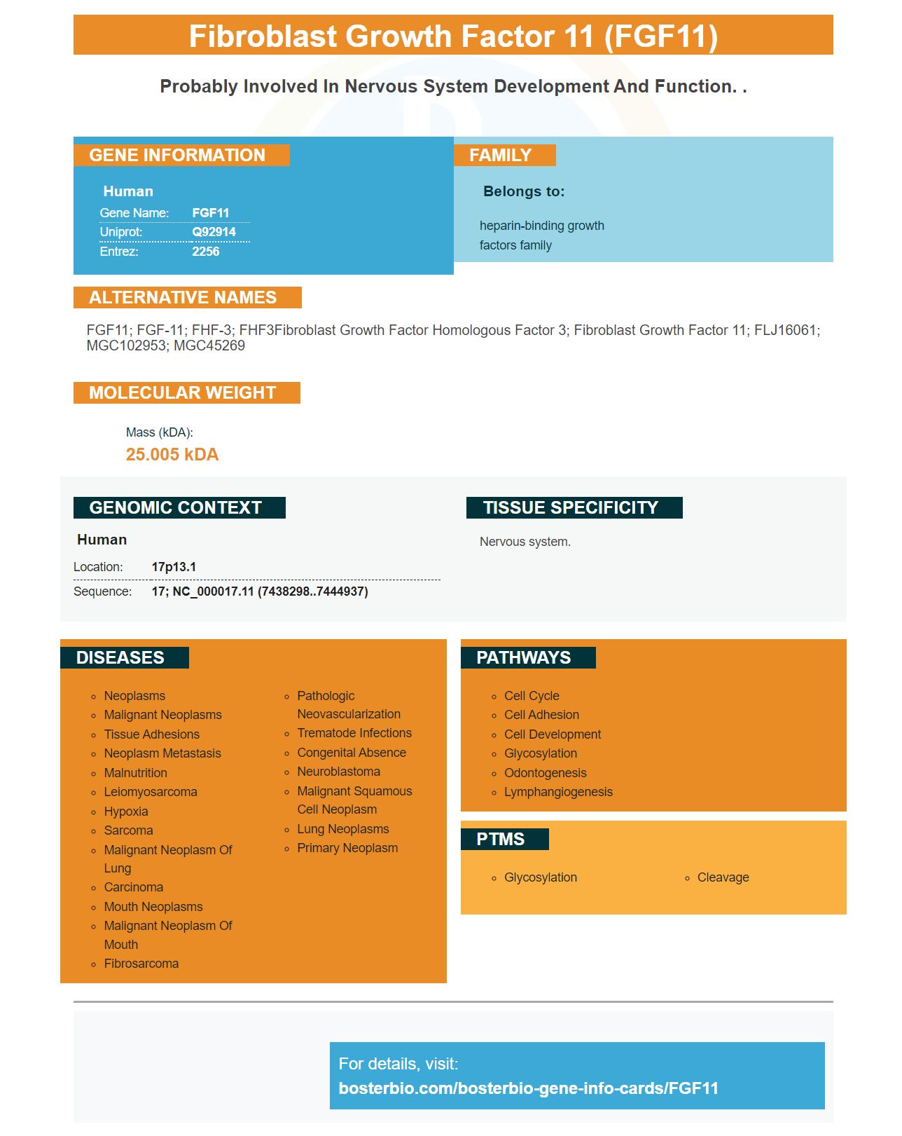
Facts about Fibroblast growth factor 11.
Human | |
---|---|
Gene Name: | FGF11 |
Uniprot: | Q92914 |
Entrez: | 2256 |
Belongs to: |
---|
heparin-binding growth factors family |
FGF11; FGF-11; FHF-3; FHF3Fibroblast growth factor homologous factor 3; fibroblast growth factor 11; FLJ16061; MGC102953; MGC45269
Mass (kDA):
25.005 kDA
Human | |
---|---|
Location: | 17p13.1 |
Sequence: | 17; NC_000017.11 (7438298..7444937) |
Nervous system.
In this article, we will take a look at Boster Bio's Anti-FGFR1 Antibody. We'll also be discussing Boster Bio's Anti-Fibroblast Activation Protein FAP. We hope you find this information useful. We also welcome your feedback about Boster Bio products. We'll update this article when we learn of new product releases!
Boster Bio's anti FGFR1 antibody is available under catalog number PA1477. This antibody reacts well with Human, Mouse, or Rat samples. It can be stored at -20°C for a year. This product contains 5mg BSA, 0.05m thimerosal and 0.015m NaN3. This antibody is available for use in the following ways:
Boster Bio Anti-Fibroblast Aktation Protein FAP Antibody detects Fibroblast Activation Protein alpha/FAP in the cytoplasm and plasma membrane of C2C12 mouse cells. Its in vivo substrates have not been determined. Boster scientists may submit their results for species or applications for credit. This offer is available for all scientists around the globe.
FAP is a type 2 transmembrane surface protein of the cell membrane that belongs to post-proline dipeptidase. It shares the highest homology with the enzyme dipeptidyl peptidase IV (DPPIV). It is expressed in fibroblasts. It is not present in normal adult tissues.
The growth of solid tumors requires the formation of a supporting stroma and blood vessel network, a process known as angiogenesis. FAP expression can be targeted for cancer and inhibits tumor formation by suppressing some aspects of angiogenesis. Targeting FAP might be an adjunctive therapy for breast cancer patients. If approved, this antibody may inhibit tumor growth and angiogenesis.
Studies were conducted using an ELISA commercial isotyping tool. The colorimetric results revealed positive reactions only for rows A and G. This indicates that the antibody binds at the same epitope 4G5. It is important to keep in mind that different antibodies could have similar banding patterns. This can make Boster Bio Antifibroblast Protein FAP antibody useful for clinical trials as well as research.
Another aspect of this invention provides a nucleic Acid encoding an antibody, or a fragment thereof, that binds with a target antigen. The antigen-binding region of the antibody is cross species reactive. However, the polynucleotide sequence which encodes binding polypeptides recognizes naturally expressed canine FAP proteins. A negative control includes the omission of the primary antibody.
In one study, mice were exposed to 5% GS (and HK) to inhibit FAP-sync expression. The number of apoptotic cell was increased by FAP-shRNA treatment. The number and percentage of apoptotic tissues in mice that were treated with 5% GS, HK, was threefold higher in these mice than those in the control groups. These results support the use Boster Bio Anti-Fibroblast Activity Protein FAP Antibody for cancer research.
A new humanized antibody called Boster Bio Anti-FGFR1 Markers can now help researchers analyze FGFR1 protein and mRNA levels in various tissues and cells. This tool allows researchers visually to detect antigens found in tissue and cells. This product can also be used in clinical trials or research. Continue reading to learn about the product's diagnostic properties. This product has been designed for use in cell culture, tissue samples, and immunohistochemistry (IHC).
Many suppliers can sell antibodies against the FGFR1 proteins. Boster Bio Anti FGFR1 antibodies, among others, target human, mouse and rat protein. This antibody is 91.9 kilodaltons (in mass). Antibodies to FGFR1 are often called BFGFR, CD331, CEK, and HBGFR. These antibodies can be based either on canine or mouse orthologs.
RTPCR was used in 33 SCLC specimens to analyze FGFR1's mRNA. To indicate the level of mRNA expression, CT values were used. The CT values were scored as 1+, 2+, or 3+, and samples with at least 10% positive tumor cells were considered FGFR1 IHC-positive. Similar results were observed for the clinical characteristics of patients with cancer.
As an effective therapeutic target, FGFR1-mediated signaling is a promising tool for treating cancer. The protein triggers a signal transmission cascade that regulates cell survival, proliferation, and growth. The gene is located at chromosome 8p12 where it is found. Mutations in the gene are associated with Antley Bixler syndrome and Jackson-Weiss disorder, among other things.
PMID: 8790420 by Smallwood P.M., et al. Fibroblast growth factor (FGF) homologous factors: new members of the FGF family implicated in nervous system development.