This website uses cookies to ensure you get the best experience on our website.
- Table of Contents
2 Q&As
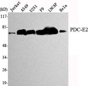
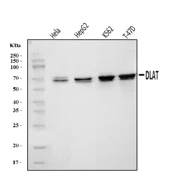
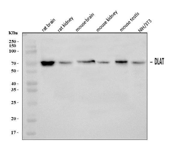
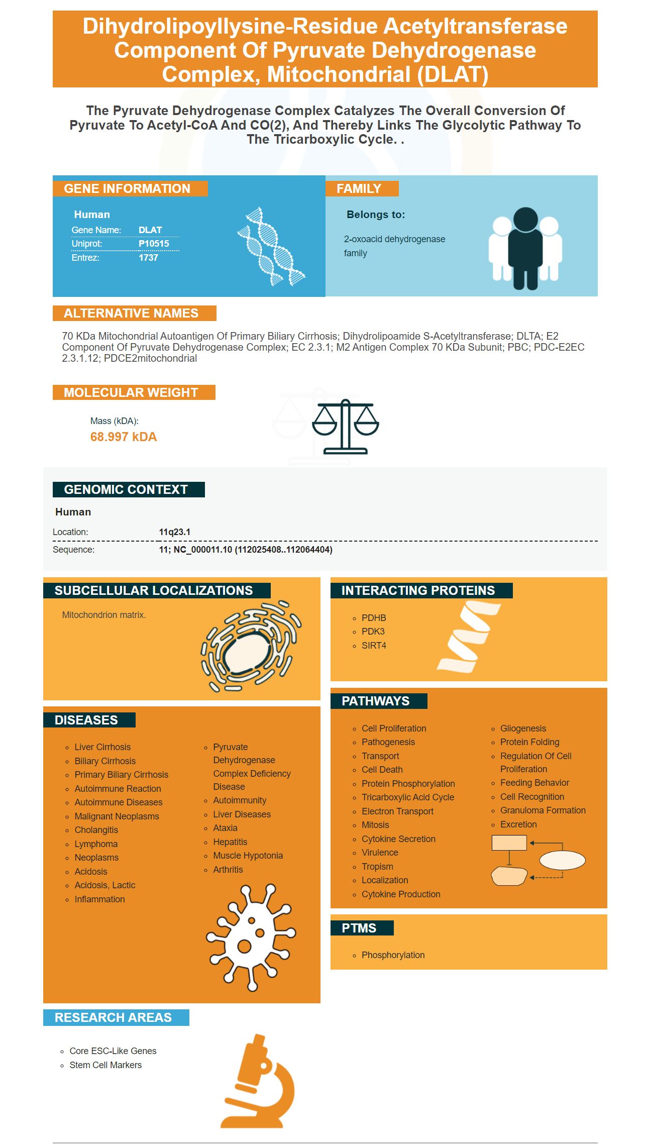
Facts about Dihydrolipoyllysine-residue acetyltransferase component of pyruvate dehydrogenase complex, mitochondrial.
Human | |
---|---|
Gene Name: | DLAT |
Uniprot: | P10515 |
Entrez: | 1737 |
Belongs to: |
---|
2-oxoacid dehydrogenase family |
70 kDa mitochondrial autoantigen of primary biliary cirrhosis; dihydrolipoamide S-acetyltransferase; DLTA; E2 component of pyruvate dehydrogenase complex; EC 2.3.1; M2 antigen complex 70 kDa subunit; PBC; PDC-E2EC 2.3.1.12; PDCE2mitochondrial
Mass (kDA):
68.997 kDA
Human | |
---|---|
Location: | 11q23.1 |
Sequence: | 11; NC_000011.10 (112025408..112064404) |
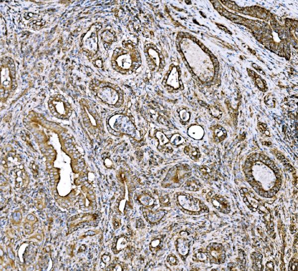
Mitochondrion matrix.
Boster Bio founder Steven Boster created his first product in 1993. He quickly became known as "the man who brought science in the toilet" after he had developed hundreds of primary antibody products. Boster was China's largest catalog antibody company by end of the 1990s. Boster uses trade secrets to create high-sensitivity ELISA kit. Here are a few of his most popular products.
The Gal-3 in situ hybridization detection kit, manufactured by Wuhan Boster Bio-engineering Co., Ltd., PR China, is used for in situ hybridization. The samples were washed with x2 salt citrate at 37°C. After blocking for 30 minutes, biotinylated mouse anti-digoxin (Biotinylated Mouse Anti-digoxin) was added. The samples were incubated at 37degC over 1 h, washed again, and the colours were developed using DAB. Positive and negative control samples were used as controls. Light microscopy was employed to observe positive and negative signals.
The discovery of high-affinity primary antibodies for GPC3 in HCC cells has significant therapeutic implications, since these antibodies can recognize a broad variety of cell surface-associated proteins. These antibodies also detect low expression of GPC3 in ovarian clear cell cancer and melanoma cell lines. The development of high-affinity antibodies to GPC3 is a broad approach to identify therapeutic antibodies against cancer.
First, prepare an appropriate solution comprising 50-100ul primary antibody and DLAT marker. The solution should be kept from light and placed in a plastic container. It is recommended to store it at 4 degrees Celsius. The second step is spinning the slides. After spinning, add PBS and let them sit for 30 minutes. Replace the buffer after this period, and then store them in plastic holders so that they are safe from the risk of light exposure.
There are a variety of issues that arise when detecting specific antigens. It can be difficult to tell the difference between two antigens, especially if they belong to the same species. Fortunately, it is possible to identify subtype-specific secondary antibodies that avoid the double-staining issue. Another alternative is to label the primary antibody with the use of a fluorescent dye. The fluorescent dye can be purchased from Molecular Probes. It removes one round of signal amplification. This method is particularly useful for proteins that are abundant in tissues.
The process of making the fusion proteins is more complicated than purchasing peptides. However this method is more precise in producing effective antibodies. It is important that you choose a manufacturer that offers enough fusion proteins to test the affinity purification process as well as depletion and depletion. The DLAT marker is a biomarker with high specificity, is able to distinguish high-affinity primary antibodies and low-affinity secondary antibody.
The GPC3 protein has a native form and is found on the cell surface in both the native and heparan sulfate forms. Utilizing DLAT as a marker for GPC3 mAbs will aid in the development of new treatments. It is a powerful tool for researching diseases such as Alzheimer's and cancer. It can also be used to identify antigens in the blood.
The process of determining whether the antigen present is in fact present must include fixation. Based on the type and the severity of the antibody, the conditions for fixing may differ. Fixation conditions differ based on the type of antibody as well as the tissue of interest. Other options for fixing include chemical permeabilization and collagenase treatment. The final fixation method will differ based on the antigen that is targeted and its accessibility.
PMID: 3174635 by Coppel R.L., et al. Primary structure of the human M2 mitochondrial autoantigen of primary biliary cirrhosis: dihydrolipoamide acetyltransferase.
PMID: 3191998 by Thekkumkara T.J., et al. Nucleotide sequence of a cDNA for the dihydrolipoamide acetyltransferase component of human pyruvate dehydrogenase complex.