This website uses cookies to ensure you get the best experience on our website.
- Table of Contents
Facts about Catenin delta-1.
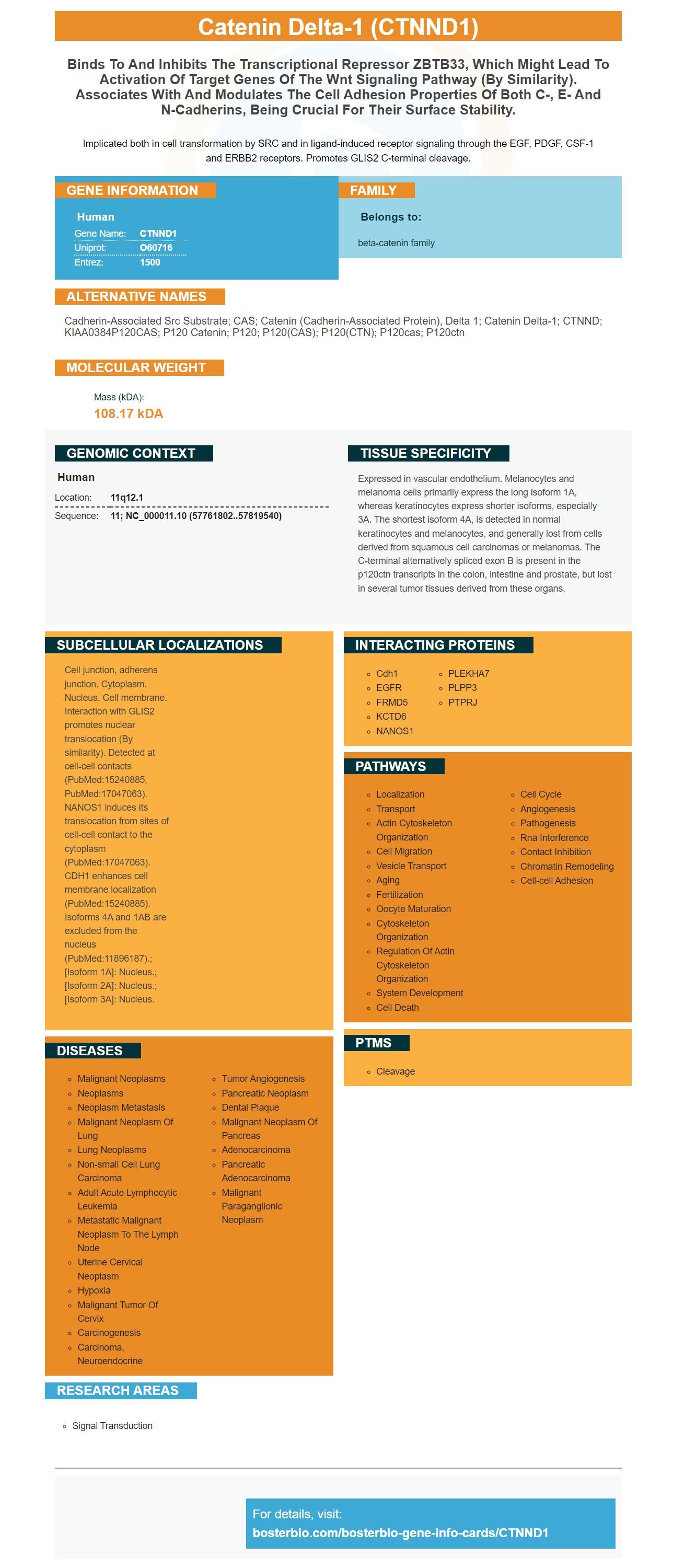
Implicated both in cell transformation by SRC and in ligand-induced receptor signaling through the EGF, PDGF, CSF-1 and ERBB2 receptors. Promotes GLIS2 C-terminal cleavage.
Human | |
---|---|
Gene Name: | CTNND1 |
Uniprot: | O60716 |
Entrez: | 1500 |
Belongs to: |
---|
beta-catenin family |
Cadherin-associated Src substrate; CAS; catenin (cadherin-associated protein), delta 1; catenin delta-1; CTNND; KIAA0384P120CAS; p120 catenin; p120; p120(CAS); p120(CTN); p120cas; p120ctn
Mass (kDA):
108.17 kDA
Human | |
---|---|
Location: | 11q12.1 |
Sequence: | 11; NC_000011.10 (57761802..57819540) |
Expressed in vascular endothelium. Melanocytes and melanoma cells primarily express the long isoform 1A, whereas keratinocytes express shorter isoforms, especially 3A. The shortest isoform 4A, is detected in normal keratinocytes and melanocytes, and generally lost from cells derived from squamous cell carcinomas or melanomas. The C-terminal alternatively spliced exon B is present in the p120ctn transcripts in the colon, intestine and prostate, but lost in several tumor tissues derived from these organs.
Cell junction, adherens junction. Cytoplasm. Nucleus. Cell membrane. Interaction with GLIS2 promotes nuclear translocation (By similarity). Detected at cell-cell contacts (PubMed:15240885, PubMed:17047063). NANOS1 induces its translocation from sites of cell-cell contact to the cytoplasm (PubMed:17047063). CDH1 enhances cell membrane localization (PubMed:15240885). Isoforms 4A and 1AB are excluded from the nucleus (PubMed:11896187).; [Isoform 1A]: Nucleus.; [Isoform 2A]: Nucleus.; [Isoform 3A]: Nucleus.
There is more information on the Boster Bio The Best Uses of the Anti-CTNND1 marker. Here are some helpful tips to optimize:
The CTNND1 marker is a biomarker that is widely used in research into cancer. Its sensitivity can be as low as a picogram. Boster Biologicals has ELISA kits and research antibodies that can be used to identify this biomarker. Boster Biologicals also sells other immunological reagents through its website, tebubio.org.
The Anti-GAPDH Marker in Boster Bio is a highly versatile tool for biomedical research. It allows researchers to detect GAPDH across a variety of species. It is made up of GAPDH, a marker proteins located on the surface of three species of Mycoplasma. These are Mycoplasma pneumoniae, M. hyopneumoniae and M. suis. Scientists can make use of this marker to gain knowledge about the role played by GAPDH in the Leishmania infection.
GAPDH is a surface-located molecule that binds to plasminogen. This binding enhances the targeting of proteolysis as well as aids in the dispersal of M. hyorhinis. This marker can be used to identify a variety of microorganisms. Below are the details of this test. Once you have the test, you can start your research. It will be out soon.
Boster Bio's Anti-GAPDH marker has been proven to block cervical cancer cell proliferation and induce apoptosis. This suggests that this protein may serve as a receptor for the Bt parasporal proteins that stimulate the death of cancerous cells. If it is identified as the receptor for Bt18 this would be invaluable in our understanding of the pharmacological mechanism of Bt18.
The Anti-GAPDH Antibody found in Boster Bio is labeled with the specific GAPDH marker for the M. Hyorhinis. This product reacts to GAPDH from mice, rats, and humans. The Anti-GAPDH Marker found in Boster Bio has five micrograms of BSA per mL of the product. Our anti-GAPDH antibody can be used to detect GAPDH in a variety tests.
This enzyme is present in all organs and tissues and is crucial for glycolysis. It is also involved in various illnesses, including Alzheimer's as well as cancer. It can be detected by immunofluorescence, Western Blot, and ELISA. The Anti-GAPDH Marker in Boster Bio is a potent tool for researchers studying GAPDH in various areas. These tests provide vital details on the function of the enzyme in various tissues and conditions.
The GAPDH gene is highly conserved across different M. hyorhinis strains, and the Anti-GAPDH marker in Boster Bio was developed from this gene. The GAPDH gene was made into an expression vector (pET-28a) and expressed in E. coli BL21 cells. Western Blotting using an anti-His antibody and SDS-PAGE were used to verify whether the gene was successfully recombinant.
Indirect immunofluorescence was employed to create the Anti-GAPDH marker. This test identifies binding between rGAPDH protein and cell membrane proteins. Cell membrane proteins were prepared by preparing membranes with a commercial Membrane and Cytosol Protein Extraction Kit. The cells were then incubated for two hours at 37 degC with 100 mg of protein in the media. A negative control was also used.
Anti-GAPDH antibodies block GAPDH from the plasma membrane of CEM-SS cells. This antibody was also used prior to treatment using the Bt18 parasporal protein. After the treatment with the Bt18 parasporal protein, Bt18 cells showed a decrease in fluorescence signal. This suggests that the Anti-GAPDH anti-GAPDH antibody may reduce the leukemic cell's binding.
Flow procedures provide a range of options. Boster Bio optimization techniques, guides and tools can help you optimize your experiments. Here are a few examples. You can also check out the most frequently asked questions about CTNND1 markers to assist you in optimizing your experiments. Boster Bio also offers flow procedures and isotype controls. Here are some of the answers to the most frequently-asked questions asked by customers.
PMID: 9653641 by Keirsebilck A., et al. Molecular cloning of the human p120ctn catenin gene (CTNND1): expression of multiple alternatively spliced isoforms.
PMID: 7623846 by Kim L., et al. The cytoplasmic tyrosine kinase FER is associated with the catenin- like substrate pp120 and is activated by growth factors.