This website uses cookies to ensure you get the best experience on our website.
- Table of Contents
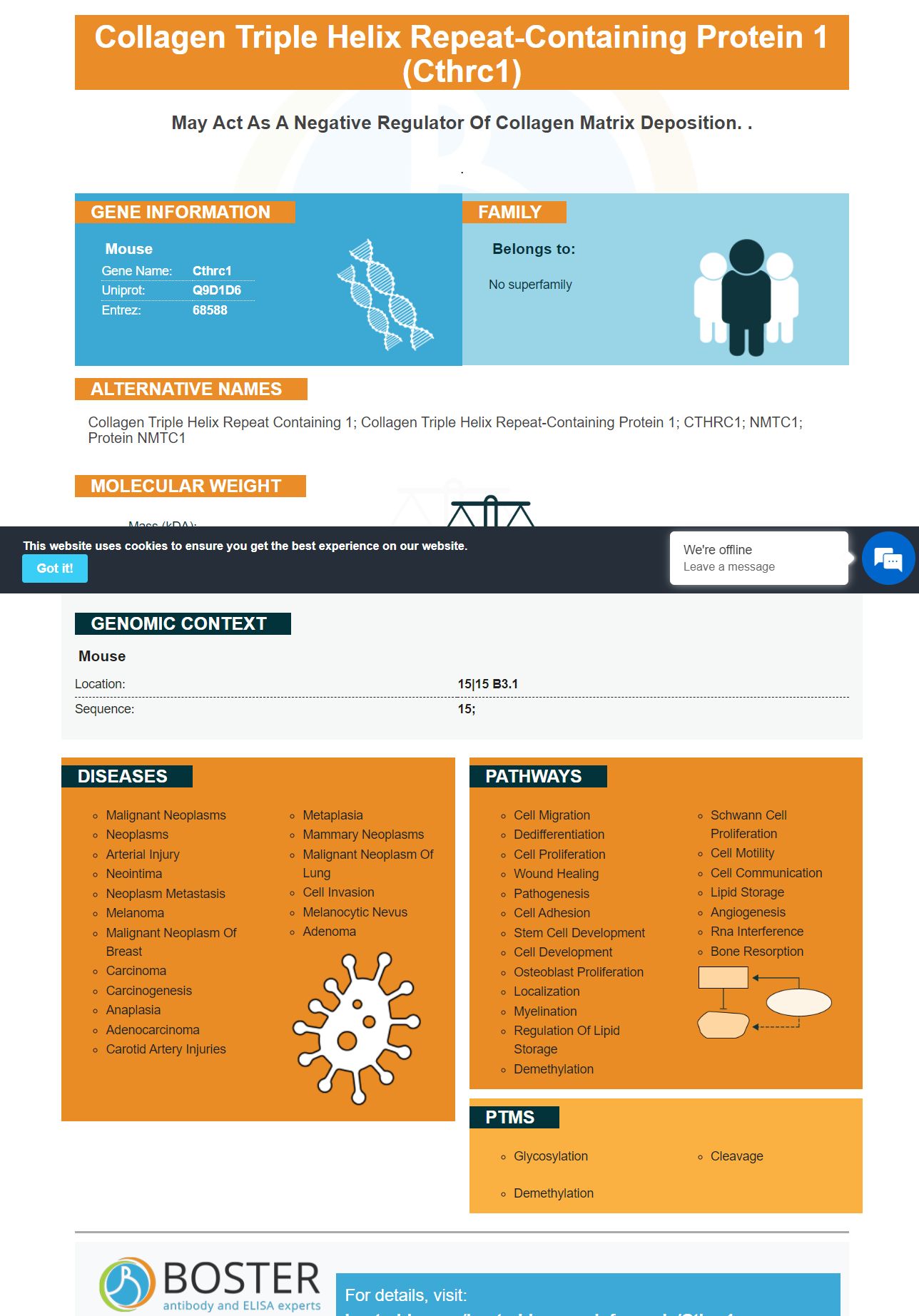
Facts about Collagen triple helix repeat-containing protein 1.
.
Mouse | |
---|---|
Gene Name: | Cthrc1 |
Uniprot: | Q9D1D6 |
Entrez: | 68588 |
Belongs to: |
---|
No superfamily |
collagen triple helix repeat containing 1; collagen triple helix repeat-containing protein 1; CTHRC1; NMTC1; Protein NMTC1
Mass (kDA):
26.46 kDA
Mouse | |
---|---|
Location: | 15|15 B3.1 |
Sequence: | 15; |
There are many biological assays which make use of antibodies to identify Cthrc1. These antibodies react with a variety of animal proteins and can be monoclonal or polyclonal. Boster Bio has developed antibodies that recognize Cthrc1 specifically in rabbits and mouse. In this article, we'll explore how Cthrc1 functions in the body, and discuss its importance in clinical practice.
The CTHRC1 member of the TGFb superfamily is the CTHRC1 marker. It activates FZD1, Smad2/3, and as well as regulates TGFb and the signaling p21. CTHRC1 is also involved in the progression of cancer by playing a role in the canonical Wnt/b-catenin and PCP noncanonical pathways. The tumor cells caused by CTHRC1 overexpression of the gene could encourage metastasis and growth of cancerous cells.
CTHRC1 is a complex molecular mechanisms. However it has been established that CTHRC1 expression is linked to a variety of aspects of EOC. CTHRC1 increase in expression leads to a significant reduction of E-cadherin, and also activates the Wnt/bcaten pathway. This is a typical biomarker for EMT. CTHRC1 is involved in tumor metastasis and inhibits the production of extracellular matrix, which in turn affects the microenvironment around the tumor.
In addition to its role in tumor metastasis CTHRC1 has been demonstrated to regulate the expression of Integrin B2/B3. In addition to its role in regulating integrins, CTHRC1 increases the activity of phosphorylated EK/AKT and the expression of VEGF as a key growth factor in the growth of tumors. CTHRC1 regulates integrin B/B2 as well as p53.
Plasma levels of CTHRC1 are significantly different in RA patients than in healthy controls. Additionally, elevated levels of CTHRC1 could help diagnose RA. However there is still a need for more research to confirm this association. Here are some specifics about the clinical significance of this marker. These findings have significant clinical implications. We will explore the importance of CTHRC1 in RA research. Further reading: Clinical importance of CTHRC1 in RA research
The tumor's buds express the CTHRC1 gene. Recent studies have linked CTHRC1 expression with epithelial-mesenchymal transition (EMT) in human cancers. Tumor budding is believed to be to be a morphological manifestation of EMT and an early event in metastatic processes. It is a biomarker for colorectal cancer and can be used to predict the future. However, the process of tumor growth is difficult to determine.
In the cytoplasms of CHO cells, and in the porcine aorta, antibodies against Cthrc1 were observed. This study compared blood levels of Cthrc1 to the levels of chronic hepatitis B carriers, asymptomatic carriers and HBV-induced liver fibrosis. However these results do NOT suggest a definitive correlation between clinical and hepatic fibrosis.
Patients with RA, OA, or ReA were significantly more likely to have higher plasma CTHRC1 levels. This marker could have a practical value in detecting RA, but more research is needed to confirm its usefulness. In addition being a sensitive marker, CTHRC1 was observed to be linked to other markers, including ACPA, the IL-8 gene, and IFNg. We will present the methodology and discuss the practical benefits.
CTHRC1 expression analysis was conducted using the Oncomine and TIMER databases. We utilized Spearman's correlation analysis to evaluate gene expression information from TIMER and UALCAN. R version 3.6.3 was used to analyze gene expression, GSEA and GO analyses. Table S1 presents the results of these studies.
CTHRC1 mRNA levels are associated with the development of colorectal carcinoma. The CTHRC1 marker can be used to identify those suffering from autoimmune disorders. CTHRC1 may play a role in tumor angiogenesis as well as the immune response to the tumor microenvironment. It could also be a factor in tumor invasion and metastasis.
This gene is expressed in the lung tissue of mice and IPF patients. Its ubiquitination blockades activation of CTHRC1/HIF-1a and increases the level of b-catenin. Activated b-catenin is crucial for the survival and differentiation of LFs. This protein is also a repressor for the CTHRC1/HIF-1a axis. These functions could be the primary reason NEDD4L is a great research target for drug discovery.
Using a b-catenin-treated NEDD4L-treated LF first, we determined the amount of NEDD4L. The molecular markers for NEDD4L were identified by staining cells of HEK293 with the protease inhibitor cocktail. After centrifugation, the lysate was incubated for 30 minutes at 4°C with a ProteinA/G Sepharose column. The cells were then incubated for 30 minutes with antibodies against NEDD4L or b-catenin.
In addition to its function in controlling NEDD4L, CTHRC1 has been implicated in pulmonary fibrosis. Patients with IPF are more likely to have fibrosis when they have high levels of CTHRC1. The expression of NEDD4L and CTHRC1 were not positively correlated in IPF. Hypoxia-inducible factors-1a have been linked to IPF progression and treatment.
Boster Bio's CTHRC1 has been identified as a unique marker for DNA hypomethylation in human cancer cells. Patients with a high concentration of CTHRC1 are enriched in B cells and CD4+ memory T cells, and CD8+ T-cells. Additionally, these patients are more enriched in NK T-cells and Treg T cells. While there isn't definitive evidence for the role of CTHRC1 in DNA hypomethylation, it may be one of the primary indicators of a patient's prognosis.
The high expression of CTHRC1 has been linked to positive prognosis among patients suffering from COAD however, its clinical value as a marker has been limited. The strict criteria for inclusion and the size of the sample make it difficult to predict its value in the context of heterogeneous conditions. However the CTHRC1 marker for DNA hypomethylation provides a promising new tool to aid in the diagnosis and treatment of COAD.
In addition to being an excellent diagnostic tool for DNA hypomethylation, CTHRC1 has important functions in cancer progression. It regulates the external environment which includes the E6/E7 POU2F1 and focal adhesionkinase signaling pathways. It stimulates macrophages that resemble M2-like cells and myometrial invasion. It also stimulates the integrin-Akt pathway. Although the functions of CTHRC1 in cancer aren't entirely understood, it is clear that CTHRC1 has multiple roles in the regulation of tumor microenvironment.
To measure CTHRC1 expression in cells Boster Bio's method uses the use of antibodies that recognize the changes in DNA. The antibodies employed in the Boster Bio assay react with Cthrc1 in numerous animal samples. As a result, the CTHRC1 alterations were found to have a significant effect on cell proliferation, morphogenesis and the formation of collagen trimers and also the lumen of the reticulum endoplasmic.
The TCGA-COAD dataset used to create the CTHRC1 gene expression dataset comprised data from 480 patients with COAD and 41 healthy tissues. GSEA analysis revealed several activated signaling pathways based on CTHRC1 gene expression. CTHRC1 expression was enhanced in gene sets involved with Ga signaling, GPCR binding, neutrophil degranulation, and cytoskeletal tumourigenesis for example.
In addition to DNA methylation the CTHRC1 gene also regulates b-catenin, which is an important regulator of the CTHRC1/HIF-1a axis. This protein binds to the promoter region CTHRC1 to block cell proliferation and the invasiveness of LFs. It also inhibits the pro-inflammatory p53 protein, as well as NEDD4L.
The expression of CTHRC1 is linked to its role in the development of pulmonary the fibrosis. We performed a transwell assay to study the role of CTHRC1 for pulmonary fibrosis. The overexpression of CTHRC1 within LFs resulted in an increase in expression of HIF-1a, CTHRC1, and NEDD4L. This suggests that NEDD4L regulates HIF-1a and CTHRC1 expression in pulmonary fibrosis.
The current study revealed that peribronchial and alveolar alveolar fibroblasts were high in CTHRC1 marker, Col1a1 as well as ACTA2. We also found that Mcam+ cells had low engraftment potential. In addition, we found that all the cells engrafted into pulmonary fibrosis were upregulated for Col1a1 and Cthrc1 which could indicate their involvement in the progress of fibrosis.
Antibodies against Cthrc1 are extensively utilized in biological tests. Cthrc1 can be detected in animal specimens, as well as monoclonal antibodies. To achieve this, Boster Bio uses rabbit and mouse for the development of antibodies to the CTHRC1 protein. These antibodies are highly sensitive to human cells and are highly specific. The CTHRC1 marker has a significant negative correlation with boster Bio.
PMID: 16141072 by Carninci P., et al. The transcriptional landscape of the mammalian genome.
PMID: 19468303 by Church D.M., et al. Lineage-specific biology revealed by a finished genome assembly of the mouse.