This website uses cookies to ensure you get the best experience on our website.
- Table of Contents
1 Citations 7 Q&As
Facts about Baculoviral IAP repeat-containing protein 7.
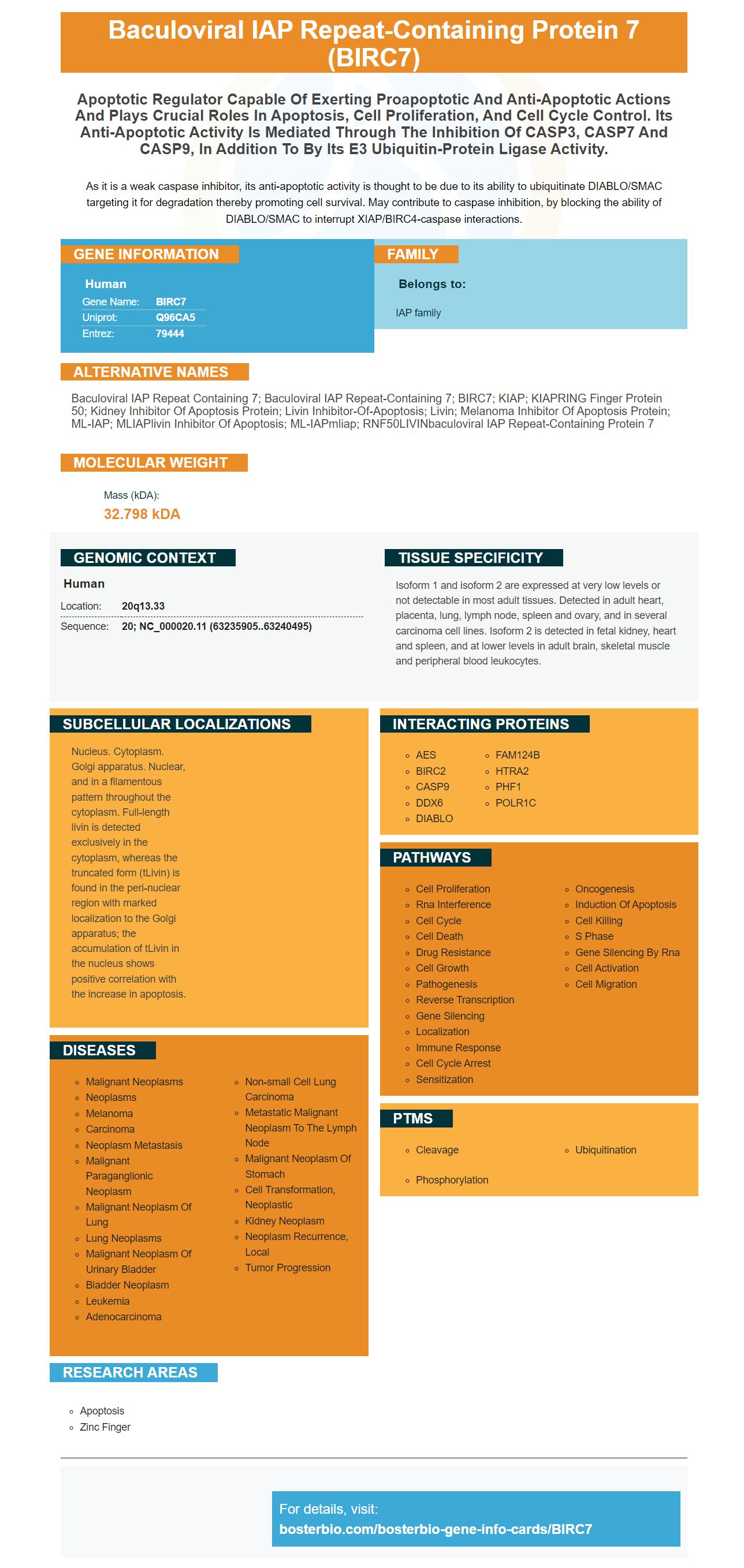
As it is a weak caspase inhibitor, its anti-apoptotic activity is thought to be due to its ability to ubiquitinate DIABLO/SMAC targeting it for degradation thereby promoting cell survival. May contribute to caspase inhibition, by blocking the ability of DIABLO/SMAC to interrupt XIAP/BIRC4-caspase interactions.
Human | |
---|---|
Gene Name: | BIRC7 |
Uniprot: | Q96CA5 |
Entrez: | 79444 |
Belongs to: |
---|
IAP family |
baculoviral IAP repeat containing 7; baculoviral IAP repeat-containing 7; BIRC7; KIAP; KIAPRING finger protein 50; Kidney inhibitor of apoptosis protein; livin inhibitor-of-apoptosis; Livin; Melanoma inhibitor of apoptosis protein; ML-IAP; MLIAPlivin inhibitor of apoptosis; ML-IAPmliap; RNF50LIVINbaculoviral IAP repeat-containing protein 7
Mass (kDA):
32.798 kDA
Human | |
---|---|
Location: | 20q13.33 |
Sequence: | 20; NC_000020.11 (63235905..63240495) |
Isoform 1 and isoform 2 are expressed at very low levels or not detectable in most adult tissues. Detected in adult heart, placenta, lung, lymph node, spleen and ovary, and in several carcinoma cell lines. Isoform 2 is detected in fetal kidney, heart and spleen, and at lower levels in adult brain, skeletal muscle and peripheral blood leukocytes.
Nucleus. Cytoplasm. Golgi apparatus. Nuclear, and in a filamentous pattern throughout the cytoplasm. Full-length livin is detected exclusively in the cytoplasm, whereas the truncated form (tLivin) is found in the peri-nuclear region with marked localization to the Golgi apparatus; the accumulation of tLivin in the nucleus shows positive correlation with the increase in apoptosis.
The BIRC7 marker is a member of the inhibitor of apoptosis protein (IAP) family that inhibits autophagy and promotes synergistic anti-tumor efficacy in PTC cells. This gene is expressed by Nthy-ori-3-1 cells and is applicable to scientists worldwide. Read on to learn more about this marker and how to use it to make your life better!
BIRC7 is a member of the IAP family. It is responsible for regulating a variety of biological processes, including apoptosis and autophagy. Its domains are zinc finger proteins and invariably contain three cysteines and one histidine. Although its function is not completely understood, it is thought to be involved in protein-protein interactions. It was first identified in the baculoviral protein, but has since been characterized in yeast, worms, insects, and mammalian cells. BIRC7 is also associated with the genetic disorder spinal muscular atrophy.
BIRC7 is part of a family of proteins called IAPs. These proteins are pivotal regulators of apoptosis and contribute to cancer formation. They control apoptosis by affecting various cell death signaling pathways. This protein is also associated with oncogenesis, and dysregulation of IAPs may be a cause of cancer.
These proteins were first identified in baculoviruses. Their role in promoting viral propagation is believed to be related to inhibiting apoptosis in the host insect cell. But their function in cellular inhibition of apoptosis is not fully understood. Researchers used public datasets and bioinformatics tools to examine the expression levels of IAPs in NSCLC patients. They also used gene expression profiling interactive analysis and Kaplan-Meier Plotter database to determine whether they were associated with a poor prognosis.
The IAP protein family includes genes such as BIRC7, which is a member of the TBRF gene. BIRC1 and BIRC2 are positively correlated with BIRC7, and BIRC3 and BIRC5 are negatively correlated with BIRC7. These proteins interact with other IAP genes to promote cell survival.
BIRC7 inhibits autophagy, leading to EMT-like changes in PCT cells. These cells have decreased levels of E-cadherin, N-cadherin, Vimentin, Snail, and ERCC2. In addition, the BIRC7 KD cell line showed a pronounced increase in RFP-positive red puncta.
In addition to inhibiting autophagy, BIRC7 also enhances migration and invasion of PTC cells. The effects of this effect were confirmed by western blotting using 48 pairs of PTC and control tissue samples. Using matrigel invasion and wound healing assays, BIRC7 overexpression or knockdown cells increased their migration and invasion capacity. Moreover, the knockdown and overexpression of BIRC7 significantly reduced the number of PTC cells.
BIRC7 represses Akt/mTOR signaling and autophagy in PTC cells. In a study using K-1 cells, BIRC7 inhibited the expression of PI3K and Akt. It also increased P62, LC3B, and p-mTOR. Although the inhibitor of BIRC7 inhibits autophagy, the results suggest that BIRC7 may be involved in the progression of papillary thyroid cancer.
Furthermore, BIRC7 represses the activity of FGFR2-IIIb, a protein that is associated with autophagy. Moreover, it also participates in tumour progression. As part of the autophagy pathway, DNAJB1 inhibits the activity of apoptosomes, and is associated with the essential tumour suppressor gene p53.
The researchers collected 187 cases of PTC from the Department of Thyroid Surgery at the Harbin Medical University. These patients were divided into two groups. Forty-three patients with PTC had lymph node metastasis, while the remaining patients did not. The samples were snap-frozen and stored at -80degC for qRT-PCR analyses. Additionally, the PTC tumor samples were compared with normal thyroid follicular tissue specimens to identify differences in autophagy. The samples were analyzed by two endocrine pathologists.
The pharmacological effect of BIRC7 on PTC cells has been confirmed in an experimental lung metastasis model. BIRC7 knockdown cells show a significant reduction in the formation of pulmonary nodules. Furthermore, cells transfected with the BIRC7 KD virus vector showed a significant decrease in the number of lung tumors. Furthermore, these cells exhibited reduced levels of E-cadherin, N-cadherin, Vimentin, and Snail.
These results highlight the importance of BIRC7 in the development of cancer. Tumor metastasis relies on the progression of cancer cells through several steps. One such step is epithelial-to-mesenchymal transition (EMT), which involves the loss of characteristic epithelial properties such as adhesion and motility. Moreover, BIRC7 confers increased migratory potential and ability to invade surrounding tissues.
The anti-BIRC7 antibody was derived from two sources. The P62 antibody was supplied by the Abcam company and the BECN1 antibody by Cell Signaling Technology. The other two anti-cadherin antibodies were supplied by Biosmedi Co., which is located in Shanghai, China. All antibodies were synthesized in a volume of 10 mL using a Superscript III First-Strand Synthesis System. The primers used for BIRC7 and b-actin were 5'-CATGTGTCTAGGC-3'.
Boster Bio BIRC7 enhanced the anti-tumor activity of PTC cells by enhancing their ability to interact with tumor-associated proteins. This enhanced anti-tumor activity is beneficial for cancer patients, but it is also important for promoting healthy cell growth. These compounds are also beneficial for patients with weakened immune systems.
The expression of BIRC7 is not high in Nthy-ori-3-1 cells, but it is elevated in other cell lines, including those from PTC. This protein is barely expressed in normal tissues, but is remarkably elevated in PTC tissue. Because of its role in PTC progression, inhibitors of BIRC7 have been studied in preclinical settings. In addition to demonstrating the potential therapeutic utility of these inhibitors, BIRC7 may also be useful for research on tumor progression.
To determine whether BIRC7 is expressed in Nthsy-ori-3-1 cells, we performed a cell proliferation assay. Cells were treated with different concentrations of PLX4032 for 24 h. The proportion of surviving cells was determined using the XTT cell proliferation assay. We then determined the IC50 of PLX4032 using Nthy-ori-3-1 cells and BCPAP cells.
In addition to detecting BIRC7 expression, we investigated lipid peroxidation using the MDA assay. The MDA levels of Nthy-ori-3-1 cells exposed to CdCl2 were significantly increased. In addition, quercetin 5 mM attenuated this effect. This result demonstrates that the ER is failing to reverse the damaging effect of a toxic environment.
This mutation stabilizes the NAF-1 cluster and results in an increase in mitochondrial iron and reactive oxygen species. It also reduces cellular tolerance to oxidative stress. Therefore, BIRC7 may be a viable target for treatment of breast cancer patients with high levels of NAF-1 expression. If the 3Cys-1His cluster can be stabilized, drugs targeting the tumor might be effective in reducing tumor size and increasing cellular sensitivity to oxidative stress.
BIRC7 is an essential regulator of autophagy, a conserved process required for cell growth and homeostasis. However, the BIRC7 protein is also involved in tumor progression, as it controls autophagic processes and regulates cellular migration and invasion. Recently, researchers have shown that silencing BIRC7 increases autophagic activity and sensitizes cancer cells to chemotherapy.
The suppression of autophagy by BIRC7 results in EMT-like changes in PCT cells. BIRC7 KD cells are found to contain lower levels of E-cadherin and N-cadherin, and to express low levels of Vimentin and Snail. Moreover, these cells are more likely to exhibit the BIRC7 KD-associated EMT phenotype, which suggests that they are predisposed to tumor development.
Interestingly, the BIRC7 protein is required for the progression of PTC and is essential for tumor growth. The expression of BIRC7 in cancer cells is high, making it an attractive therapeutic target. But it is not known exactly what function BIRC7 plays in cancer. Researchers are attempting to find ways to inhibit its activity and improve the disease's prognosis.
BIRC7 expression is related to EMT, a critical step in the progression of tumor growth. Knocking down BIRC7 decreased mesenchymal markers, such as snail, while overexpression of the protein had the opposite effect. The results suggest that BIRC7 inhibits autophagy in BIRC7 KD cells.
To assess whether the Boster Bio BIRC7 marker inhibited autophagy in BIRC7 KL cells, researchers isolated total RNA from PTC cells. Total RNA was assessed by agarose gel electrophoresis to determine integrity of the RNA. From this, 1 mg of cDNA was synthesized in 10 mL using the Superscript III First-Strand Synthesis System. qRT-PCR was performed using BIRC7-specific primers (5'-CATGTATCCAGGTGTATCG-3').
PMID: 11162435 by Lin J.-H., et al. KIAP, a novel member of the inhibitor of apoptosis protein family.
PMID: 11322947 by Ashhab Y., et al. Two splicing variants of a new inhibitor of apoptosis gene with different biological properties and tissue distribution pattern.
*More publications can be found for each product on its corresponding product page