This website uses cookies to ensure you get the best experience on our website.
- Table of Contents

17 Q&As


Facts about Adenosine deaminase.

Modulates signaling by extracellular adenosine, and so leads indirectly to cellular signaling events. Its interaction with DPP4 modulates lymphocyte- epithelial cell adhesion (PubMed:11772392).
| Human | |
|---|---|
| Gene Name: | ADA |
| Uniprot: | P00813 |
| Entrez: | 100 |

| Belongs to: |
|---|
| metallo-dependent hydrolases superfamily |

ADA; ADA1; adenine deaminase; Adenosine aminohydrolase; Adenosine Deaminase; EC 3.5.4.4
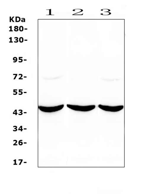
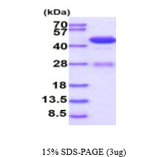
Mass (kDA):
40.764 kDA

| Human | |
|---|---|
| Location: | 20q13.12 |
| Sequence: | 20; NC_000020.11 (44619519..44651758, complement) |
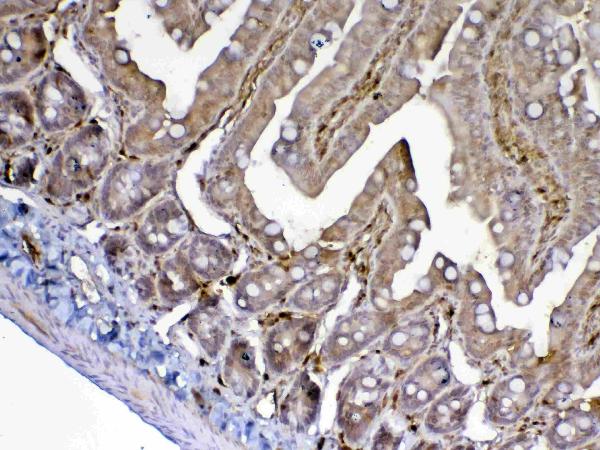
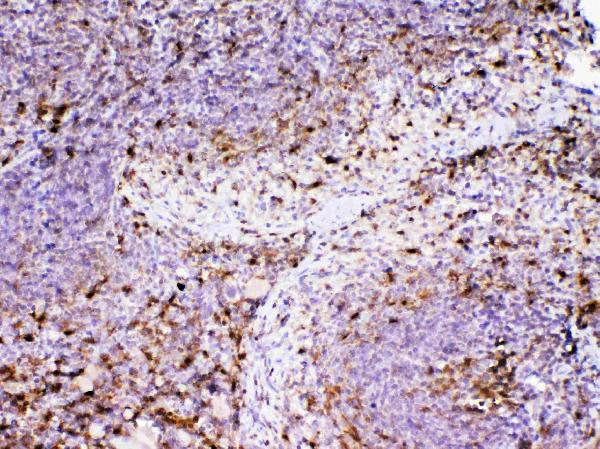
Found in all tissues, occurs in large amounts in T-lymphocytes (PubMed:20959412). Expressed at the time of weaning in gastrointestinal tissues.
Cell membrane; Peripheral membrane protein; Extracellular side. Cell junction. Cytoplasmic vesicle lumen. Cytoplasm. Lysosome. Colocalized with DPP4 at the cell surface.


In this Boster Bio review we will look at the ADA Marker, Boster Bio's IHC protocol and Steven's background. If you are planning to make use of the Marker We highly recommend that you read the FAQ page first. It is also worth checking out the resources on Boster Bio. These documents can be shared at no cost by researchers and educators, provided they provide a link to the source.
The ADA Marker Guide: The Best Uses includes a detailed procedure, illustrations, suggested solutions for troubleshooting, and reagents. The guide also includes detailed details on how to prepare IHC samples. It offers detailed information on the different methods for analysing samples as well as the most effective ones to use for each study. For the ADA Marker, Boster Bio is a good option.
The ADA Marker is used for identification of accessible facilities and services. The American Disabilities Act prohibits discrimination against disabled people in a variety of areas, including employment, transportation, and local and government-run sites. The ADA Marker can also be an excellent way to identify the severity of tuberculosis or to rule out the illness. Here are a few of the most frequently used applications of the ADA Marker.
Portable display elements are a perfect example of this rule, since they are often transported into temporary spaces for events. While they don't need to be in compliance with ADA Standards, they should be situated close to an accessible road and allow for wheelchairs and scooters. In addition to this there should be signs on the sides that clearly show that they are ADA-compliant. It is important to remember, however, that the ADA Marker does not replace accessible facilities.
Tissue samples are sourced from a variety of sources such as biopsy, surgery and animal models. Autopsy samples can only be collected after the animal has been dead for at least two hours. This makes them less suitable for IHC. This kind of tissue requires more time for fixation and preservation than other tissues, since the antigens may denature or disappear after two hours. To determine antigens, the tissue sections must be removed as soon as possible.
Blocking background staining is the initial step in the IHC protocol. Triton X-100 is used to destroy any endogenous substances and also to alter the structure of the membrane. It is important to remember that endogenous tissues typically contain fluorescent molecules which can cause false positive results. Before proceeding with the IHC procedure it is imperative to scrutinize the tissue section for any endogenous background.
The IHC protocol includes the crucial step of fixing the tissue. This will ensure that you save as much antigen as you can. The ideal situation is that the tissue sample is fixed in order to maintain the smallest amount of structure. Furthermore, fixing should ensure the integrity of cell morphology and the readable structure of the tissue. Fixing too much tissue can reduce the antigenicity of the tissue and lead to false negative results. Follow the manufacturer's directions to correct the fixation of tissue.
After the sample is fixed, the primary antibody should be reduced by Incubation Buffer. After the primary antibody has been diluted in Incubation Buffer it should be incubated in the sample for at minimum 4 hours. The ideal incubation time for R&D Systems antibodies are overnight at 2-8 degC. The overnight staining is critical to ensure that the antibody is specific enough the antibody to tissue targets while reducing background staining non-specific to the target. It is possible to alter certain parameters to optimize the process for your specific system.
PMID: 6090454 by Daddona P.E., et al. Human adenosine deaminase. cDNA and complete primary amino acid sequence.
PMID: 6546794 by Wiginton D.A., et al. Sequence of human adenosine deaminase cDNA including the coding region and a small intron.