Click image to see more details
-

-
-
-
-
+1
Product Info Summary
| SKU: | A03364-1 |
|---|---|
| Size: | 100 μg/vial |
| Reactive Species: | Human, Mouse, Rat |
| Host: | Rabbit |
| Application: | ELISA, IHC, WB |
Customers Who Bought This Also Bought

Product info
Product Name
Anti-Vitamin D Binding protein/GC Antibody Picoband™
View all Vitamin D BP Antibodies
SKU/Catalog Number
A03364-1
Size
100 μg/vial
Form
Lyophilized
Description
Boster Bio Anti-Vitamin D Binding protein/GC Antibody Picoband™ catalog # A03364-1. Tested in ELISA, IHC, WB applications. This antibody reacts with Human, Mouse, Rat.
Storage & Handling
Store at -20˚C for one year from date of receipt. After reconstitution, at 4˚C for one month. It can also be aliquotted and stored frozen at -20˚C for six months. Avoid repeated freeze-thaw cycles.
Cite This Product
Anti-Vitamin D Binding protein/GC Antibody Picoband™ (Boster Biological Technology, Pleasanton CA, USA, Catalog # A03364-1)
Host
Rabbit
Contents
Each vial contains 4mg Trehalose, 0.9mg NaCl, 0.2mg Na2HPO4, 0.05mg NaN3.
Clonality
Polyclonal
Isotype
Rabbit IgG
Immunogen
E. coli-derived human Vitamin D Binding protein recombinant protein (Position: L17-E256).
*Blocking peptide can be purchased. Costs vary based on immunogen length. Contact us for pricing.
Cross-reactivity
No cross-reactivity with other proteins.
Reactive Species
A03364-1 is reactive to GC in Human, Mouse, Rat
Applications
A03364-1 is guaranteed for ELISA, IHC, WB Boster Guarantee
Observed Molecular Weight
53 kDa
Calculated molecular weight
Background of Vitamin D BP
Vitamin D-binding protein, also/originally known as gc-globulin (group-specific component), is a protein that in humans is encoded by the GC gene.The protein encoded by this gene belongs to the albumin gene family. It is a multifunctional protein found in plasma, ascitic fluid, cerebrospinal fluid and on the surface of many cell types. It binds to vitamin D and its plasma metabolites and transports them to target tissues.
Antibody Validation
Boster validates all antibodies on WB, IHC, ICC, Immunofluorescence, and ELISA with known positive control and negative samples to ensure specificity and high affinity, including thorough antibody incubations.
Assay dilution & Images
Reconstitution
Add 0.2ml of distilled water will yield a concentration of 500ug/ml.
Assay Dilutions Recommendation
The recommendations below provide a starting point for assay optimization. The actual working concentration varies and should be decided by the user.
Western blot, 0.1-0.5μg/ml
Immunohistochemistry (Paraffin-embedded Section), 0.5-1μg/ml
Direct ELISA, 0.1-0.5μg/ml
Validation Images & Assay Conditions
Click image to see more details
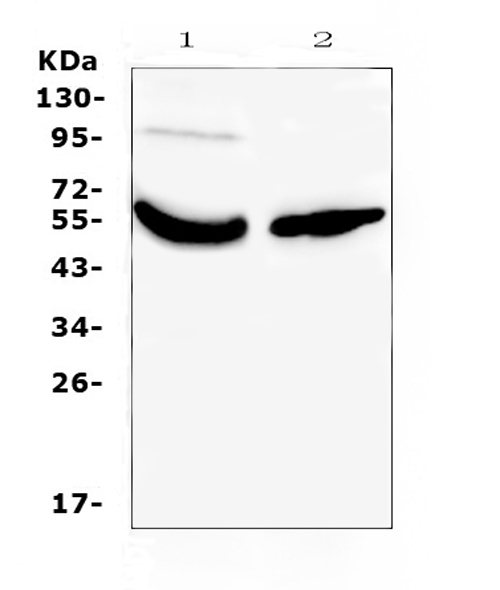
Figure 1. Western blot analysis of Vitamin D Binding protein using anti-Vitamin D Binding protein antibody (A03364-1).
Electrophoresis was performed on a 5-20% SDS-PAGE gel at 70V (Stacking gel) / 90V (Resolving gel) for 2-3 hours. The sample well of each lane was loaded with 50ug of sample under reducing conditions.
Lane 1: human placenta tissue lysates,
Lane 2: human A431 whole cell lysates.
After Electrophoresis, proteins were transferred to a Nitrocellulose membrane at 150mA for 50-90 minutes. Blocked the membrane with 5% Non-fat Milk/ TBS for 1.5 hour at RT. The membrane was incubated with rabbit anti-Vitamin D Binding protein antigen affinity purified polyclonal antibody (Catalog # A03364-1) at 0.5 ug/mL overnight at 4 then washed with TBS-0.1%Tween 3 times with 5 minutes each and probed with a goat anti-rabbit IgG-HRP secondary antibody at a dilution of 1:10000 for 1.5 hour at RT. The signal is developed using an Enhanced Chemiluminescent detection (ECL) kit (Catalog # EK1002) with Tanon 5200 system. A specific band was detected for Vitamin D Binding protein at approximately 53KD. The expected band size for Vitamin D Binding protein is at 53KD.

Click image to see more details
Figure 2. IHC analysis of Vitamin D Binding protein using anti-Vitamin D Binding protein antibody (A03364-1).
Vitamin D Binding protein was detected in paraffin-embedded section of human liver cancer tissue. Heat mediated antigen retrieval was performed in citrate buffer (pH6, epitope retrieval solution) for 20 mins. The tissue section was blocked with 10% goat serum. The tissue section was then incubated with 1ug/ml rabbit anti-Vitamin D Binding protein Antibody (A03364-1) overnight at 4 Biotinylated goat anti-rabbit IgG was used as secondary antibody and incubated for 30 minutes at 37 The tissue section was developed using Strepavidin-Biotin-Complex (SABC)(Catalog # SA1022) with DAB as the chromogen.

Click image to see more details
Figure 3. IHC analysis of Vitamin D Binding protein using anti-Vitamin D Binding protein antibody (A03364-1).
Vitamin D Binding protein was detected in paraffin-embedded section of human lung cancer tissue. Heat mediated antigen retrieval was performed in citrate buffer (pH6, epitope retrieval solution) for 20 mins. The tissue section was blocked with 10% goat serum. The tissue section was then incubated with 1ug/ml rabbit anti-Vitamin D Binding protein Antibody (A03364-1) overnight at 4 Biotinylated goat anti-rabbit IgG was used as secondary antibody and incubated for 30 minutes at 37 The tissue section was developed using Strepavidin-Biotin-Complex (SABC)(Catalog # SA1022) with DAB as the chromogen.

Click image to see more details
Figure 4. IHC analysis of Vitamin D Binding protein using anti-Vitamin D Binding protein antibody (A03364-1).
Vitamin D Binding protein was detected in paraffin-embedded section of mouse liver tissue. Heat mediated antigen retrieval was performed in citrate buffer (pH6, epitope retrieval solution) for 20 mins. The tissue section was blocked with 10% goat serum. The tissue section was then incubated with 1ug/ml rabbit anti-Vitamin D Binding protein Antibody (A03364-1) overnight at 4 Biotinylated goat anti-rabbit IgG was used as secondary antibody and incubated for 30 minutes at 37 The tissue section was developed using Strepavidin-Biotin-Complex (SABC)(Catalog # SA1022) with DAB as the chromogen.

Click image to see more details
Figure 5. IHC analysis of Vitamin D Binding protein using anti-Vitamin D Binding protein antibody (A03364-1).
Vitamin D Binding protein was detected in paraffin-embedded section of human rectal cancer tissue. Heat mediated antigen retrieval was performed in citrate buffer (pH6, epitope retrieval solution) for 20 mins. The tissue section was blocked with 10% goat serum. The tissue section was then incubated with 1ug/ml rabbit anti-Vitamin D Binding protein Antibody (A03364-1) overnight at 4 Biotinylated goat anti-rabbit IgG was used as secondary antibody and incubated for 30 minutes at 37 The tissue section was developed using Strepavidin-Biotin-Complex (SABC)(Catalog # SA1022) with DAB as the chromogen.
Protein Target Info & Infographic
Gene/Protein Information For GC (Source: Uniprot.org, NCBI)
Gene Name
GC
Full Name
Vitamin D-binding protein
Weight
Superfamily
ALB/AFP/VDB family
Alternative Names
DBP; DBP/GC; DBP-MAF; DBPVDBG; EC 6.3.1.5; Gc; gc-globulin; Gc-MAF; GRD3; group-specific component (vitamin D binding protein); Group-specific component; hDBP; VDB; VDBG; VDBP; Vitamin D BP; vitamin D-binding alpha-globulin; vitamin D-binding protein GC DBP, DBP-maf, DBP/GC, GRD3, Gc-MAF, GcMAF, HEL-S-51, VDB, VDBG, VDBP GC vitamin D binding protein vitamin D-binding protein|epididymis secretory protein Li 51|gc protein-derived macrophage activating factor|gc-globulin|group-specific component (vitamin D binding protein)|vitamin D-binding alpha-globulin|vitamin D-binding protein-macrophage activating factor
*If product is indicated to react with multiple species, protein info is based on the gene entry specified above in "Species".For more info on GC, check out the GC Infographic

We have 30,000+ of these available, one for each gene! Check them out.
In this infographic, you will see the following information for GC: database IDs, superfamily, protein function, synonyms, molecular weight, chromosomal locations, tissues of expression, subcellular locations, post-translational modifications, and related diseases, research areas & pathways. If you want to see more information included, or would like to contribute to it and be acknowledged, please contact [email protected].
Specific Publications For Anti-Vitamin D Binding protein/GC Antibody Picoband™ (A03364-1)
Hello CJ!
No publications found for A03364-1
*Do you have publications using this product? Share with us and receive a reward. Ask us for more details.
Recommended Resources
Here are featured tools and databases that you might find useful.
- Boster's Pathways Library
- Protein Databases
- Bioscience Research Protocol Resources
- Data Processing & Analysis Software
- Photo Editing Software
- Scientific Literature Resources
- Research Paper Management Tools
- Molecular Biology Software
- Primer Design Tools
- Bioinformatics Tools
- Phylogenetic Tree Analysis
Customer Reviews
Have you used Anti-Vitamin D Binding protein/GC Antibody Picoband™?
Submit a review and receive an Amazon gift card.
- $30 for a review with an image
0 Reviews For Anti-Vitamin D Binding protein/GC Antibody Picoband™
Customer Q&As
Have a question?
Find answers in Q&As, reviews.
Can't find your answer?
Submit your question
4 Customer Q&As for Anti-Vitamin D Binding protein/GC Antibody Picoband™
Question
Is this A03364-1 anti-Vitamin D Binding protein/GC antibody reactive to the isotypes of GC?
Verified Customer
Verified customer
Asked: 2019-06-26
Answer
The immunogen of A03364-1 anti-Vitamin D Binding protein/GC antibody is E. coli-derived human Vitamin D Binding protein recombinant protein (Position: L17-E256). Could you tell me which isotype you are interested in so I can help see if the immunogen is part of this isotype?
Boster Scientific Support
Answered: 2019-06-26
Question
Will A03364-1 anti-Vitamin D Binding protein/GC antibody work on parafin embedded sections? If so, which fixation method do you recommend we use (PFA, paraformaldehyde, other)?
Verified Customer
Verified customer
Asked: 2019-05-06
Answer
It shows on the product datasheet, A03364-1 anti-Vitamin D Binding protein/GC antibody as been validated on WB. It is best to use PFA for fixation because it has better tissue penetration ability. PFA needs to be prepared fresh before use. Long term stored PFA turns into formalin, as the PFA molecules congregate and become formalin.
Boster Scientific Support
Answered: 2019-05-06
Question
I was wanting to use your anti-Vitamin D Binding protein/GC antibody for WB for rat liver on frozen tissues, but I want to know if it has been validated for this particular application. Has this antibody been validated and is this antibody a good choice for rat liver identification?
D. Krishna
Verified customer
Asked: 2013-10-02
Answer
It shows on the product datasheet, A03364-1 anti-Vitamin D Binding protein/GC antibody has been validated for ELISA, IHC, WB on human, mouse, rat tissues. We have an innovator award program that if you test this antibody and show it works in rat liver in IHC-frozen, you can get your next antibody for free.
Boster Scientific Support
Answered: 2013-10-02
Question
We appreciate helping with my inquiry over the phone. Here are the WB image, lot number and protocol we used for liver using anti-Vitamin D Binding protein/GC antibody A03364-1. Let me know if you need anything else.
O. Zhao
Verified customer
Asked: 2013-01-17
Answer
We appreciate the data. You have provided everything we needed. Our lab team are working to resolve your inquiry as quickly as possible, and we appreciate your patience and understanding! Please let me know if there is anything you need in the meantime.
Boster Scientific Support
Answered: 2013-01-17
