Click image to see more details
-

-
-
-
-
+11
Product Info Summary
| SKU: | A05017 |
|---|---|
| Size: | 100 µg/vial |
| Reactive Species: | Human, Mouse, Rat |
| Host: | Rabbit |
| Application: | ELISA, Flow Cytometry, IF, IHC, ICC, WB |
Customers Who Bought This Also Bought


Product info
Product Name
Anti-Stanniocalcin 2/STC2 Antibody Picoband™
View all Stanniocalcin 2/STC-2 Antibodies
SKU/Catalog Number
A05017
Size
100 µg/vial
Form
Lyophilized
Description
Boster Bio Anti-Stanniocalcin 2/STC2 Antibody Picoband™ catalog # A09658. Tested in WB, IHC, ICC/IF, IF, FCM, ELISA applications. This antibody reacts with Human, Mouse, Rat.
Storage & Handling
At -20°C for one year from date of receipt. After reconstitution, at 4°C for one month. It can also be aliquotted and stored frozen at -20°C for six months. Avoid repeated freezing and thawing.
Cite This Product
Anti-Stanniocalcin 2/STC2 Antibody Picoband™ (Boster Biological Technology, Pleasanton CA, USA, Catalog # A05017)
Host
Rabbit
Contents
Each vial contains 4 mg Trehalose, 0.9 mg NaCl, 0.2 mg Na2HPO4.
Clonality
Polyclonal
Isotype
IgG
Immunogen
E.coli-derived human Stanniocalcin 2/STC2 recombinant protein (Position: A51-Q226). Human STC2 shares 90.3% and 89.8% amino acid (aa) sequence identity with mouse and rat STC2, respectively.
*Blocking peptide can be purchased. Costs vary based on immunogen length. Contact us for pricing.
Cross-reactivity
No cross reactivity with other proteins.
Reactive Species
A05017 is reactive to STC2 in Human, Mouse, Rat
Applications
A05017 is guaranteed for ELISA, Flow Cytometry, IF, IHC, ICC, WB Boster Guarantee
Observed Molecular Weight
33 kDa
Calculated molecular weight
52588 MW
Background of Stanniocalcin 2/STC-2
Stanniocalcin-2 is a protein that in humans is encoded by the STC2 gene. This gene encodes a secreted, homodimeric glycoprotein that is expressed in a wide variety of tissues and may have autocrine or paracrine functions. The encoded protein has 10 of its 15 cysteine residues conserved among stanniocalcin family members and is phosphorylated by casein kinase 2 exclusively on its serine residues. Its C-terminus contains a cluster of histidine residues which may interact with metal ions. The protein may play a role in the regulation of renal and intestinal calcium and phosphate transport, cell metabolism, or cellular calcium/phosphate homeostasis. Constitutive overexpression of human stanniocalcin 2 in mice resulted in pre- and postnatal growth restriction, reduced bone and skeletal muscle growth, and organomegaly. Expression of this gene is induced by estrogen and altered in some breast cancers.
Antibody Validation
Boster validates all antibodies on WB, IHC, ICC, Immunofluorescence, and ELISA with known positive control and negative samples to ensure specificity and high affinity, including thorough antibody incubations.
Assay dilution & Images
Reconstitution
Adding 0.2 ml of distilled water will yield a concentration of 500 µg/ml.
Assay Dilutions Recommendation
The recommendations below provide a starting point for assay optimization. The actual working concentration varies and should be decided by the user.
Western blot, 0.25-0.5 µg/ml, Human, Mouse, Rat
Immunohistochemistry, 2-5 µg/ml, Human
Immunocytochemistry/Immunofluorescence, 5 µg/ml, Human
Immunofluorescence, 5 µg/ml, Human
Flow Cytometry (Fixed), 1-3 µg /1x106 cells, Human
ELISA, 0.1-0.5 µg/ml, Human
Validation Images & Assay Conditions
Click image to see more details
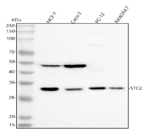
Figure 1. Western blot analysis of Stanniocalcin 2/STC2 using anti-Stanniocalcin 2/STC2 antibody (A05017).
Electrophoresis was performed on a 5-20% SDS-PAGE gel at 70V (Stacking gel) / 90V (Resolving gel) for 2-3 hours. The sample well of each lane was loaded with 30 ug of sample under reducing conditions.
Lane 1: human MCF-7 whole cell lysates,
Lane 2: human Caco-2 whole cell lysates,
Lane 3: rat PC-12 whole cell lysates,
Lane 4: mouse Raw264.7 whole cell lysates.
After electrophoresis, proteins were transferred to a nitrocellulose membrane at 150 mA for 50-90 minutes. Blocked the membrane with 5% non-fat milk/TBS for 1.5 hour at RT. The membrane was incubated with rabbit anti-Stanniocalcin 2/STC2 antigen affinity purified polyclonal antibody (Catalog # A05017) at 0.5 μg/mL overnight at 4°C, then washed with TBS-0.1%Tween 3 times with 5 minutes each and probed with a goat anti-rabbit IgG-HRP secondary antibody at a dilution of 1:5000 for 1.5 hour at RT. The signal is developed using an Enhanced Chemiluminescent detection (ECL) kit (Catalog # EK1002) with Tanon 5200 system. A specific band was detected for Stanniocalcin 2/STC2 at approximately 33 kDa. The expected band size for Stanniocalcin 2/STC2 is at 33 kDa.

Click image to see more details
Figure 2. IHC analysis of Stanniocalcin 2/STC2 using anti-Stanniocalcin 2/STC2 antibody (A05017).
Stanniocalcin 2/STC2 was detected in a paraffin-embedded section of human breast cancer tissue. Heat mediated antigen retrieval was performed in EDTA buffer (pH 8.0, epitope retrieval solution). The tissue section was blocked with 10% goat serum. The tissue section was then incubated with 2 μg/ml rabbit anti-Stanniocalcin 2/STC2 Antibody (A05017) overnight at 4°C. Peroxidase Conjugated Goat Anti-rabbit IgG was used as secondary antibody and incubated for 30 minutes at 37°C. The tissue section was developed using HRP Conjugated Rabbit IgG Super Vision Assay Kit (Catalog # SV0002) with DAB as the chromogen.

Click image to see more details
Figure 3. IHC analysis of Stanniocalcin 2/STC2 using anti-Stanniocalcin 2/STC2 antibody (A05017).
Stanniocalcin 2/STC2 was detected in a paraffin-embedded section of human breast cancer tissue. Heat mediated antigen retrieval was performed in EDTA buffer (pH 8.0, epitope retrieval solution). The tissue section was blocked with 10% goat serum. The tissue section was then incubated with 2 μg/ml rabbit anti-Stanniocalcin 2/STC2 Antibody (A05017) overnight at 4°C. Peroxidase Conjugated Goat Anti-rabbit IgG was used as secondary antibody and incubated for 30 minutes at 37°C. The tissue section was developed using HRP Conjugated Rabbit IgG Super Vision Assay Kit (Catalog # SV0002) with DAB as the chromogen.

Click image to see more details
Figure 4. IHC analysis of Stanniocalcin 2/STC2 using anti-Stanniocalcin 2/STC2 antibody (A05017).
Stanniocalcin 2/STC2 was detected in a paraffin-embedded section of human lung squamous cell carcinoma tissue. Heat mediated antigen retrieval was performed in EDTA buffer (pH 8.0, epitope retrieval solution). The tissue section was blocked with 10% goat serum. The tissue section was then incubated with 2 μg/ml rabbit anti-Stanniocalcin 2/STC2 Antibody (A05017) overnight at 4°C. Peroxidase Conjugated Goat Anti-rabbit IgG was used as secondary antibody and incubated for 30 minutes at 37°C. The tissue section was developed using HRP Conjugated Rabbit IgG Super Vision Assay Kit (Catalog # SV0002) with DAB as the chromogen.

Click image to see more details
Figure 5. IHC analysis of Stanniocalcin 2/STC2 using anti-Stanniocalcin 2/STC2 antibody (A05017).
Stanniocalcin 2/STC2 was detected in a paraffin-embedded section of human lung squamous cell carcinoma tissue. Heat mediated antigen retrieval was performed in EDTA buffer (pH 8.0, epitope retrieval solution). The tissue section was blocked with 10% goat serum. The tissue section was then incubated with 2 μg/ml rabbit anti-Stanniocalcin 2/STC2 Antibody (A05017) overnight at 4°C. Peroxidase Conjugated Goat Anti-rabbit IgG was used as secondary antibody and incubated for 30 minutes at 37°C. The tissue section was developed using HRP Conjugated Rabbit IgG Super Vision Assay Kit (Catalog # SV0002) with DAB as the chromogen.

Click image to see more details
Figure 6. IHC analysis of Stanniocalcin 2/STC2 using anti-Stanniocalcin 2/STC2 antibody (A05017).
Stanniocalcin 2/STC2 was detected in a paraffin-embedded section of human placenta tissue. Heat mediated antigen retrieval was performed in EDTA buffer (pH 8.0, epitope retrieval solution). The tissue section was blocked with 10% goat serum. The tissue section was then incubated with 2 μg/ml rabbit anti-Stanniocalcin 2/STC2 Antibody (A05017) overnight at 4°C. Peroxidase Conjugated Goat Anti-rabbit IgG was used as secondary antibody and incubated for 30 minutes at 37°C. The tissue section was developed using HRP Conjugated Rabbit IgG Super Vision Assay Kit (Catalog # SV0002) with DAB as the chromogen.

Click image to see more details
Figure 7. IHC analysis of Stanniocalcin 2/STC2 using anti-Stanniocalcin 2/STC2 antibody (A05017).
Stanniocalcin 2/STC2 was detected in a paraffin-embedded section of human placenta tissue. Heat mediated antigen retrieval was performed in EDTA buffer (pH 8.0, epitope retrieval solution). The tissue section was blocked with 10% goat serum. The tissue section was then incubated with 2 μg/ml rabbit anti-Stanniocalcin 2/STC2 Antibody (A05017) overnight at 4°C. Peroxidase Conjugated Goat Anti-rabbit IgG was used as secondary antibody and incubated for 30 minutes at 37°C. The tissue section was developed using HRP Conjugated Rabbit IgG Super Vision Assay Kit (Catalog # SV0002) with DAB as the chromogen.

Click image to see more details
Figure 8. IHC analysis of Stanniocalcin 2/STC2 using anti-Stanniocalcin 2/STC2 antibody (A05017).
Stanniocalcin 2/STC2 was detected in a paraffin-embedded section of human rectum adenocarcinoma tissue. Heat mediated antigen retrieval was performed in EDTA buffer (pH 8.0, epitope retrieval solution). The tissue section was blocked with 10% goat serum. The tissue section was then incubated with 2 μg/ml rabbit anti-Stanniocalcin 2/STC2 Antibody (A05017) overnight at 4°C. Peroxidase Conjugated Goat Anti-rabbit IgG was used as secondary antibody and incubated for 30 minutes at 37°C. The tissue section was developed using HRP Conjugated Rabbit IgG Super Vision Assay Kit (Catalog # SV0002) with DAB as the chromogen.

Click image to see more details
Figure 9. IHC analysis of Stanniocalcin 2/STC2 using anti-Stanniocalcin 2/STC2 antibody (A05017).
Stanniocalcin 2/STC2 was detected in a paraffin-embedded section of human rectum adenocarcinoma tissue. Heat mediated antigen retrieval was performed in EDTA buffer (pH 8.0, epitope retrieval solution). The tissue section was blocked with 10% goat serum. The tissue section was then incubated with 2 μg/ml rabbit anti-Stanniocalcin 2/STC2 Antibody (A05017) overnight at 4°C. Peroxidase Conjugated Goat Anti-rabbit IgG was used as secondary antibody and incubated for 30 minutes at 37°C. The tissue section was developed using HRP Conjugated Rabbit IgG Super Vision Assay Kit (Catalog # SV0002) with DAB as the chromogen.

Click image to see more details
Figure 10. IHC analysis of Stanniocalcin 2/STC2 using anti-Stanniocalcin 2/STC2 antibody (A05017).
Stanniocalcin 2/STC2 was detected in a paraffin-embedded section of human testicular seminoma tissue. Heat mediated antigen retrieval was performed in EDTA buffer (pH 8.0, epitope retrieval solution). The tissue section was blocked with 10% goat serum. The tissue section was then incubated with 2 μg/ml rabbit anti-Stanniocalcin 2/STC2 Antibody (A05017) overnight at 4°C. Peroxidase Conjugated Goat Anti-rabbit IgG was used as secondary antibody and incubated for 30 minutes at 37°C. The tissue section was developed using HRP Conjugated Rabbit IgG Super Vision Assay Kit (Catalog # SV0002) with DAB as the chromogen.

Click image to see more details
Figure 11. IHC analysis of Stanniocalcin 2/STC2 using anti-Stanniocalcin 2/STC2 antibody (A05017).
Stanniocalcin 2/STC2 was detected in a paraffin-embedded section of human testicular seminoma tissue. Heat mediated antigen retrieval was performed in EDTA buffer (pH 8.0, epitope retrieval solution). The tissue section was blocked with 10% goat serum. The tissue section was then incubated with 2 μg/ml rabbit anti-Stanniocalcin 2/STC2 Antibody (A05017) overnight at 4°C. Peroxidase Conjugated Goat Anti-rabbit IgG was used as secondary antibody and incubated for 30 minutes at 37°C. The tissue section was developed using HRP Conjugated Rabbit IgG Super Vision Assay Kit (Catalog # SV0002) with DAB as the chromogen.

Click image to see more details
Figure 12. IF analysis of Stanniocalcin 2/STC2 using anti-Stanniocalcin 2/STC2 antibody (A05017).
Stanniocalcin 2/STC2 was detected in an immunocytochemical section of PC-3 cells. Enzyme antigen retrieval was performed using IHC enzyme antigen retrieval reagent (AR0022) for 15 mins. The cells were blocked with 10% goat serum. And then incubated with 5 μg/mL rabbit anti-Stanniocalcin 2/STC2 Antibody (A05017) overnight at 4°C. Cy3 Conjugated Goat Anti-Rabbit IgG (BA1032) was used as secondary antibody at 1:500 dilution and incubated for 30 minutes at 37°C. The section was counterstained with DAPI. Visualize using a fluorescence microscope and filter sets appropriate for the label used.

Click image to see more details
Figure 13. Flow Cytometry analysis of MCF-7 cells using anti-Stanniocalcin 2/STC2 antibody (A05017).
Overlay histogram showing MCF-7 cells stained with A05017 (Blue line). The cells were fixed with 4% paraformaldehyde and blocked with 10% normal goat serum. And then incubated with rabbit anti-Stanniocalcin 2/STC2 Antibody (A05017, 1 μg/1x106 cells) for 30 min at 20°C. DyLight®488 conjugated goat anti-rabbit IgG (BA1127, 5-10 μg/1x106 cells) was used as secondary antibody for 30 minutes at 20°C. Isotype control antibody (Green line) was rabbit IgG (1 μg/1x106) used under the same conditions. Unlabelled sample (Red line) was also used as a control.

Click image to see more details
Figure 14. IF analysis of Stanniocalcin 2/STC2 using anti-Stanniocalcin 2/STC2 antibody (A05017).
Stanniocalcin 2/STC2 was detected in a paraffin-embedded section of human breast cancer tissue. Heat mediated antigen retrieval was performed in EDTA buffer (pH 8.0, epitope retrieval solution). The tissue section was blocked with 10% goat serum. The tissue section was then incubated with 5 μg/mL rabbit anti-Stanniocalcin 2/STC2 Antibody (A05017) overnight at 4°C. DyLight®488 Conjugated Goat Anti-Rabbit IgG (BA1127) was used as secondary antibody at 1:500 dilution and incubated for 30 minutes at 37°C. The section was counterstained with DAPI. Visualize using a fluorescence microscope and filter sets appropriate for the label used.

Click image to see more details
Figure 15. IF analysis of Stanniocalcin 2/STC2 using anti-Stanniocalcin 2/STC2 antibody (A05017).
Stanniocalcin 2/STC2 was detected in a paraffin-embedded section of human lung cancer tissue. Heat mediated antigen retrieval was performed in EDTA buffer (pH 8.0, epitope retrieval solution). The tissue section was blocked with 10% goat serum. The tissue section was then incubated with 5 μg/mL rabbit anti-Stanniocalcin 2/STC2 Antibody (A05017) overnight at 4°C. DyLight®488 Conjugated Goat Anti-Rabbit IgG (BA1127) was used as secondary antibody at 1:500 dilution and incubated for 30 minutes at 37°C. The section was counterstained with DAPI. Visualize using a fluorescence microscope and filter sets appropriate for the label used.
Protein Target Info & Infographic
Gene/Protein Information For STC2 (Source: Uniprot.org, NCBI)
Gene Name
STC2
Full Name
Stanniocalcin-2
Weight
52588 MW
Superfamily
stanniocalcin family
Alternative Names
Stanniocalcin 2; stanniocalcin-2; STC2; STC-2; STC-2Stanniocalcin-related protein; STC-related protein; STCRP STC2 STC-2, STCRP stanniocalcin 2 stanniocalcin-2|STC-related protein|stanniocalcin-related protein
*If product is indicated to react with multiple species, protein info is based on the gene entry specified above in "Species".For more info on STC2, check out the STC2 Infographic

We have 30,000+ of these available, one for each gene! Check them out.
In this infographic, you will see the following information for STC2: database IDs, superfamily, protein function, synonyms, molecular weight, chromosomal locations, tissues of expression, subcellular locations, post-translational modifications, and related diseases, research areas & pathways. If you want to see more information included, or would like to contribute to it and be acknowledged, please contact [email protected].
Specific Publications For Anti-Stanniocalcin 2/STC2 Antibody Picoband™ (A05017)
Hello CJ!
No publications found for A05017
*Do you have publications using this product? Share with us and receive a reward. Ask us for more details.
Recommended Resources
Here are featured tools and databases that you might find useful.
- Boster's Pathways Library
- Protein Databases
- Bioscience Research Protocol Resources
- Data Processing & Analysis Software
- Photo Editing Software
- Scientific Literature Resources
- Research Paper Management Tools
- Molecular Biology Software
- Primer Design Tools
- Bioinformatics Tools
- Phylogenetic Tree Analysis
Customer Reviews
Have you used Anti-Stanniocalcin 2/STC2 Antibody Picoband™?
Submit a review and receive an Amazon gift card.
- $30 for a review with an image
0 Reviews For Anti-Stanniocalcin 2/STC2 Antibody Picoband™
Customer Q&As
Have a question?
Find answers in Q&As, reviews.
Can't find your answer?
Submit your question
