Click image to see more details
-

-
-
-
-
+3
Product Info Summary
| SKU: | A04392-1 |
|---|---|
| Size: | 100 µg/vial |
| Reactive Species: | Human |
| Host: | Rabbit |
| Application: | ELISA, IF, IHC, WB |
Customers Who Bought This Also Bought

Product info
Product Name
Anti-RFX1 Antibody Picoband™
SKU/Catalog Number
A04392-1
Size
100 µg/vial
Form
Lyophilized
Description
Boster Bio Anti-RFX1 Antibody Picoband™ catalog # A04392-1. Tested in ELISA, IF, IHC, WB applications. This antibody reacts with Human.
Storage & Handling
At -20°C for one year from date of receipt. After reconstitution, at 4°C for one month. It can also be aliquotted and stored frozen at -20°C for six months. Avoid repeated freezing and thawing.
Cite This Product
Anti-RFX1 Antibody Picoband™ (Boster Biological Technology, Pleasanton CA, USA, Catalog # A04392-1)
Host
Rabbit
Contents
Each vial contains 4 mg Trehalose, 0.9 mg NaCl, 0.2 mg Na2HPO4.
Clonality
Polyclonal
Isotype
Rabbit IgG
Immunogen
E.coli-derived human RFX1 recombinant protein (Position: A48-P977).
*Blocking peptide can be purchased. Costs vary based on immunogen length. Contact us for pricing.
Cross-reactivity
No cross-reactivity with other proteins.
Reactive Species
A04392-1 is reactive to RFX1 in Human
Applications
A04392-1 is guaranteed for ELISA, IF, IHC, WB Boster Guarantee
Observed Molecular Weight
135 kDa
Calculated molecular weight
Background of RFX1
MHC class II regulatory factor RFX1 is a protein that, in humans, is encoded by the RFX1 gene located on the short arm of chromosome 19. This gene encodes a member of the regulatory factor X (RFX) family of transcription factors, which are characterized by a winged-helix DNA-binding domain. The encoded transcription factor contains an N-terminal activation domain and a C-terminal repression domain, and may activate or repress target gene expression depending on cellular context. This transcription factor has been shown to regulate a wide variety of genes involved in immunity and cancer, including the MHC class II genes and genes that may be involved in cancer progression. This gene exhibits altered expression in glioblastoma and the autoimmune disease systemic lupus erythematosis (SLE).
Antibody Validation
Boster validates all antibodies on WB, IHC, ICC, Immunofluorescence, and ELISA with known positive control and negative samples to ensure specificity and high affinity, including thorough antibody incubations.
Assay dilution & Images
Reconstitution
Adding 0.2 ml of distilled water will yield a concentration of 500 µg/ml.
Assay Dilutions Recommendation
The recommendations below provide a starting point for assay optimization. The actual working concentration varies and should be decided by the user.
Western blot, 0.25-0.5 µg/ml, Human
Immunohistochemistry(Paraffin-embedded Section), 2-5 µg/ml, Human
Immunofluorescence, 5 µg/ml, Human
Direct ELISA, 0.1-0.5 µg/ml, Human
Validation Images & Assay Conditions
Click image to see more details
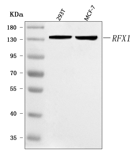
Figure 1. Western blot analysis of RFX1 using anti-RFX1 antibody (A04392-1).
Electrophoresis was performed on a 5-20% SDS-PAGE gel at 70V (Stacking gel) / 90V (Resolving gel) for 2-3 hours. The sample well of each lane was loaded with 30 ug of sample under reducing conditions.
Lane 1: human 293T whole cell lysates,
Lane 2: human MCF-7 whole cell lysates.
After electrophoresis, proteins were transferred to a nitrocellulose membrane at 150 mA for 50-90 minutes. Blocked the membrane with 5% non-fat milk/TBS for 1.5 hour at RT. The membrane was incubated with rabbit anti-RFX1 antigen affinity purified polyclonal antibody (Catalog # A04392-1) at 0.5 μg/mL overnight at 4°C, then washed with TBS-0.1%Tween 3 times with 5 minutes each and probed with a goat anti-rabbit IgG-HRP secondary antibody at a dilution of 1:5000 for 1.5 hour at RT. The signal is developed using an Enhanced Chemiluminescent detection (ECL) kit (Catalog # EK1002) with Tanon 5200 system. A specific band was detected for RFX1 at approximately 135 kDa. The expected band size for RFX1 is at 105 kDa.

Click image to see more details
Figure 2. IHC analysis of RFX1 using anti-RFX1 antibody (A04392-1).
RFX1 was detected in a paraffin-embedded section of Diffuse large B-cell lymphoma of human intestine tissue. Heat mediated antigen retrieval was performed in EDTA buffer (pH 8.0, epitope retrieval solution). The tissue section was blocked with 10% goat serum. The tissue section was then incubated with 2 μg/ml rabbit anti-RFX1 Antibody (A04392-1) overnight at 4°C. Peroxidase Conjugated Goat Anti-rabbit IgG was used as secondary antibody and incubated for 30 minutes at 37°C. The tissue section was developed using HRP Conjugated Rabbit IgG Super Vision Assay Kit (Catalog # SV0002) with DAB as the chromogen.

Click image to see more details
Figure 3. IHC analysis of RFX1 using anti-RFX1 antibody (A04392-1).
RFX1 was detected in a paraffin-embedded section of human adrenocortical adenoma tissue. Heat mediated antigen retrieval was performed in EDTA buffer (pH 8.0, epitope retrieval solution). The tissue section was blocked with 10% goat serum. The tissue section was then incubated with 2 μg/ml rabbit anti-RFX1 Antibody (A04392-1) overnight at 4°C. Peroxidase Conjugated Goat Anti-rabbit IgG was used as secondary antibody and incubated for 30 minutes at 37°C. The tissue section was developed using HRP Conjugated Rabbit IgG Super Vision Assay Kit (Catalog # SV0002) with DAB as the chromogen.

Click image to see more details
Figure 4. IHC analysis of RFX1 using anti-RFX1 antibody (A04392-1).
RFX1 was detected in a paraffin-embedded section of human Gastric adenocarcinoma tissue. Heat mediated antigen retrieval was performed in EDTA buffer (pH 8.0, epitope retrieval solution). The tissue section was blocked with 10% goat serum. The tissue section was then incubated with 2 μg/ml rabbit anti-RFX1 Antibody (A04392-1) overnight at 4°C. Peroxidase Conjugated Goat Anti-rabbit IgG was used as secondary antibody and incubated for 30 minutes at 37°C. The tissue section was developed using HRP Conjugated Rabbit IgG Super Vision Assay Kit (Catalog # SV0002) with DAB as the chromogen.

Click image to see more details
Figure 5. IHC analysis of RFX1 using anti-RFX1 antibody (A04392-1).
RFX1 was detected in a paraffin-embedded section of human lung adenocarcinoma tissue. Heat mediated antigen retrieval was performed in EDTA buffer (pH 8.0, epitope retrieval solution). The tissue section was blocked with 10% goat serum. The tissue section was then incubated with 2 μg/ml rabbit anti-RFX1 Antibody (A04392-1) overnight at 4°C. Peroxidase Conjugated Goat Anti-rabbit IgG was used as secondary antibody and incubated for 30 minutes at 37°C. The tissue section was developed using HRP Conjugated Rabbit IgG Super Vision Assay Kit (Catalog # SV0002) with DAB as the chromogen.

Click image to see more details
Figure 6. IHC analysis of RFX1 using anti-RFX1 antibody (A04392-1).
RFX1 was detected in a paraffin-embedded section of human testicular seminoma tissue. Heat mediated antigen retrieval was performed in EDTA buffer (pH 8.0, epitope retrieval solution). The tissue section was blocked with 10% goat serum. The tissue section was then incubated with 2 μg/ml rabbit anti-RFX1 Antibody (A04392-1) overnight at 4°C. Peroxidase Conjugated Goat Anti-rabbit IgG was used as secondary antibody and incubated for 30 minutes at 37°C. The tissue section was developed using HRP Conjugated Rabbit IgG Super Vision Assay Kit (Catalog # SV0002) with DAB as the chromogen.

Click image to see more details
Figure 7. IF analysis of RFX1 using anti-RFX1 antibody (A04392-1).
RFX1 was detected in a paraffin-embedded section of human esophageal squamous cell carcinoma tissue. Heat mediated antigen retrieval was performed in EDTA buffer (pH 8.0, epitope retrieval solution). The tissue section was blocked with 10% goat serum. The tissue section was then incubated with 5 μg/mL rabbit anti-RFX1 Antibody (A04392-1) overnight at 4°C. DyLight®550 Conjugated Goat Anti-Rabbit IgG (BA1135) was used as secondary antibody at 1:500 dilution and incubated for 30 minutes at 37°C. Visualize using a fluorescence microscope and filter sets appropriate for the label used.
Protein Target Info & Infographic
Gene/Protein Information For RFX1 (Source: Uniprot.org, NCBI)
Gene Name
RFX1
Full Name
MHC class II regulatory factor RFX1
Weight
Superfamily
RFX family
Alternative Names
EFC; EF-C; Enhancer factor C; MHC class II regulatory factor RFX; MHC class II regulatory factor RFX1; Regulatory factor X 1; regulatory factor X, 1 (influences HLA class II expression); RFX; trans-acting regulatory factor 1; Transcription factor RFX1 RFX1 EFC, RFX regulatory factor X1 MHC class II regulatory factor RFX1|MHC class II regulatory factor RFX|enhancer factor C|regulatory factor X, 1 (influences HLA class II expression)|trans-acting regulatory factor 1|transcription factor RFX1
*If product is indicated to react with multiple species, protein info is based on the gene entry specified above in "Species".For more info on RFX1, check out the RFX1 Infographic

We have 30,000+ of these available, one for each gene! Check them out.
In this infographic, you will see the following information for RFX1: database IDs, superfamily, protein function, synonyms, molecular weight, chromosomal locations, tissues of expression, subcellular locations, post-translational modifications, and related diseases, research areas & pathways. If you want to see more information included, or would like to contribute to it and be acknowledged, please contact [email protected].
Specific Publications For Anti-RFX1 Antibody Picoband™ (A04392-1)
Hello CJ!
No publications found for A04392-1
*Do you have publications using this product? Share with us and receive a reward. Ask us for more details.
Recommended Resources
Here are featured tools and databases that you might find useful.
- Boster's Pathways Library
- Protein Databases
- Bioscience Research Protocol Resources
- Data Processing & Analysis Software
- Photo Editing Software
- Scientific Literature Resources
- Research Paper Management Tools
- Molecular Biology Software
- Primer Design Tools
- Bioinformatics Tools
- Phylogenetic Tree Analysis
Customer Reviews
Have you used Anti-RFX1 Antibody Picoband™?
Submit a review and receive an Amazon gift card.
- $30 for a review with an image
0 Reviews For Anti-RFX1 Antibody Picoband™
Customer Q&As
Have a question?
Find answers in Q&As, reviews.
Can't find your answer?
Submit your question
