Click image to see more details
-

-
-
-
-
+20
Product Info Summary
| SKU: | A00863 |
|---|---|
| Size: | 100 μg/vial |
| Reactive Species: | Human, Monkey, Mouse, Rat |
| Host: | Rabbit |
| Application: | ELISA, Flow Cytometry, IHC, WB |
Customers Who Bought This Also Bought


Product info
Product Name
Anti-Osteoprotegerin/TNFRSF11B Antibody Picoband™
View all Osteoprotegerin/TNFRSF11B Antibodies
SKU/Catalog Number
A00863
Size
100 μg/vial
Form
Lyophilized
Description
Boster Bio Anti-Osteoprotegerin/TNFRSF11B Antibody Picoband™ catalog # A00863. Tested in ELISA, Flow Cytometry, IHC, WB applications. This antibody reacts with Human, Monkey, Mouse, Rat.
Storage & Handling
Store at -20˚C for one year from date of receipt. After reconstitution, at 4˚C for one month. It can also be aliquotted and stored frozen at -20˚C for six months. Avoid repeated freeze-thaw cycles.
Cite This Product
Anti-Osteoprotegerin/TNFRSF11B Antibody Picoband™ (Boster Biological Technology, Pleasanton CA, USA, Catalog # A00863)
Host
Rabbit
Contents
Each vial contains 4mg Trehalose, 0.9mg NaCl, 0.2mg Na2HPO4, 0.05mg NaN3.
Clonality
Polyclonal
Isotype
Rabbit IgG
Immunogen
E.coli-derived human Osteoprotegerin/TNFRSF11B recombinant protein (Position: Q247-R296).
*Blocking peptide can be purchased. Costs vary based on immunogen length. Contact us for pricing.
Cross-reactivity
No cross-reactivity with other proteins.
Reactive Species
A00863 is reactive to TNFRSF11B in Human, Monkey, Mouse, Rat
Applications
A00863 is guaranteed for ELISA, Flow Cytometry, IHC, WB Boster Guarantee
Observed Molecular Weight
55 kDa
Calculated molecular weight
34632 MW
Background of Osteoprotegerin/TNFRSF11B
Tumor necrosis factor receptor superfamily member 11B (TNFRSF11B), also known as OPG, is a protein that in humans is encoded by the TNFRSF11B gene. OPG is a cytokine receptor, and a member of the tumor necrosis factor (TNF) receptor superfamily. By analysis of radiation hybrids, TNFRSF11B gene was mapped to chromosome 8q24. OPG is a decoy receptor for the receptor activator of nuclear factor kappa B ligand (RANKL). By binding RANKL, OPG inhibits nuclear kappa B (NF-κB) which is a central and rapid acting transcription factor for immune-related genes, and a key regulator of inflammation, innate immunity, and cell survival and differentiation. OPG binding to RANKL on osteoblast/stromal cells, blocks the RANKL-RANK ligand interaction between osteoblast/stromal cells and ostepclast precursors.
Antibody Validation
Boster validates all antibodies on WB, IHC, ICC, Immunofluorescence, and ELISA with known positive control and negative samples to ensure specificity and high affinity, including thorough antibody incubations.
Assay dilution & Images
Reconstitution
Add 0.2ml of distilled water will yield a concentration of 500ug/ml.
Assay Dilutions Recommendation
The recommendations below provide a starting point for assay optimization. The actual working concentration varies and should be decided by the user.
Western blot, 0.25-0.5μg/ml, Human, Monkey, Mouse, Rat
Immunohistochemistry (Paraffin-embedded Section), 0.5-1μg/ml, Human, Mouse, Rat, By Heat
Flow Cytometry (Fixed), 1-3μg/1x106 cells, Human
Direct ELISA, 0.1-0.5μg/ml, Human
Validation Images & Assay Conditions
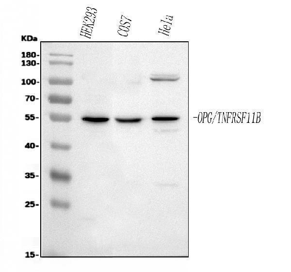
Click image to see more details
Figure 1. Western blot analysis of TNFRSF11B using anti-TNFRSF11B antibody (A00863).
Electrophoresis was performed on a 5-20% SDS-PAGE gel at 70V (Stacking gel) / 90V (Resolving gel) for 2-3 hours. The sample well of each lane was loaded with 30 ug of sample under reducing conditions.
Lane 1: human HEK293 whole cell lysates,
Lane 2: monkey COS-7 whole cell lysates,
Lane 3: human Hela whole cell lysates.
After electrophoresis, proteins were transferred to a nitrocellulose membrane at 150 mA for 50-90 minutes. Blocked the membrane with 5% non-fat milk/TBS for 1.5 hour at RT. The membrane was incubated with rabbit anti-TNFRSF11B antigen affinity purified polyclonal antibody (Catalog # A00863) at 0.5 μg/mL overnight at 4°C, then washed with TBS-0.1%Tween 3 times with 5 minutes each and probed with a goat anti-rabbit IgG-HRP secondary antibody at a dilution of 1:5000 for 1.5 hour at RT. The signal is developed using an Enhanced Chemiluminescent detection (ECL) kit (Catalog # EK1002) with Tanon 5200 system. A specific band was detected for TNFRSF11B at approximately 55 kDa. The expected band size for TNFRSF11B is at 46 kDa.

Click image to see more details
Figure 2. IHC analysis of TNFRSF11B using anti-TNFRSF11B antibody (A00863).
TNFRSF11B was detected in paraffin-embedded section of human appendicitis tissues. Heat mediated antigen retrieval was performed in citrate buffer (pH6, epitope retrieval solution) for 20 mins. The tissue section was blocked with 10% goat serum. The tissue section was then incubated with 1μg/ml rabbit anti-TNFRSF11B Antibody (A00863) overnight at 4°C. Biotinylated goat anti-rabbit IgG was used as secondary antibody and incubated for 30 minutes at 37°C. The tissue section was developed using Strepavidin-Biotin-Complex (SABC)(Catalog # SA1022) with DAB as the chromogen.

Click image to see more details
Figure 3. IHC analysis of TNFRSF11B using anti-TNFRSF11B antibody (A00863).
TNFRSF11B was detected in paraffin-embedded section of human endometrial carcinoma tissues. Heat mediated antigen retrieval was performed in citrate buffer (pH6, epitope retrieval solution) for 20 mins. The tissue section was blocked with 10% goat serum. The tissue section was then incubated with 1μg/ml rabbit anti-TNFRSF11B Antibody (A00863) overnight at 4°C. Biotinylated goat anti-rabbit IgG was used as secondary antibody and incubated for 30 minutes at 37°C. The tissue section was developed using Strepavidin-Biotin-Complex (SABC)(Catalog # SA1022) with DAB as the chromogen.

Click image to see more details
Figure 4. IHC analysis of TNFRSF11B using anti-TNFRSF11B antibody (A00863).
TNFRSF11B was detected in paraffin-embedded section of human glioma tissues. Heat mediated antigen retrieval was performed in citrate buffer (pH6, epitope retrieval solution) for 20 mins. The tissue section was blocked with 10% goat serum. The tissue section was then incubated with 1μg/ml rabbit anti-TNFRSF11B Antibody (A00863) overnight at 4°C. Biotinylated goat anti-rabbit IgG was used as secondary antibody and incubated for 30 minutes at 37°C. The tissue section was developed using Strepavidin-Biotin-Complex (SABC)(Catalog # SA1022) with DAB as the chromogen.

Click image to see more details
Figure 5. IHC analysis of TNFRSF11B using anti-TNFRSF11B antibody (A00863).
TNFRSF11B was detected in paraffin-embedded section of human glioma tissues. Heat mediated antigen retrieval was performed in citrate buffer (pH6, epitope retrieval solution) for 20 mins. The tissue section was blocked with 10% goat serum. The tissue section was then incubated with 1μg/ml rabbit anti-TNFRSF11B Antibody (A00863) overnight at 4°C. Biotinylated goat anti-rabbit IgG was used as secondary antibody and incubated for 30 minutes at 37°C. The tissue section was developed using Strepavidin-Biotin-Complex (SABC)(Catalog # SA1022) with DAB as the chromogen.

Click image to see more details
Figure 6. IHC analysis of TNFRSF11B using anti-TNFRSF11B antibody (A00863).
TNFRSF11B was detected in paraffin-embedded section of human liver cancer tissues. Heat mediated antigen retrieval was performed in citrate buffer (pH6, epitope retrieval solution) for 20 mins. The tissue section was blocked with 10% goat serum. The tissue section was then incubated with 1μg/ml rabbit anti-TNFRSF11B Antibody (A00863) overnight at 4°C. Biotinylated goat anti-rabbit IgG was used as secondary antibody and incubated for 30 minutes at 37°C. The tissue section was developed using Strepavidin-Biotin-Complex (SABC)(Catalog # SA1022) with DAB as the chromogen.

Click image to see more details
Figure 7. IHC analysis of TNFRSF11B using anti-TNFRSF11B antibody (A00863).
TNFRSF11B was detected in paraffin-embedded section of human liver cancer tissues. Heat mediated antigen retrieval was performed in citrate buffer (pH6, epitope retrieval solution) for 20 mins. The tissue section was blocked with 10% goat serum. The tissue section was then incubated with 1μg/ml rabbit anti-TNFRSF11B Antibody (A00863) overnight at 4°C. Biotinylated goat anti-rabbit IgG was used as secondary antibody and incubated for 30 minutes at 37°C. The tissue section was developed using Strepavidin-Biotin-Complex (SABC)(Catalog # SA1022) with DAB as the chromogen.

Click image to see more details
Figure 8. IHC analysis of TNFRSF11B using anti-TNFRSF11B antibody (A00863). TNFRSF11B was detected in paraffin-embedded section of human Lung cancer tissues. Heat mediated antigen retrieval was performed in citrate buffer (pH6, epitope retrieval solution) for 20 mins. The tissue section was blocked with 10% goat serum. The tissue section was then incubated with 1μg/ml rabbit anti-TNFRSF11B Antibody (A00863) overnight at 4°C. Biotinylated goat anti-rabbit IgG was used as secondary antibody and incubated for 30 minutes at 37°C. The tissue section was developed using Strepavidin-Biotin-Complex (SABC)(Catalog # SA1022) with DAB as the chromogen.

Click image to see more details
Figure 9. IHC analysis of TNFRSF11B using anti-TNFRSF11B antibody (A00863). TNFRSF11B was detected in paraffin-embedded section of human Lung cancer tissues. Heat mediated antigen retrieval was performed in citrate buffer (pH6, epitope retrieval solution) for 20 mins. The tissue section was blocked with 10% goat serum. The tissue section was then incubated with 1μg/ml rabbit anti-TNFRSF11B Antibody (A00863) overnight at 4°C. Biotinylated goat anti-rabbit IgG was used as secondary antibody and incubated for 30 minutes at 37°C. The tissue section was developed using Strepavidin-Biotin-Complex (SABC)(Catalog # SA1022) with DAB as the chromogen.

Click image to see more details
Figure 10. IHC analysis of TNFRSF11B using anti-TNFRSF11B antibody (A00863).
TNFRSF11B was detected in paraffin-embedded section of human Lung cancer tissues. Heat mediated antigen retrieval was performed in citrate buffer (pH6, epitope retrieval solution) for 20 mins. The tissue section was blocked with 10% goat serum. The tissue section was then incubated with 1μg/ml rabbit anti-TNFRSF11B Antibody (A00863) overnight at 4°C. Biotinylated goat anti-rabbit IgG was used as secondary antibody and incubated for 30 minutes at 37°C. The tissue section was developed using Strepavidin-Biotin-Complex (SABC)(Catalog # SA1022) with DAB as the chromogen.

Click image to see more details
Figure 11. IHC analysis of TNFRSF11B using anti-TNFRSF11B antibody (A00863).
TNFRSF11B was detected in paraffin-embedded section of human oesophagus squama cancer tissues. Heat mediated antigen retrieval was performed in citrate buffer (pH6, epitope retrieval solution) for 20 mins. The tissue section was blocked with 10% goat serum. The tissue section was then incubated with 1μg/ml rabbit anti-TNFRSF11B Antibody (A00863) overnight at 4°C. Biotinylated goat anti-rabbit IgG was used as secondary antibody and incubated for 30 minutes at 37°C. The tissue section was developed using Strepavidin-Biotin-Complex (SABC)(Catalog # SA1022) with DAB as the chromogen.

Click image to see more details
Figure 12. IHC analysis of TNFRSF11B using anti-TNFRSF11B antibody (A00863).
TNFRSF11B was detected in paraffin-embedded section of rat brain tissues. Heat mediated antigen retrieval was performed in citrate buffer (pH6, epitope retrieval solution) for 20 mins. The tissue section was blocked with 10% goat serum. The tissue section was then incubated with 1μg/ml rabbit anti-TNFRSF11B Antibody (A00863) overnight at 4°C. Biotinylated goat anti-rabbit IgG was used as secondary antibody and incubated for 30 minutes at 37°C. The tissue section was developed using Strepavidin-Biotin-Complex (SABC)(Catalog # SA1022) with DAB as the chromogen.

Click image to see more details
Figure 13. IHC analysis of TNFRSF11B using anti-TNFRSF11B antibody (A00863).
TNFRSF11B was detected in paraffin-embedded section of rat brain tissues. Heat mediated antigen retrieval was performed in citrate buffer (pH6, epitope retrieval solution) for 20 mins. The tissue section was blocked with 10% goat serum. The tissue section was then incubated with 1μg/ml rabbit anti-TNFRSF11B Antibody (A00863) overnight at 4°C. Biotinylated goat anti-rabbit IgG was used as secondary antibody and incubated for 30 minutes at 37°C. The tissue section was developed using Strepavidin-Biotin-Complex (SABC)(Catalog # SA1022) with DAB as the chromogen.

Click image to see more details
Figure 14. IHC analysis of TNFRSF11B using anti-TNFRSF11B antibody (A00863).
TNFRSF11B was detected in paraffin-embedded section of human Cholangiocarcinoma tissues. Heat mediated antigen retrieval was performed in citrate buffer (pH6, epitope retrieval solution) for 20 mins. The tissue section was blocked with 10% goat serum. The tissue section was then incubated with 1μg/ml rabbit anti-TNFRSF11B Antibody (A00863) overnight at 4°C. Biotinylated goat anti-rabbit IgG was used as secondary antibody and incubated for 30 minutes at 37°C. The tissue section was developed using Strepavidin-Biotin-Complex (SABC)(Catalog # SA1022) with DAB as the chromogen.

Click image to see more details
Figure 15. IHC analysis of TNFRSF11B using anti-TNFRSF11B antibody (A00863).
TNFRSF11B was detected in paraffin-embedded section of human mammary cancer tissues. Heat mediated antigen retrieval was performed in citrate buffer (pH6, epitope retrieval solution) for 20 mins. The tissue section was blocked with 10% goat serum. The tissue section was then incubated with 1μg/ml rabbit anti-TNFRSF11B Antibody (A00863) overnight at 4°C. Biotinylated goat anti-rabbit IgG was used as secondary antibody and incubated for 30 minutes at 37°C. The tissue section was developed using Strepavidin-Biotin-Complex (SABC)(Catalog # SA1022) with DAB as the chromogen.

Click image to see more details
Figure 16. IHC analysis of TNFRSF11B using anti-TNFRSF11B antibody (A00863).
TNFRSF11B was detected in paraffin-embedded section of human mammary cancer tissues. Heat mediated antigen retrieval was performed in citrate buffer (pH6, epitope retrieval solution) for 20 mins. The tissue section was blocked with 10% goat serum. The tissue section was then incubated with 1μg/ml rabbit anti-TNFRSF11B Antibody (A00863) overnight at 4°C. Biotinylated goat anti-rabbit IgG was used as secondary antibody and incubated for 30 minutes at 37°C. The tissue section was developed using Strepavidin-Biotin-Complex (SABC)(Catalog # SA1022) with DAB as the chromogen.

Click image to see more details
Figure 17. IHC analysis of TNFRSF11B using anti-TNFRSF11B antibody (A00863).
TNFRSF11B was detected in paraffin-embedded section of human placenta tissues. Heat mediated antigen retrieval was performed in citrate buffer (pH6, epitope retrieval solution) for 20 mins. The tissue section was blocked with 10% goat serum. The tissue section was then incubated with 1μg/ml rabbit anti-TNFRSF11B Antibody (A00863) overnight at 4°C. Biotinylated goat anti-rabbit IgG was used as secondary antibody and incubated for 30 minutes at 37°C. The tissue section was developed using Strepavidin-Biotin-Complex (SABC)(Catalog # SA1022) with DAB as the chromogen.

Click image to see more details
Figure 18. IHC analysis of TNFRSF11B using anti-TNFRSF11B antibody (A00863).
TNFRSF11B was detected in paraffin-embedded section of human rectal cancer tissues. Heat mediated antigen retrieval was performed in citrate buffer (pH6, epitope retrieval solution) for 20 mins. The tissue section was blocked with 10% goat serum. The tissue section was then incubated with 1μg/ml rabbit anti-TNFRSF11B Antibody (A00863) overnight at 4°C. Biotinylated goat anti-rabbit IgG was used as secondary antibody and incubated for 30 minutes at 37°C. The tissue section was developed using Strepavidin-Biotin-Complex (SABC)(Catalog # SA1022) with DAB as the chromogen.

Click image to see more details
Figure 19. IHC analysis of TNFRSF11B using anti-TNFRSF11B antibody (A00863).
TNFRSF11B was detected in paraffin-embedded section of human rectal cancer tissues. Heat mediated antigen retrieval was performed in citrate buffer (pH6, epitope retrieval solution) for 20 mins. The tissue section was blocked with 10% goat serum. The tissue section was then incubated with 1μg/ml rabbit anti-TNFRSF11B Antibody (A00863) overnight at 4°C. Biotinylated goat anti-rabbit IgG was used as secondary antibody and incubated for 30 minutes at 37°C. The tissue section was developed using Strepavidin-Biotin-Complex (SABC)(Catalog # SA1022) with DAB as the chromogen.

Click image to see more details
Figure 20. IHC analysis of TNFRSF11B using anti-TNFRSF11B antibody (A00863).
TNFRSF11B was detected in paraffin-embedded section of human tonsil tissues. Heat mediated antigen retrieval was performed in citrate buffer (pH6, epitope retrieval solution) for 20 mins. The tissue section was blocked with 10% goat serum. The tissue section was then incubated with 1μg/ml rabbit anti-TNFRSF11B Antibody (A00863) overnight at 4°C. Biotinylated goat anti-rabbit IgG was used as secondary antibody and incubated for 30 minutes at 37°C. The tissue section was developed using Strepavidin-Biotin-Complex (SABC)(Catalog # SA1022) with DAB as the chromogen.

Click image to see more details
Figure 21. IHC analysis of TNFRSF11B using anti-TNFRSF11B antibody (A00863).
TNFRSF11B was detected in paraffin-embedded section of human tonsil tissues. Heat mediated antigen retrieval was performed in citrate buffer (pH6, epitope retrieval solution) for 20 mins. The tissue section was blocked with 10% goat serum. The tissue section was then incubated with 1μg/ml rabbit anti-TNFRSF11B Antibody (A00863) overnight at 4°C. Biotinylated goat anti-rabbit IgG was used as secondary antibody and incubated for 30 minutes at 37°C. The tissue section was developed using Strepavidin-Biotin-Complex (SABC)(Catalog # SA1022) with DAB as the chromogen.

Click image to see more details
Figure 22. IHC analysis of TNFRSF11B using anti-TNFRSF11B antibody (A00863).
TNFRSF11B was detected in paraffin-embedded section of mouse brain tissues. Heat mediated antigen retrieval was performed in citrate buffer (pH6, epitope retrieval solution) for 20 mins. The tissue section was blocked with 10% goat serum. The tissue section was then incubated with 1μg/ml rabbit anti-TNFRSF11B Antibody (A00863) overnight at 4°C. Biotinylated goat anti-rabbit IgG was used as secondary antibody and incubated for 30 minutes at 37°C. The tissue section was developed using Strepavidin-Biotin-Complex (SABC)(Catalog # SA1022) with DAB as the chromogen.

Click image to see more details
Figure 23. IHC analysis of TNFRSF11B using anti-TNFRSF11B antibody (A00863).
TNFRSF11B was detected in paraffin-embedded section of human appendicitis tissues. Heat mediated antigen retrieval was performed in citrate buffer (pH6, epitope retrieval solution) for 20 mins. The tissue section was blocked with 10% goat serum. The tissue section was then incubated with 1μg/ml rabbit anti-TNFRSF11B Antibody (A00863) overnight at 4°C. Biotinylated goat anti-rabbit IgG was used as secondary antibody and incubated for 30 minutes at 37°C. The tissue section was developed using Strepavidin-Biotin-Complex (SABC)(Catalog # SA1022) with DAB as the chromogen.

Click image to see more details
Figure 24. Flow Cytometry analysis of U20S cells using anti-TNFRSF11B antibody (A00863).
Overlay histogram showing U20S cells stained with A00863 (Blue line).The cells were blocked with 10% normal goat serum. And then incubated with rabbit anti-TNFRSF11B Antibody (A00863,1μg/1x106 cells) for 30 min at 20°C. DyLight®488 conjugated goat anti-rabbit IgG (BA1127, 5-10μg/1x106 cells) was used as secondary antibody for 30 minutes at 20°C. Isotype control antibody (Green line) was rabbit IgG (1μg/1x106) used under the same conditions. Unlabelled sample (Red line) was also used as a control.
Protein Target Info & Infographic
Gene/Protein Information For TNFRSF11B (Source: Uniprot.org, NCBI)
Gene Name
TNFRSF11B
Full Name
Tumor necrosis factor receptor superfamily member 11B
Weight
34632 MW
Alternative Names
OCIF; OCIFMGC29565; OPGtumor necrosis factor receptor superfamily member 11B; Osteoclastogenesis inhibitory factor; Osteoprotegerin; TNFRSF11B; TR1; tumor necrosis factor receptor superfamily, member 11b TNFRSF11B OCIF, OPG, PDB5, TR1 TNF receptor superfamily member 11b tumor necrosis factor receptor superfamily member 11B|osteoclastogenesis inhibitory factor|osteoprotegerin|tumor necrosis factor receptor superfamily, member 11b
*If product is indicated to react with multiple species, protein info is based on the gene entry specified above in "Species".For more info on TNFRSF11B, check out the TNFRSF11B Infographic

We have 30,000+ of these available, one for each gene! Check them out.
In this infographic, you will see the following information for TNFRSF11B: database IDs, superfamily, protein function, synonyms, molecular weight, chromosomal locations, tissues of expression, subcellular locations, post-translational modifications, and related diseases, research areas & pathways. If you want to see more information included, or would like to contribute to it and be acknowledged, please contact [email protected].
Specific Publications For Anti-Osteoprotegerin/TNFRSF11B Antibody Picoband™ (A00863)
Hello CJ!
A00863 has been cited in 5 publications:
*The publications in this section are manually curated by our staff scientists. They may differ from Bioz's machine gathered results. Both are accurate. If you find a publication citing this product but is missing from this list, please let us know we will issue you a thank-you coupon.
Anti-osteoporotic activity of puerarin 6″-O-xyloside on ovariectomized mice and its potential mechanism
Alendronate-loaded hydroxyapatite-TiO2 nanotubes for improved bone formation in osteoporotic rabbits
Effects of Phytoestrogen α-ZAL and Mechanical Stimulation on Proliferation, Osteoblastic Differentiation, and OPG/RANKL Expression in MC3T3-E1 Pre-Osteoblasts
Tang Y, Sun F, Li X, Zhou Y, Yin S, Zhou X. J Endod. 2011 Dec;37(12):1653-8. Doi: 10.1016/J.Joen.2011.08.015. Epub 2011 Oct 2. Porphyromonas Endodontalis Lipopolysaccharides Induce Rankl By Mouse Osteoblast In A Way Different From That Of Escheric...
Zhao H, Ning Ll, Wang Zy, Li Ht, Qiao D, Yao Y, Qin Hl. Cell Biochem Biophys. 2014 Nov;70(2):1097-104. Doi: 10.1007/S12013-014-0028-Z. Calcitonin Gene-Related Peptide Inhibits Osteolytic Factors Induced By Osteoblast In Co-Culture System With Brea...
Recommended Resources
Here are featured tools and databases that you might find useful.
- Boster's Pathways Library
- Protein Databases
- Bioscience Research Protocol Resources
- Data Processing & Analysis Software
- Photo Editing Software
- Scientific Literature Resources
- Research Paper Management Tools
- Molecular Biology Software
- Primer Design Tools
- Bioinformatics Tools
- Phylogenetic Tree Analysis
Customer Reviews
Have you used Anti-Osteoprotegerin/TNFRSF11B Antibody Picoband™?
Submit a review and receive an Amazon gift card.
- $30 for a review with an image
0 Reviews For Anti-Osteoprotegerin/TNFRSF11B Antibody Picoband™
Customer Q&As
Have a question?
Find answers in Q&As, reviews.
Can't find your answer?
Submit your question
1 Customer Q&As for Anti-Osteoprotegerin/TNFRSF11B Antibody Picoband™
Question
We are currently using anti-Osteoprotegerin/TNFRSF11B antibody A00863 for rat tissue, and we are content with the IHC-P results. The species of reactivity given in the datasheet says human, mouse, rat. Is it true that the antibody can work on bovine tissues as well?
Verified Customer
Verified customer
Asked: 2020-01-16
Answer
The anti-Osteoprotegerin/TNFRSF11B antibody (A00863) has not been validated for cross reactivity specifically with bovine tissues, though there is a good chance of cross reactivity. We have an innovator award program that if you test this antibody and show it works in bovine you can get your next antibody for free. Please contact me if I can help you with anything.
Boster Scientific Support
Answered: 2020-01-16
