Click image to see more details
Product Info Summary
| SKU: | A00909 |
|---|---|
| Size: | 100 μg/vial |
| Reactive Species: | Human, Mouse, Rat |
| Host: | Rabbit |
| Application: | ELISA, Flow Cytometry, WB |
Customers Who Bought This Also Bought

Product info
Product Name
Anti-Orai1 Antibody
SKU/Catalog Number
A00909
Size
100 μg/vial
Form
Lyophilized
Description
Boster Bio Anti-Orai1 Antibody catalog # A00909. Tested in ELISA, Flow Cytometry, WB applications. This antibody reacts with Human, Mouse, Rat.
Storage & Handling
Store at -20˚C for one year from date of receipt. After reconstitution, at 4˚C for one month. It can also be aliquotted and stored frozen at -20˚C for six months. Avoid repeated freeze-thaw cycles.
Cite This Product
Anti-Orai1 Antibody (Boster Biological Technology, Pleasanton CA, USA, Catalog # A00909)
Host
Rabbit
Contents
Each vial contains 4mg Trehalose, 0.9mg NaCl, 0.2mg Na2HPO4, 0.05mg NaN3.
Clonality
Polyclonal
Isotype
Rabbit IgG
Immunogen
E.coli-derived human Orai1 recombinant protein (Position: A49-A301).
*Blocking peptide can be purchased. Costs vary based on immunogen length. Contact us for pricing.
Cross-reactivity
No cross-reactivity with other proteins.
Reactive Species
A00909 is reactive to ORAI1 in Human, Mouse, Rat
Applications
A00909 is guaranteed for ELISA, Flow Cytometry, WB Boster Guarantee
Observed Molecular Weight
39 kDa
Calculated molecular weight
Background of ORAI1
ORAI1 (ORAI calcium release-activated calcium modulator 1), also known as CRACM1, TMEM142A, Calcium release-activated calcium channel protein 1, Protein orai-1, Transmembrane protein 142A, FLJ14466, is a calcium selectiveion channelthat in humans is encoded by the ORAI1 gene. Orai1 channels play an important role in the activation of T-lymphocytes. The loss of function mutation of Orai1 causes severe combined immunodeficiency (SCID) in humans. The mammalian orai family has two additional homologs, orai2 and orai3. Orai proteins share no homology with any other ion channel family of any other known proteins. They have 4 transmembrane domains and form tetramers.Prakriya et al. showed that ORAI1 is a PM protein, and that CRAC channel function is sensitive to mutation of 2 conserved acidic residues in the transmembrane segments. Glu106-to-asp (E106D) and glu190-to-gln (E190Q) substitutions in transmembrane helices 1 and 3, respectively, diminished calcium ion influx, increased current carried by monovalent cations, and rendered the channel permeable to cesium ion.Prakriya et al. showed that ORAI1 is a PM protein, and that CRAC channel function is sensitive to mutation of 2 conserved acidic residues in the transmembrane segments.
Antibody Validation
Boster validates all antibodies on WB, IHC, ICC, Immunofluorescence, and ELISA with known positive control and negative samples to ensure specificity and high affinity, including thorough antibody incubations.
Assay dilution & Images
Reconstitution
Add 0.2ml of distilled water will yield a concentration of 500ug/ml.
Assay Dilutions Recommendation
The recommendations below provide a starting point for assay optimization. The actual working concentration varies and should be decided by the user.
Western blot, 0.25-0.5μg/ml
Flow Cytometry (Fixed), 1-3μg/1x106 cells
Direct ELISA, 0.1-0.5μg/ml
Validation Images & Assay Conditions
Click image to see more details
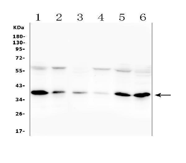
Figure 1. Western blot analysis of ORAI1 using anti-ORAI1 antibody (A00909).
Electrophoresis was performed on a 5-20% SDS-PAGE gel at 70V (Stacking gel) / 90V (Resolving gel) for 2-3 hours. The sample well of each lane was loaded with 50ug of sample under reducing conditions.
Lane 1: human A375 whole cell lysates,
Lane 2: human Jurka whole cell lysates,
Lane 3: human A549 whole cell lysates,
Lane 4: human A431 whole cell lysates,
Lane 5: human HepG2 whole cell lysates,
Lane 6: human K562 whole cell lysates,
After Electrophoresis, proteins were transferred to a Nitrocellulose membrane at 150mA for 50-90 minutes. Blocked the membrane with 5% Non-fat Milk/ TBS for 1.5 hour at RT. The membrane was incubated with rabbit anti-ORAI1 antigen affinity purified polyclonal antibody (Catalog # A00909) at 0.5 μg/mL overnight at 4°C, then washed with TBS-0.1%Tween 3 times with 5 minutes each and probed with a goat anti-rabbit IgG-HRP secondary antibody at a dilution of 1:10000 for 1.5 hour at RT. The signal is developed using an Enhanced Chemiluminescent detection (ECL) kit (Catalog # EK1002) with Tanon 5200 system. A specific band was detected for ORAI1 at approximately 39KD. The expected band size for ORAI1 is at 33KD.

Click image to see more details
Figure 2. Flow Cytometry analysis of CACO-2 cells using anti-ORAI1 antibody (A00909).
Overlay histogram showing CACO-2 cells stained with A00909 (Blue line).The cells were blocked with 10% normal goat serum. And then incubated with rabbit anti-ORAI1 Antibody (A00909,1μg/1x106 cells) for 30 min at 20°C. DyLight®488 conjugated goat anti-rabbit IgG (BA1127, 5-10μg/1x106 cells) was used as secondary antibody for 30 minutes at 20°C. Isotype control antibody (Green line) was rabbit IgG (1μg/1x106) used under the same conditions. Unlabelled sample (Red line) was also used as a control.

Click image to see more details
Figure 3. Flow Cytometry analysis of A549 cells using anti-ORAI1 antibody (A00909).
Overlay histogram showing A549 cells stained with A00909 (Blue line).The cells were blocked with 10% normal goat serum. And then incubated with rabbit anti-ORAI1 Antibody (A00909,1μg/1x106 cells) for 30 min at 20°C. DyLight®488 conjugated goat anti-rabbit IgG (BA1127, 5-10μg/1x106 cells) was used as secondary antibody for 30 minutes at 20°C. Isotype control antibody (Green line) was rabbit IgG (1μg/1x106) used under the same conditions. Unlabelled sample (Red line) was also used as a control.
Protein Target Info & Infographic
Gene/Protein Information For ORAI1 (Source: Uniprot.org, NCBI)
Gene Name
ORAI1
Full Name
Calcium release-activated calcium channel protein 1
Weight
Superfamily
Orai family
Alternative Names
CRACM1; CRACM1ORAT1; FLJ14466; ORAI calcium release-activated calcium modulator 1; Orai1; ORAT1; Protein orai-1; TMEM142A; TMEM142Acalcium release-activated calcium modulator 1; Transmembrane protein 142Acalcium release-activated calcium channel protein 1 ORAI1 CRACM1, IMD9, ORAT1, TAM2, TMEM142A ORAI calcium release-activated calcium modulator 1 calcium release-activated calcium channel protein 1|calcium release-activated calcium modulator 1|protein orai-1|transmembrane protein 142A
*If product is indicated to react with multiple species, protein info is based on the gene entry specified above in "Species".For more info on ORAI1, check out the ORAI1 Infographic

We have 30,000+ of these available, one for each gene! Check them out.
In this infographic, you will see the following information for ORAI1: database IDs, superfamily, protein function, synonyms, molecular weight, chromosomal locations, tissues of expression, subcellular locations, post-translational modifications, and related diseases, research areas & pathways. If you want to see more information included, or would like to contribute to it and be acknowledged, please contact [email protected].
Specific Publications For Anti-Orai1 Antibody (A00909)
Hello CJ!
No publications found for A00909
*Do you have publications using this product? Share with us and receive a reward. Ask us for more details.
Recommended Resources
Here are featured tools and databases that you might find useful.
- Boster's Pathways Library
- Protein Databases
- Bioscience Research Protocol Resources
- Data Processing & Analysis Software
- Photo Editing Software
- Scientific Literature Resources
- Research Paper Management Tools
- Molecular Biology Software
- Primer Design Tools
- Bioinformatics Tools
- Phylogenetic Tree Analysis
Customer Reviews
Have you used Anti-Orai1 Antibody?
Submit a review and receive an Amazon gift card.
- $30 for a review with an image
0 Reviews For Anti-Orai1 Antibody
Customer Q&As
Have a question?
Find answers in Q&As, reviews.
Can't find your answer?
Submit your question
6 Customer Q&As for Anti-Orai1 Antibody
Question
We have observed staining in human cervix carcinoma. Do you have any suggestions? Is anti-Orai1 antibody supposed to stain cervix carcinoma positively?
A. Wu
Verified customer
Asked: 2020-01-06
Answer
Based on literature cervix carcinoma does express ORAI1. Based on Uniprot.org, ORAI1 is expressed in muscle of leg, bone marrow, ovary, prostate uterus, mammary gland, cervix carcinoma, liver, among other tissues. Regarding which tissues have ORAI1 expression, here are a few articles citing expression in various tissues:
Bone marrow, Ovary, Prostate, and Uterus, Pubmed ID: 15489334
Cervix carcinoma, Pubmed ID: 18669648
Liver, Pubmed ID: 24275569
Mammary gland, Pubmed ID: 14702039
Boster Scientific Support
Answered: 2020-01-06
Question
Would A00909 work on mouse and rat samples?
Verified customer
Asked: 2019-09-12
Answer
We tested the Anti-Orai1 Antibody (A00909) on mouse and rat samples and it didn't work well.
Boster Scientific Support
Answered: 2019-09-12
Question
We are currently using anti-Orai1 antibody A00909 for human tissue, and we are content with the WB results. The species of reactivity given in the datasheet says human. Is it likely that the antibody can work on zebrafish tissues as well?
Verified Customer
Verified customer
Asked: 2019-08-28
Answer
The anti-Orai1 antibody (A00909) has not been validated for cross reactivity specifically with zebrafish tissues, though there is a good chance of cross reactivity. We have an innovator award program that if you test this antibody and show it works in zebrafish you can get your next antibody for free. Please contact me if I can help you with anything.
Boster Scientific Support
Answered: 2019-08-28
Question
Our lab were happy with the WB result of your anti-Orai1 antibody. However we have seen positive staining in prostate uterus cell membrane using this antibody. Is that expected? Could you tell me where is ORAI1 supposed to be expressed?
Verified Customer
Verified customer
Asked: 2019-07-08
Answer
According to literature, prostate uterus does express ORAI1. Generally ORAI1 expresses in cell membrane. Regarding which tissues have ORAI1 expression, here are a few articles citing expression in various tissues:
Bone marrow, Ovary, Prostate, and Uterus, Pubmed ID: 15489334
Cervix carcinoma, Pubmed ID: 18669648
Liver, Pubmed ID: 24275569
Mammary gland, Pubmed ID: 14702039
Boster Scientific Support
Answered: 2019-07-08
Question
My lab would like using your anti-Orai1 antibody for calcium ion import studies. Has this antibody been tested with western blotting on a549 whole cell lysates? We would like to see some validation images before ordering.
Verified Customer
Verified customer
Asked: 2018-03-08
Answer
I appreciate your inquiry. This A00909 anti-Orai1 antibody is tested on human a549, a549 whole cell lysates, a431 whole cell lysates, hepg2 whole cell lysates, k562 whole cell lysates. It is guaranteed to work for ELISA, Flow Cytometry, WB in human. Our Boster guarantee will cover your intended experiment even if the sample type has not been be directly tested.
Boster Scientific Support
Answered: 2018-03-08
Question
We bought anti-Orai1 antibody for Flow Cytometry on mammary gland a few years ago. I am using human, and We are going to use the antibody for WB next. you antibody examining mammary gland as well as muscle of leg in our next experiment. Do you have any suggestion on which antibody would work the best for WB?
D. Zhang
Verified customer
Asked: 2014-03-03
Answer
I looked at the website and datasheets of our anti-Orai1 antibody and it appears that A00909 has been validated on human in both Flow Cytometry and WB. Thus A00909 should work for your application. Our Boster satisfaction guarantee will cover this product for WB in human even if the specific tissue type has not been validated. We do have a comprehensive range of products for WB detection and you can check out our website bosterbio.com to find out more information about them.
Boster Scientific Support
Answered: 2014-03-03
