Click image to see more details
-

-
-
-
-
+3
Product Info Summary
| SKU: | A01776-3 |
|---|---|
| Size: | 100 μg/vial |
| Reactive Species: | Human, Mouse, Rat |
| Host: | Rabbit |
| Application: | ELISA, Flow Cytometry, IHC, WB |
Customers Who Bought This Also Bought

Product info
Product Name
Anti-KPNA2 Antibody Picoband™
View all Importin alpha 2/KPNA2 Antibodies
SKU/Catalog Number
A01776-3
Size
100 μg/vial
Form
Lyophilized
Description
Boster Bio Anti-KPNA2 Antibody Picoband™ catalog # A01776-3. Tested in ELISA, Flow Cytometry, IHC, WB applications. This antibody reacts with Human, Mouse, Rat.
Storage & Handling
At -20°C for one year from date of receipt. After reconstitution, at 4°C for one month. It can also be aliquotted and stored frozen at -20°C for six months. Avoid repeated freezing and thawing.
Cite This Product
Anti-KPNA2 Antibody Picoband™ (Boster Biological Technology, Pleasanton CA, USA, Catalog # A01776-3)
Host
Rabbit
Contents
Each vial contains 4 mg Trehalose, 0.9 mg NaCl, 0.2 mg Na2HPO4.
Clonality
Polyclonal
Isotype
Rabbit IgG
Immunogen
E.coli-derived human KPNA2 recombinant protein (Position: R51-F529).
*Blocking peptide can be purchased. Costs vary based on immunogen length. Contact us for pricing.
Cross-reactivity
No cross-reactivity with other proteins.
Reactive Species
A01776-3 is reactive to KPNA2 in Human, Mouse, Rat
Applications
A01776-3 is guaranteed for ELISA, Flow Cytometry, IHC, WB Boster Guarantee
Observed Molecular Weight
58 kDa
Calculated molecular weight
57862 MW
Background of Importin alpha 2/KPNA2
Importin subunit alpha-2 is a protein that in humans is encoded by the KPNA2 gene. The import of proteins into the nucleus is a process that involves at least 2 steps. The first is an energy-independent docking of the protein to the nuclear envelope and the second is an energy-dependent translocation through the nuclear pore complex. Imported proteins require a nuclear localization sequence (NLS) which generally consists of a short region of basic amino acids or 2 such regions spaced about 10 amino acids apart. Proteins involved in the first step of nuclear import have been identified in different systems. These include the Xenopus protein importin and its yeast homolog, SRP1 (a suppressor of certain temperature-sensitive mutations of RNA polymerase I in Saccharomyces cerevisiae), which bind to the NLS. KPNA2 protein interacts with the NLSs of DNA helicase Q1 and SV40 T antigen and may be involved in the nuclear transport of proteins. KPNA2 also may play a role in V(D)J recombination.
Antibody Validation
Boster validates all antibodies on WB, IHC, ICC, Immunofluorescence, and ELISA with known positive control and negative samples to ensure specificity and high affinity, including thorough antibody incubations.
Assay dilution & Images
Reconstitution
Adding 0.2 ml of distilled water will yield a concentration of 500 μg/ml.
Assay Dilutions Recommendation
The recommendations below provide a starting point for assay optimization. The actual working concentration varies and should be decided by the user.
Western blot, 0.1-0.25 μg/ml, Human, Mouse, Rat
Immunohistochemistry(Paraffin-embedded Section), 2-5 μg/ml, Human, Rat
Flow Cytometry (Fixed), 1-3 μg/1x106 cells, Human
Direct ELISA, 0.1-0.5 μg/ml, Human
Validation Images & Assay Conditions
Click image to see more details
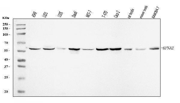
Figure 1. Western blot analysis of KPNA2 using anti-KPNA2 antibody (A01776-3).
Electrophoresis was performed on a 5-20% SDS-PAGE gel at 70V (Stacking gel) / 90V (Resolving gel) for 2-3 hours. The sample well of each lane was loaded with 30 ug of sample under reducing conditions.
Lane 1: human A549 whole cell lysates,
Lane 2: human U251 whole cell lysates,
Lane 3: human U20S whole cell lysates,
Lane 4: human Daudi whole cell lysates,
Lane 5: human MCF-7 whole cell lysates,
Lane 6: human T-47D whole cell lysates,
Lane 7: human Caco-2 whole cell lysates,
Lane 8: rat testis tissue lysates,
Lane 9: mouse testis tissue lysates,
Lane 10: mouse RAW264.7 whole cell lysates.
After electrophoresis, proteins were transferred to a nitrocellulose membrane at 150 mA for 50-90 minutes. Blocked the membrane with 5% non-fat milk/TBS for 1.5 hour at RT. The membrane was incubated with rabbit anti-KPNA2 antigen affinity purified polyclonal antibody (Catalog # A01776-3) at 0.25 μg/mL overnight at 4°C, then washed with TBS-0.1%Tween 3 times with 5 minutes each and probed with a goat anti-rabbit IgG-HRP secondary antibody at a dilution of 1:5000 for 1.5 hour at RT. The signal is developed using an Enhanced Chemiluminescent detection (ECL) kit (Catalog # EK1002) with Tanon 5200 system. A specific band was detected for KPNA2 at approximately 58 kDa. The expected band size for KPNA2 is at 58 kDa.

Click image to see more details
Figure 2. IHC analysis of KPNA2 using anti-KPNA2 antibody (A01776-3).
KPNA2 was detected in a paraffin-embedded section of rat testis tissue. Heat mediated antigen retrieval was performed in EDTA buffer (pH 8.0, epitope retrieval solution). The tissue section was blocked with 10% goat serum. The tissue section was then incubated with 2 μg/ml rabbit anti-KPNA2 Antibody (A01776-3) overnight at 4°C. Peroxidase Conjugated Goat Anti-rabbit IgG was used as secondary antibody and incubated for 30 minutes at 37°C. The tissue section was developed using HRP Conjugated Rabbit IgG Super Vision Assay Kit (Catalog # SV0002) with DAB as the chromogen.

Click image to see more details
Figure 3. IHC analysis of KPNA2 using anti-KPNA2 antibody (A01776-3).
KPNA2 was detected in a paraffin-embedded section of human adenocarcinoma of the right colon tissue. Heat mediated antigen retrieval was performed in EDTA buffer (pH 8.0, epitope retrieval solution). The tissue section was blocked with 10% goat serum. The tissue section was then incubated with 2 μg/ml rabbit anti-KPNA2 Antibody (A01776-3) overnight at 4°C. Peroxidase Conjugated Goat Anti-rabbit IgG was used as secondary antibody and incubated for 30 minutes at 37°C. The tissue section was developed using HRP Conjugated Rabbit IgG Super Vision Assay Kit (Catalog # SV0002) with DAB as the chromogen.

Click image to see more details
Figure 4. IHC analysis of KPNA2 using anti-KPNA2 antibody (A01776-3).
KPNA2 was detected in a paraffin-embedded section of human lymphoma tissue. Heat mediated antigen retrieval was performed in EDTA buffer (pH 8.0, epitope retrieval solution). The tissue section was blocked with 10% goat serum. The tissue section was then incubated with 2 μg/ml rabbit anti-KPNA2 Antibody (A01776-3) overnight at 4°C. Peroxidase Conjugated Goat Anti-rabbit IgG was used as secondary antibody and incubated for 30 minutes at 37°C. The tissue section was developed using HRP Conjugated Rabbit IgG Super Vision Assay Kit (Catalog # SV0002) with DAB as the chromogen.

Click image to see more details
Figure 5. IHC analysis of KPNA2 using anti-KPNA2 antibody (A01776-3).
KPNA2 was detected in a paraffin-embedded section of human lung cancer tissue. Heat mediated antigen retrieval was performed in EDTA buffer (pH 8.0, epitope retrieval solution). The tissue section was blocked with 10% goat serum. The tissue section was then incubated with 2 μg/ml rabbit anti-KPNA2 Antibody (A01776-3) overnight at 4°C. Peroxidase Conjugated Goat Anti-rabbit IgG was used as secondary antibody and incubated for 30 minutes at 37°C. The tissue section was developed using HRP Conjugated Rabbit IgG Super Vision Assay Kit (Catalog # SV0002) with DAB as the chromogen.

Click image to see more details
Figure 6. IHC analysis of KPNA2 using anti-KPNA2 antibody (A01776-3).
KPNA2 was detected in a paraffin-embedded section of human tonsil tissue. Heat mediated antigen retrieval was performed in EDTA buffer (pH 8.0, epitope retrieval solution). The tissue section was blocked with 10% goat serum. The tissue section was then incubated with 2 μg/ml rabbit anti-KPNA2 Antibody (A01776-3) overnight at 4°C. Peroxidase Conjugated Goat Anti-rabbit IgG was used as secondary antibody and incubated for 30 minutes at 37°C. The tissue section was developed using HRP Conjugated Rabbit IgG Super Vision Assay Kit (Catalog # SV0002) with DAB as the chromogen.

Click image to see more details
Figure 7. Flow Cytometry analysis of U87 cells using anti-KPNA2 antibody (A01776-3).
Overlay histogram showing U87 cells stained with A01776-3 (Blue line). To facilitate intracellular staining, cells were fixed with 4% paraformaldehyde and permeabilized with permeabilization buffer. The cells were blocked with 10% normal goat serum. And then incubated with rabbit anti-KPNA2 Antibody (A01776-3, 1 μg/1x106 cells) for 30 min at 20°C. DyLight®488 conjugated goat anti-rabbit IgG (BA1127, 5-10 μg/1x106 cells) was used as secondary antibody for 30 minutes at 20°C. Isotype control antibody (Green line) was rabbit IgG (1 μg/1x106) used under the same conditions. Unlabelled sample without incubation with primary antibody and secondary antibody (Red line) was used as a blank control.
Protein Target Info & Infographic
Gene/Protein Information For KPNA2 (Source: Uniprot.org, NCBI)
Gene Name
KPNA2
Full Name
Importin subunit alpha-1
Weight
57862 MW
Superfamily
importin alpha family
Alternative Names
Importin alpha 1; Importin alpha 2; importin-alpha-P1; IPOA1; karyopherin alpha 2 (RAG cohort 1, importin alpha 1); Karyopherin subunit alpha-2; KPNA2; Pendulin; QIP2; QIP2importin alpha 2; RAG cohort 1; RAG cohort protein 1; RCH1; RCH1importin subunit alpha-2; SRP1; SRP1alpha; SRP1-alpha KPNA2 IPOA1, QIP2, RCH1, SRP1-alpha, SRP1alpha karyopherin subunit alpha 2 importin subunit alpha-1|RAG cohort protein 1|importin subunit alpha-2|importin-alpha-P1|karyopherin alpha 2 (RAG cohort 1, importin alpha 1)|pendulin
*If product is indicated to react with multiple species, protein info is based on the gene entry specified above in "Species".For more info on KPNA2, check out the KPNA2 Infographic

We have 30,000+ of these available, one for each gene! Check them out.
In this infographic, you will see the following information for KPNA2: database IDs, superfamily, protein function, synonyms, molecular weight, chromosomal locations, tissues of expression, subcellular locations, post-translational modifications, and related diseases, research areas & pathways. If you want to see more information included, or would like to contribute to it and be acknowledged, please contact [email protected].
Specific Publications For Anti-KPNA2 Antibody Picoband™ (A01776-3)
Hello CJ!
No publications found for A01776-3
*Do you have publications using this product? Share with us and receive a reward. Ask us for more details.
Recommended Resources
Here are featured tools and databases that you might find useful.
- Boster's Pathways Library
- Protein Databases
- Bioscience Research Protocol Resources
- Data Processing & Analysis Software
- Photo Editing Software
- Scientific Literature Resources
- Research Paper Management Tools
- Molecular Biology Software
- Primer Design Tools
- Bioinformatics Tools
- Phylogenetic Tree Analysis
Customer Reviews
Have you used Anti-KPNA2 Antibody Picoband™?
Submit a review and receive an Amazon gift card.
- $30 for a review with an image
0 Reviews For Anti-KPNA2 Antibody Picoband™
Customer Q&As
Have a question?
Find answers in Q&As, reviews.
Can't find your answer?
Submit your question
