This website uses cookies to ensure you get the best experience on our website.
- Table of Contents
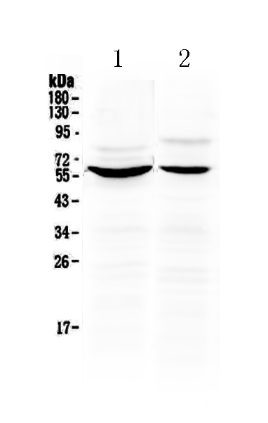
Facts about Importin subunit alpha-1.
Docking of the importin/substrate complex to the nuclear pore complex (NPC) is mediated by KPNB1 through binding to nucleoporin FxFG repeats and the complex is subsequently translocated through the pore by an energy requiring, Ran- dependent mechanism. At the nucleoplasmic side of the NPC, Ran binds to importin-beta and the three components separate and importin-alpha and -beta are re-exported in the nucleus into the cytoplasm where GTP hydrolysis releases Ran from importin.
Human | |
---|---|
Gene Name: | KPNA2 |
Uniprot: | P52292 |
Entrez: | 3838 |
Belongs to: |
---|
importin alpha family |
Importin alpha 1; Importin alpha 2; importin-alpha-P1; IPOA1; karyopherin alpha 2 (RAG cohort 1, importin alpha 1); Karyopherin subunit alpha-2; KPNA2; Pendulin; QIP2; QIP2importin alpha 2; RAG cohort 1; RAG cohort protein 1; RCH1; RCH1importin subunit alpha-2; SRP1; SRP1alpha; SRP1-alpha
Mass (kDA):
57.862 kDA
Human | |
---|---|
Location: | 17q24.2 |
Sequence: | 17; NC_000017.11 (68035735..68046859) |
Expressed ubiquitously.
Cytoplasm. Nucleus.
If you're looking for a high-affinity KPNA2 marker antibody, Boster offers a large library of antibodies that have been validated for Western Blotting, Immunohistochemistry, and ELISA. The primary antibodies that Boster offers have been validated for a variety of applications and are extensively employed in research. Boster antibodies are frequently cited in scientific circles and have a proven track record of great success.
KPNA2 expression in human tissues has been shown as a correlation with the presence of Hepatocellular carcinoma. The expression of KPNA2 is involved in the regulation of the cell cycle as well as DNA replication. These findings indicate that KPNA2 could be a viable diagnostic biomarker to detect HCC. The markers are particularly useful for the evaluation of tumors and tumor cells of the kidney.
A study showed that the nuclear KPNA2 expression was associated with the extent of tumor, lymphode metastasis and clinical stage in 179 GC samples. This finding is consistent with the previous results demonstrating an unambiguous correlation between KPNA2 expression and the presence of GC cells. This study could turn out to be an important therapeutic target for GC. However, it is emphasized that there are many problems related to the expression of GC.
In addition to the characteristics associated with tumors This marker also interacts with a variety of macromolecules and proteins. These interactions are associated with cell maintenance, transcriptional regulation and carcinogenesis, as well as aberrant differentiation pathways, and immune response. These interactions have resulted in abnormalities in the pathophysiology due to malfunctioning of the nuclear transportation system. Therefore, blocking this mechanism of export and import into the nuclear system is a promising therapeutic intervention. Numerous inhibitors of nuclear import/export have been studied for their antiviral as well as cancer properties.
In addition to its properties related to tumors, KPNA2 also mediates radioresistance. In addition, siRNA-treated MDA-MB-231 cells significantly reduced the rate of death caused by radiation. Western blot analysis revealed increased levels of apoptotic markers including PARP and cleaved Caspase-3. Furthermore, depletion of KPNA2 reduced the expression of pBRCA1.
KPNA2 expression may also have an effect on the proliferation of cells or radioresistance. BRCA1 is a nuclear importer when it is hyperphosphorylated by several kinases including ATM and Chk2. In the case of radiation-induced KPNA2 the pBRCA1 levels decrease and remains in the nucleus for up to 50 hours after radiation. It is, however, possible that IR-induced KPNA2 is a factor in BRCA1 nuclear localization.
Recent research suggests that KPNA2 may be a biomarker that could be used to identify various kinds of malignancies. It can regulate the growth of cells and also affect the progression of tumors. Its presence has been linked with poor prognosis in breast and ovarian cancers, and has recently been associated with an unfavourable outcome in Esophageal Squamous Cell Carcinoma. This protein has not been extensively investigated in GC.
Boster Bio's KPNA2 markers allow researchers to quickly analyze samples without having to devote time to secondary antibody conjugation. Dual labeling of specimens allows researchers to ask more questions by using both antibodies. This provides more robust answers and contextual data. Boster Bio's KPNA2 antibody marker for primary antibodies provides numerous advantages over other markers. Here are some of the most compelling reasons to use this marker in your research.
To analyze the immunofluorescence signal of the secondary antibody we used U87 cells transfected the marker. The cells were then cultured for 24 hours. The cells were fixed using 44% paraformaldehyde, and then permeated with 0.2% Triton X100 for 5 minutes. The cells were then labeled with the primary antibodies using a chemiluminescence detection device.
The KPNA2 protein regulates the localization of the target and the transport. It also transports cargo proteins to the nucleus. This metabolic aspect has been associated with cancer. Particularly, high levels of KPNA2 are associated with less favorable outcomes in cancer patients. Biomarkers for cancer have been identified through studying the protein's role as a biomarker.
After getting a negative control for boster Bio's KPNA2 marker, a set of glioblastoma cell lines was fixed in 4% paraformaldehyde and subjected to incubation with a similar antibody. The primary antibody was then applied to cells and the second antibody was added to the cells to allow staining. To detect KPNA2 expression cells were stained with Alexa Fluor 594-labeled secondary antibodies.
Boster Bio's KPNA2 markers can detect autophagy in cancerous cells and can also reveal important biological processes. For example, it is vital to comprehend the role of KPNA2 in the field of cancer research. KPNA2 plays a crucial role in autophagy regulation and suppression of cell migration, which could provide better treatments for cancer.
The KPNA2 marker has recently emerged as a promising biomarker candidate for esophageal squamous-cell carcinoma. Its expression is linked to cell proliferation and a poor prognosis in gastric cancer, according to a new study published in Tumour Biology. In the study, Rachidi SM and colleagues found that KPNA2 was a consistent, uniform marker for poor prognosis.
The study consisted of 24 studies that covered 6164 patients who had solid tumors. In each study the KPNA2 expression level was correlated with time to progression of the tumor. The median sample size was 177 and the range was between 47 and 1494. The major sources of the study's data were Japan, Germany, and China. The study included patients with an array of tumor types and stage of cancer.
The study showed that KPNA2 expression is linked to a lower overall survival, a higher stage, and a more International Society of Urologic Pathology grade. This is in contrast to papillary RCC. These findings weren't confirmed through multivariate Cox regression analysis. The study suggests that KPNA2 could be a significant predictor for survival in papillary RCC. KPNA2 is therefore a significant indicator of HCC.
In addition to its prognostic use, the KPNA2 marker can also be used as an option for treating colon cancer. This discovery could result in the development of new treatments for this kind of cancer. A number of publications have been published in the journal of clinical and experimental cancer research on KPNA2 in colon carcinoma. While KPNA2 is a promising prognostic indicator, its potential for treating cancer is immense.
Research has demonstrated that KPNA2 overexpression is associated with poor outcomes in breast, colorectal and gastric cancer. These results may have been affected by the small impact. Additionally, the overexpression of the marker in certain cancer types could lead to more aggressive clinical features and lower overall survival. In addition, the research also showed a significant association with poor outcome in the Cancer Genome Atlas cohorts.
PMID: 7754385 by Weis K., et al. Identification of hSRP1 alpha as a functional receptor for nuclear localization sequences.
PMID: 11735022 by Doerr S., et al. Genomic structure of karyopherin alpha2 (KPNA2) within a low-copy repeat on chromosome 17q23-q24 and mutation analysis in patients with Russell-Silver syndrome.