Click image to see more details
Product Info Summary
| SKU: | A00388-4 |
|---|---|
| Size: | 100 μg/vial |
| Reactive Species: | Human |
| Host: | Rabbit |
| Application: | ELISA, Flow Cytometry, WB |
Customers Who Bought This Also Bought

Product info
Product Name
Anti-ERCC1 Antibody Picoband®
SKU/Catalog Number
A00388-4
Size
100 μg/vial
Form
Lyophilized
Description
Boster Bio Anti-ERCC1 Antibody Picoband® catalog # A00388-4. Tested in ELISA, Flow Cytometry, WB applications. This antibody reacts with Human. The brand Picoband indicates this is a premium antibody that guarantees superior quality, high affinity, and strong signals with minimal background in Western blot applications. Only our best-performing antibodies are designated as Picoband, ensuring unmatched performance.
Storage & Handling
At -20°C for one year from date of receipt. After reconstitution, at 4°C for one month. It can also be aliquotted and stored frozen at -20°C for six months. Avoid repeated freezing and thawing.
Cite This Product
Anti-ERCC1 Antibody Picoband® (Boster Biological Technology, Pleasanton CA, USA, Catalog # A00388-4)
Host
Rabbit
Contents
Each vial contains 4 mg Trehalose, 0.9 mg NaCl, 0.2 mg Na2HPO4.
Clonality
Polyclonal
Isotype
Rabbit IgG
Immunogen
E.coli-derived human ERCC1 recombinant protein (Position: K7-R283).
*Blocking peptide can be purchased. Costs vary based on immunogen length. Contact us for pricing.
Cross-reactivity
No cross-reactivity with other proteins.
Reactive Species
A00388-4 is reactive to ERCC1 in Human
Reconstitution
Adding 0.2 ml of distilled water will yield a concentration of 500 μg/ml.
Observed Molecular Weight
38 kDa
Calculated molecular weight
17628 MW
Background of ERCC1
DNA excision repair protein ERCC-1 is a protein that in humans is encoded by the ERCC1 gene. The product of this gene functions in the nucleotide excision repair pathway, and is required for the repair of DNA lesions such as those induced by UV light or formed by electrophilic compounds including cisplatin. The encoded protein forms a heterodimer with the XPF endonuclease (also known as ERCC4), and the heterodimeric endonuclease catalyzes the 5' incision in the process of excising the DNA lesion. The heterodimeric endonuclease is also involved in recombinational DNA repair and in the repair of inter-strand crosslinks. Mutations in this gene result in cerebrooculofacioskeletal syndrome, and polymorphisms that alter expression of this gene may play a role in carcinogenesis. Multiple transcript variants encoding different isoforms have been found for this gene. The last exon of this gene overlaps with the CD3e molecule, epsilon associated protein gene on the opposite strand.
Antibody Validation
Boster validates all antibodies on WB, IHC, ICC, Immunofluorescence, and ELISA with known positive control and negative samples to ensure specificity and high affinity, including thorough antibody incubations.
Application & Images
Applications
A00388-4 is guaranteed for ELISA, Flow Cytometry, WB Boster Guarantee
Assay Dilutions Recommendation
The recommendations below provide a starting point for assay optimization. The actual working concentration varies and should be decided by the user.
Western blot, 0.25-0.5 μg/ml, Human
Flow Cytometry (Fixed), 1-3 μg/1x106 cells, Human
ELISA, 0.1-0.5 μg/ml, Human
Positive Control
WB: human MCF-7 whole cell, human HepG2 whole cell, human Hela whole cell, human U-87MG whole cell, human A549 whole cell
FCM: RT4 cell
Validation Images & Assay Conditions
Click image to see more details
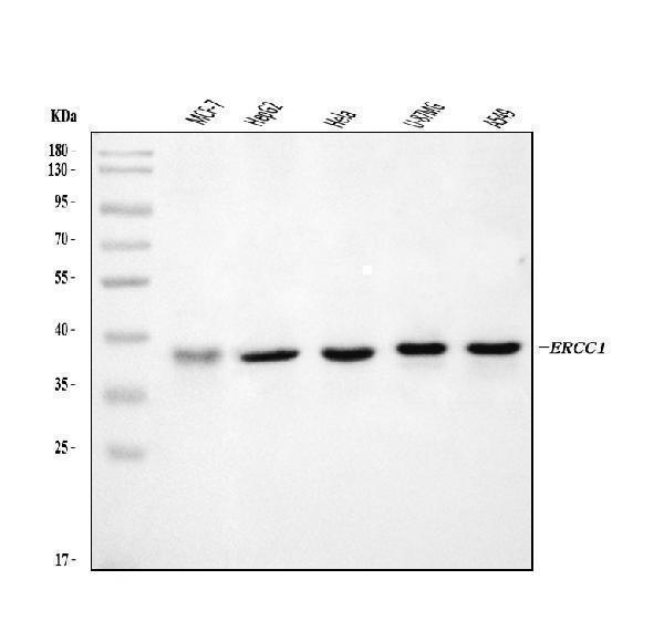
Figure 1. Western blot analysis of ERCC1 using anti-ERCC1 antibody (A00388-4).
Electrophoresis was performed on a 5-20% SDS-PAGE gel at 70V (Stacking gel) / 90V (Resolving gel) for 2-3 hours. The sample well of each lane was loaded with 30 ug of sample under reducing conditions.
Lane 1: human MCF-7 whole cell lysates,
Lane 2: human HepG2 whole cell lysates,
Lane 3: human Hela whole cell lysates,
Lane 4: human U-87MG whole cell lysates,
Lane 5: human A549 whole cell lysates.
After electrophoresis, proteins were transferred to a nitrocellulose membrane at 150 mA for 50-90 minutes. Blocked the membrane with 5% non-fat milk/TBS for 1.5 hour at RT. The membrane was incubated with rabbit anti-ERCC1 antigen affinity purified polyclonal antibody (Catalog # A00388-4) at 0.5 μg/mL overnight at 4°C, then washed with TBS-0.1%Tween 3 times with 5 minutes each and probed with a goat anti-rabbit IgG-HRP secondary antibody at a dilution of 1:5000 for 1.5 hour at RT. The signal is developed using an Enhanced Chemiluminescent detection (ECL) kit (Catalog # EK1002) with Tanon 5200 system. A specific band was detected for ERCC1 at approximately 38 kDa. The expected band size for ERCC1 is at 33 kDa.

Click image to see more details
Figure 2. Flow Cytometry analysis of RT4 cells using anti-ERCC1 antibody (A00388-4).
Overlay histogram showing RT4 cells stained with A00388-4 (Blue line). To facilitate intracellular staining, cells were fixed with 4% paraformaldehyde and permeabilized with permeabilization buffer. The cells were blocked with 10% normal goat serum. And then incubated with rabbit anti-ERCC1 Antibody (A00388-4, 1 μg/1x106 cells) for 30 min at 20°C. DyLight®488 conjugated goat anti-rabbit IgG (BA1127, 5-10 μg/1x106 cells) was used as secondary antibody for 30 minutes at 20°C. Isotype control antibody (Green line) was rabbit IgG (1 μg/1x106) used under the same conditions. Unlabelled sample without incubation with primary antibody and secondary antibody (Red line) was used as a blank control.
Protein Target Info & Infographic
Gene/Protein Information For ERCC1 (Source: Uniprot.org, NCBI)
Gene Name
ERCC1
Full Name
DNA excision repair protein ERCC-1
Weight
17628 MW
Superfamily
ERCC1/RAD10/SWI10 family
Alternative Names
DNA excision repair protein ERCC-1; ERCC1 ERCC1 COFS4, RAD10, UV20 ERCC excision repair 1, endonuclease non-catalytic subunit DNA excision repair protein ERCC-1|excision repair cross-complementation group 1|excision repair cross-complementing rodent repair deficiency, complementation group 1 (includes overlapping antisense sequence)
*If product is indicated to react with multiple species, protein info is based on the gene entry specified above in "Species".For more info on ERCC1, check out the ERCC1 Infographic

We have 30,000+ of these available, one for each gene! Check them out.
In this infographic, you will see the following information for ERCC1: database IDs, superfamily, protein function, synonyms, molecular weight, chromosomal locations, tissues of expression, subcellular locations, post-translational modifications, and related diseases, research areas & pathways. If you want to see more information included, or would like to contribute to it and be acknowledged, please contact [email protected].
Specific Publications For Anti-ERCC1 Antibody Picoband® (A00388-4)
Hello CJ!
No publications found for A00388-4
*Do you have publications using this product? Share with us and receive a reward. Ask us for more details.
Recommended Resources
Here are featured tools and databases that you might find useful.
- Boster's Pathways Library
- Protein Databases
- Bioscience Research Protocol Resources
- Data Processing & Analysis Software
- Photo Editing Software
- Scientific Literature Resources
- Research Paper Management Tools
- Molecular Biology Software
- Primer Design Tools
- Bioinformatics Tools
- Phylogenetic Tree Analysis
Customer Reviews
Have you used Anti-ERCC1 Antibody Picoband®?
Submit a review and receive an Amazon gift card.
- $30 for a review with an image
0 Reviews For Anti-ERCC1 Antibody Picoband®
Customer Q&As
Have a question?
Find answers in Q&As, reviews.
Can't find your answer?
Submit your question
