
Click image to see more details
-

-
-
-
-
+5
Product Info Summary
| SKU: | M03229-1 |
|---|---|
| Size: | 100 μg/vial |
| Reactive Species: | Human, Mouse, Rat |
| Host: | Mouse |
| Application: | Flow Cytometry, IF, IHC, ICC, WB |
Customers Who Bought This Also Bought

Product info
Product Name
Anti-Cyclophilin B PPIB Antibody Picoband® (monoclonal, 11C11)
View all Cyclophilin B Antibodies
SKU/Catalog Number
M03229-1
Size
100 μg/vial
Form
Lyophilized
Description
Boster Bio Anti-Cyclophilin B PPIB Antibody Picoband® (monoclonal, 11C11) catalog # M03229-1. Tested in Flow Cytometry, IF, IHC, ICC, WB applications. This antibody reacts with Human, Mouse, Rat. The brand Picoband indicates this is a premium antibody that guarantees superior quality, high affinity, and strong signals with minimal background in Western blot applications. Only our best-performing antibodies are designated as Picoband, ensuring unmatched performance.
Storage & Handling
Store at -20˚C for one year from date of receipt. After reconstitution, at 4˚C for one month. It can also be aliquotted and stored frozen at -20˚C for six months. Avoid repeated freeze-thaw cycles.
Cite This Product
Anti-Cyclophilin B PPIB Antibody Picoband® (monoclonal, 11C11) (Boster Biological Technology, Pleasanton CA, USA, Catalog # M03229-1)
Host
Mouse
Contents
Each vial contains 4mg Trehalose, 0.9mg NaCl, 0.2mg Na2HPO4, 0.05mg NaN3.
Clonality
Monoclonal
Clone Number
11C11
Isotype
Mouse IgG1
Immunogen
E. coli-derived human Cyclophilin B recombinant protein (Position: K158-E216).
*Blocking peptide can be purchased. Costs vary based on immunogen length. Contact us for pricing.
Cross-reactivity
No cross-reactivity with other proteins.
Reactive Species
M03229-1 is reactive to PPIB in Human, Mouse, Rat
Reconstitution
Add 0.2ml of distilled water will yield a concentration of 500μg/ml.
Observed Molecular Weight
21 kDa
Calculated molecular weight
23.743kDa
Background of Cyclophilin B
Peptidyl-prolyl cis-trans isomerase B, also known as CYPB, is an enzyme that in humans is encoded by the PPIB gene. This gene is mapped to 15q22.31. The protein encoded by this gene is a cyclosporine-binding protein and is mainly located within the endoplasmic reticulum. It is associated with the secretory pathway and released in biological fluids. This protein can bind to cells derived from T- and B-lymphocytes, and may regulate cyclosporine A-mediated immunosuppression. Variants have been identified in this protein that give rise to recessive forms of osteogenesis imperfecta.
Antibody Validation
Boster validates all antibodies on WB, IHC, ICC, Immunofluorescence, and ELISA with known positive control and negative samples to ensure specificity and high affinity, including thorough antibody incubations.
Application & Images
Applications
M03229-1 is guaranteed for Flow Cytometry, IF, IHC, ICC, WB Boster Guarantee
Assay Dilutions Recommendation
The recommendations below provide a starting point for assay optimization. The actual working concentration varies and should be decided by the user.
Western blot, 0.1-0.5μg/ml
Immunohistochemistry (Paraffin-embedded Section), 0.5-1μg/ml
Immunocytochemistry/Immunofluorescence, 2μg/ml
Flow Cytometry (Fixed), 1-3μg/1x106 cells
Positive Control
WB: human placenta tissue, U-87MG whole cell, HepG2 whole cell, Caco-2 whole cell, SW620 whole cell, PANC-1 whole cell, THP-1 whole cell, HEK293 whole cell, rat spleen tissue, rat lung tissue, mouse spleen tissue, mouse lung tissue
IHC: human oesophagus squama cancer tissue, human ovarian cancer tissue, human placenta tissue, human tonsil tissue, human lung cancer tissue
ICC: U20S cell
FCM: U20S cell
Validation Images & Assay Conditions
Click image to see more details
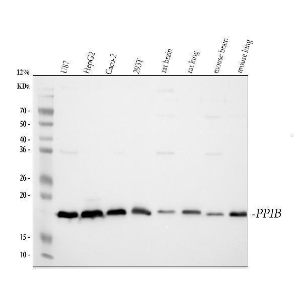
Figure 1. Western blot analysis of Cyclophilin B using anti-Cyclophilin B antibody (M03229-1).
Electrophoresis was performed on a 5-20% SDS-PAGE gel at 70V (Stacking gel) / 90V (Resolving gel) for 2-3 hours. The sample well of each lane was loaded with 50ug of sample under reducing conditions.
Lane 1: human placenta tissue lysates,
Lane 2: U-87MG whole cell lysates,
Lane 3: HepG2 whole cell lysates,
Lane 4: Caco-2 whole cell lysates,
Lane 5: SW620 whole cell lysates,
Lane 6: PANC-1 whole cell lysates,
Lane 7: THP-1 whole cell lysates,
Lane 8: HEK293 whole cell lysates.
After Electrophoresis, proteins were transferred to a Nitrocellulose membrane at 150mA for 50-90 minutes. Blocked the membrane with 5% Non-fat Milk/ TBS for 1.5 hour at RT. The membrane was incubated with mouse anti-Cyclophilin B antigen affinity purified monoclonal antibody (Catalog # M03229-1) at 0.5 μg/mL overnight at 4°C, then washed with TBS-0.1%Tween 3 times with 5 minutes each and probed with a goat anti-mouse IgG-HRP secondary antibody at a dilution of 1:5000 for 1.5 hour at RT. The signal is developed using an Enhanced Chemiluminescent detection (ECL) kit (Catalog # EK1001) with Tanon 5200 system. A specific band was detected for Cyclophilin B at approximately 21KD. The expected band size for Cyclophilin B is at 21KD.

Click image to see more details
Figure 2. Western blot analysis of Cyclophilin B using anti-Cyclophilin B antibody (M03229-1).
Electrophoresis was performed on a 5-20% SDS-PAGE gel at 70V (Stacking gel) / 90V (Resolving gel) for 2-3 hours. The sample well of each lane was loaded with 50ug of sample under reducing conditions.
Lane 1: rat spleen tissue lysates,
Lane 2: rat lung tissue lysates,
Lane 3: mouse spleen tissue lysates,
Lane 4: mouse lung tissue lysates.
After Electrophoresis, proteins were transferred to a Nitrocellulose membrane at 150mA for 50-90 minutes. Blocked the membrane with 5% Non-fat Milk/ TBS for 1.5 hour at RT. The membrane was incubated with mouse anti-Cyclophilin B antigen affinity purified monoclonal antibody (Catalog # M03229-1) at 0.5 μg/mL overnight at 4°C, then washed with TBS-0.1%Tween 3 times with 5 minutes each and probed with a goat anti-mouse IgG-HRP secondary antibody at a dilution of 1:5000 for 1.5 hour at RT. The signal is developed using an Enhanced Chemiluminescent detection (ECL) kit (Catalog # EK1001) with Tanon 5200 system. A specific band was detected for Cyclophilin B at approximately 21KD. The expected band size for Cyclophilin B is at 21KD.

Click image to see more details
Figure 3. IHC analysis of Cyclophilin B using anti-Cyclophilin B antibody (M03229-1).
Cyclophilin B was detected in paraffin-embedded section of human oesophagus squama cancer tissue. Heat mediated antigen retrieval was performed in EDTA buffer (pH8.0, epitope retrieval solution). The tissue section was blocked with 10% goat serum. The tissue section was then incubated with 1μg/ml mouse anti-Cyclophilin B Antibody (M03229-1) overnight at 4°C. Biotinylated goat anti-mouse IgG was used as secondary antibody and incubated for 30 minutes at 37°C. The tissue section was developed using Strepavidin-Biotin-Complex (SABC) (Catalog # SA1021) with DAB as the chromogen.

Click image to see more details
Figure 4. IHC analysis of Cyclophilin B using anti-Cyclophilin B antibody (M03229-1).
Cyclophilin B was detected in paraffin-embedded section of human ovarian cancer tissue. Heat mediated antigen retrieval was performed in EDTA buffer (pH8.0, epitope retrieval solution). The tissue section was blocked with 10% goat serum. The tissue section was then incubated with 1μg/ml mouse anti-Cyclophilin B Antibody (M03229-1) overnight at 4°C. Biotinylated goat anti-mouse IgG was used as secondary antibody and incubated for 30 minutes at 37°C. The tissue section was developed using Strepavidin-Biotin-Complex (SABC) (Catalog # SA1021) with DAB as the chromogen.

Click image to see more details
Figure 5. IHC analysis of Cyclophilin B using anti-Cyclophilin B antibody (M03229-1).
Cyclophilin B was detected in paraffin-embedded section of human placenta tissue. Heat mediated antigen retrieval was performed in EDTA buffer (pH8.0, epitope retrieval solution). The tissue section was blocked with 10% goat serum. The tissue section was then incubated with 1μg/ml mouse anti-Cyclophilin B Antibody (M03229-1) overnight at 4°C. Biotinylated goat anti-mouse IgG was used as secondary antibody and incubated for 30 minutes at 37°C. The tissue section was developed using Strepavidin-Biotin-Complex (SABC) (Catalog # SA1021) with DAB as the chromogen.

Click image to see more details
Figure 6. IHC analysis of Cyclophilin B using anti-Cyclophilin B antibody (M03229-1).
Cyclophilin B was detected in paraffin-embedded section of human tonsil tissue. Heat mediated antigen retrieval was performed in EDTA buffer (pH8.0, epitope retrieval solution). The tissue section was blocked with 10% goat serum. The tissue section was then incubated with 1μg/ml mouse anti-Cyclophilin B Antibody (M03229-1) overnight at 4°C. Biotinylated goat anti-mouse IgG was used as secondary antibody and incubated for 30 minutes at 37°C. The tissue section was developed using Strepavidin-Biotin-Complex (SABC) (Catalog # SA1021) with DAB as the chromogen.

Click image to see more details
Figure 7. IHC analysis of Cyclophilin B using anti-Cyclophilin B antibody (M03229-1).
Cyclophilin B was detected in paraffin-embedded section of human lung cancer tissue. Heat mediated antigen retrieval was performed in EDTA buffer (pH8.0, epitope retrieval solution). The tissue section was blocked with 10% goat serum. The tissue section was then incubated with 1μg/ml mouse anti-Cyclophilin B Antibody (M03229-1) overnight at 4°C. Biotinylated goat anti-mouse IgG was used as secondary antibody and incubated for 30 minutes at 37°C. The tissue section was developed using Strepavidin-Biotin-Complex (SABC) (Catalog # SA1021) with DAB as the chromogen.

Click image to see more details
Figure 8. IHC analysis of Cyclophilin B using anti-Cyclophilin B antibody (M03229-1).
Cyclophilin B was detected in immunocytochemical section of U20S cell. Heat mediated antigen retrieval was performed in EDTA buffer (pH8.0, epitope retrieval solution). The cells were blocked with 10% goat serum. And then incubated with 2μg/ml mouse anti-Cyclophilin B Antibody (M03229-1) overnight at 4°C. Biotin conjugated goat anti-mouse IgG (BA1001) was used as secondary antibody and incubated for 30 minutes at 37°C. The section was developed using Cy3 Conjugated Avidin (BA1037). Visualize using a fluorescence microscope and filter sets appropriate for the label used.

Click image to see more details
Figure 9. Flow Cytometry analysis of U20S cells using anti-Cyclophilin B antibody (M03229-1).
Overlay histogram showing U20S cells stained with M03229-1 (Blue line). To facilitate intracellular staining, cells were fixed with 4% paraformaldehyde and permeabilized with permeabilization buffer. The cells were blocked with 10% normal goat serum. And then incubated with mouse anti-Cyclophilin B Antibody (M03229-1,1μg/1x106 cells) for 30 min at 20°C. DyLight®488 conjugated goat anti-mouse IgG (BA1126, 5-10μg/1x106 cells) was used as secondary antibody for 30 minutes at 20°C. Isotype control antibody (Green line) was mouse IgG (1μg/1x106) used under the same conditions. Unlabelled sample without incubation with primary antibody and secondary antibody (Red line) was used as a blank control.
Protein Target Info & Infographic
Gene/Protein Information For PPIB (Source: Uniprot.org, NCBI)
Gene Name
PPIB
Full Name
Peptidyl-prolyl cis-trans isomerase B
Weight
23.743kDa
Superfamily
cyclophilin-type PPIase family
Alternative Names
Peptidyl-prolyl cis-trans isomerase B; PPIase B; CYP-S1; Cyclophilin B; Rotamase B; S-cyclophilin; SCYLP; PPIB; CYPB PPIB B, CYP-S1, CYPB, HEL-S-39, OI9, SCYLP peptidylprolyl isomerase B peptidyl-prolyl cis-trans isomerase B|PPIase B|S-cyclophilin|cyclophilin-like protein|epididymis secretory protein Li 39|peptidylprolyl isomerase B (cyclophilin B)|rotamase B
*If product is indicated to react with multiple species, protein info is based on the gene entry specified above in "Species".For more info on PPIB, check out the PPIB Infographic

We have 30,000+ of these available, one for each gene! Check them out.
In this infographic, you will see the following information for PPIB: database IDs, superfamily, protein function, synonyms, molecular weight, chromosomal locations, tissues of expression, subcellular locations, post-translational modifications, and related diseases, research areas & pathways. If you want to see more information included, or would like to contribute to it and be acknowledged, please contact [email protected].
Specific Publications For Anti-Cyclophilin B PPIB Antibody Picoband® (monoclonal, 11C11) (M03229-1)
Loading publications
Recommended Resources
Here are featured tools and databases that you might find useful.
- Boster's Pathways Library
- Protein Databases
- Bioscience Research Protocol Resources
- Data Processing & Analysis Software
- Photo Editing Software
- Scientific Literature Resources
- Research Paper Management Tools
- Molecular Biology Software
- Primer Design Tools
- Bioinformatics Tools
- Phylogenetic Tree Analysis
Customer Reviews
Have you used Anti-Cyclophilin B PPIB Antibody Picoband® (monoclonal, 11C11)?
Submit a review and receive an Amazon gift card.
- $30 for a review with an image
0 Reviews For Anti-Cyclophilin B PPIB Antibody Picoband® (monoclonal, 11C11)
Customer Q&As
Have a question?
Find answers in Q&As, reviews.
Can't find your answer?
Submit your question
6 Customer Q&As for Anti-Cyclophilin B PPIB Antibody Picoband® (monoclonal, 11C11)
Question
Is a blocking peptide available for product anti-Cyclophilin B antibody (monoclonal, 11C11) (M03229-1)?
Verified Customer
Verified customer
Asked: 2020-02-07
Answer
We do provide the blocking peptide for product anti-Cyclophilin B antibody (monoclonal, 11C11) (M03229-1). If you would like to place an order for it please contact [email protected] and make a special request.
Boster Scientific Support
Answered: 2020-02-07
Question
Please see the WB image, lot number and protocol we used for melanoma using anti-Cyclophilin B antibody (monoclonal, 11C11) M03229-1. Please let me know if you require anything else.
Verified Customer
Verified customer
Asked: 2019-07-26
Answer
Thank you very much for the data. Our lab team are working to resolve this as quickly as possible, and we appreciate your patience and understanding! You have provided everything we needed. Please let me know if there is anything you need in the meantime.
Boster Scientific Support
Answered: 2019-07-26
Question
I see that the anti-Cyclophilin B antibody (monoclonal, 11C11) M03229-1 works with Flow Cytometry, what is the protocol used to produce the result images on the product page?
Verified Customer
Verified customer
Asked: 2018-09-28
Answer
You can find protocols for Flow Cytometry on the "support/technical resources" section of our navigation menu. If you have any further questions, please send an email to [email protected]
Boster Scientific Support
Answered: 2018-09-28
Question
Do you have a BSA free version of anti-Cyclophilin B antibody (monoclonal, 11C11) M03229-1 available?
Verified Customer
Verified customer
Asked: 2018-07-27
Answer
We appreciate your recent telephone inquiry. I can confirm that some lots of this anti-Cyclophilin B antibody (monoclonal, 11C11) M03229-1 are BSA free. For now, these lots are available and we can make a BSA free formula for you free of charge. It will take 3 extra days to prepare. If you require this antibody BSA free again in future, please do not hesitate to contact me and I will be pleased to check which lots we have in stock that are BSA free.
Boster Scientific Support
Answered: 2018-07-27
Question
We appreciate helping with my inquiry over the phone. Here are the WB image, lot number and protocol we used for melanoma using anti-Cyclophilin B antibody (monoclonal, 11C11) M03229-1. Let me know if you need anything else.
Verified Customer
Verified customer
Asked: 2018-07-24
Answer
Thanks for the data. You have provided everything we needed. Our lab team are working to resolve your inquiry as quickly as possible, and we appreciate your patience and understanding! Please let me know if there is anything you need in the meantime.
Boster Scientific Support
Answered: 2018-07-24
Question
My question regarding product M03229-1, anti-Cyclophilin B antibody (monoclonal, 11C11). I was wondering if it would be possible to conjugate this antibody with biotin. I would need it to be without BSA or sodium azide. I am planning on using a buffer exchange of sodium azide with PBS only. Would there be problems for me to conjugate the antibody and store it in -20 degrees in small aliquots?
D. Yang
Verified customer
Asked: 2013-11-01
Answer
We do not recommend storing this antibody with PBS buffer only in -20 degrees. If you want to store it in -20 degrees it is best to add some cryoprotectant like glycerol. If you want carrier free M03229-1 anti-Cyclophilin B antibody (monoclonal, 11C11), we can provide it to you in a special formula with trehalose and/or glycerol. These molecules will not interfere with conjugation chemistry and provide a good level of protection for the antibody from degradation. Please be sure to specify this in your purchase order.
Boster Scientific Support
Answered: 2013-11-01
