Click image to see more details
-

-
-
-
-
+4
Product Info Summary
| SKU: | M00209-6 |
|---|---|
| Size: | 100 μg/vial |
| Reactive Species: | Human, Mouse, Rat |
| Host: | Mouse |
| Application: | Flow Cytometry, IHC, WB |
Customers Who Bought This Also Bought

Product info
Product Name
Anti-CDK1 Antibody Picoband® (monoclonal, 2G11)
SKU/Catalog Number
M00209-6
Size
100 μg/vial
Form
Lyophilized
Description
Boster Bio Anti-CDK1 Antibody Picoband® (monoclonal, 2G11) catalog # M00209-6. Tested in Flow Cytometry, IHC, WB applications. This antibody reacts with Human, Mouse, Rat. The brand Picoband indicates this is a premium antibody that guarantees superior quality, high affinity, and strong signals with minimal background in Western blot applications. Only our best-performing antibodies are designated as Picoband, ensuring unmatched performance.
Storage & Handling
Store at -20˚C for one year from date of receipt. After reconstitution, at 4˚C for one month. It can also be aliquotted and stored frozen at -20˚C for six months. Avoid repeated freeze-thaw cycles.
Cite This Product
Anti-CDK1 Antibody Picoband® (monoclonal, 2G11) (Boster Biological Technology, Pleasanton CA, USA, Catalog # M00209-6)
Host
Mouse
Contents
Each vial contains 4mg Trehalose, 0.9mg NaCl, 0.2mg Na2HPO4, 0.05mg NaN3.
Clonality
Monoclonal
Clone Number
2G11
Isotype
Mouse IgG2b
Immunogen
E.coli-derived human CDK1 recombinant protein (Position: L66-M297). Human CDK1 shares 97.8% and 98.3% amino acid (aa) sequence identity with mouse and rat CDK1, respectively.
*Blocking peptide can be purchased. Costs vary based on immunogen length. Contact us for pricing.
Cross-reactivity
No cross-reactivity with other proteins.
Reactive Species
M00209-6 is reactive to CDK1 in Human, Mouse, Rat
Reconstitution
Add 0.2ml of distilled water will yield a concentration of 500μg/ml.
Observed Molecular Weight
34 kDa
Calculated molecular weight
34.095kDa
Background of CDC2/CDK1
CDC2, Cell Division Cycle 2, is also known as CDK1 (Cyclin-dependent Kinase 1). CDC2 is a catalytic subunit of a protein kinase complex, called the M-phase promoting factor that induces entry into mitosis and is universal among eukaryotes. In HeLa cells CDC2 is the most abundant phosphotyrosine-containing protein and its phosphotyrosine content is subject to cell cycle regulation. CDC2 gene is located on chromosome 10.
Antibody Validation
Boster validates all antibodies on WB, IHC, ICC, Immunofluorescence, and ELISA with known positive control and negative samples to ensure specificity and high affinity, including thorough antibody incubations.
Application & Images
Applications
M00209-6 is guaranteed for Flow Cytometry, IHC, WB Boster Guarantee
Assay Dilutions Recommendation
The recommendations below provide a starting point for assay optimization. The actual working concentration varies and should be decided by the user.
Western blot, 0.1-0.5μg/ml
Immunohistochemistry (Paraffin-embedded Section), 0.5-1μg/ml
Flow Cytometry (Fixed), 1-3μg/1x106 cells
Positive Control
WB: human HEK293 whole cell, human A549 whole cell, human HepG2 whole cell, human THP-1 whole cell, human PANC-1 whole cell, human SW620 whole cell, rat RH35 whole cell, mouse NIH/3T3 whole cell,
IHC: human colon cancer tissue, human tonsil tissue, human lung cancer tissue, mouse testis tissue, rat testis tissue
FCM: PC-3 cell, U20S cell
Validation Images & Assay Conditions
Click image to see more details
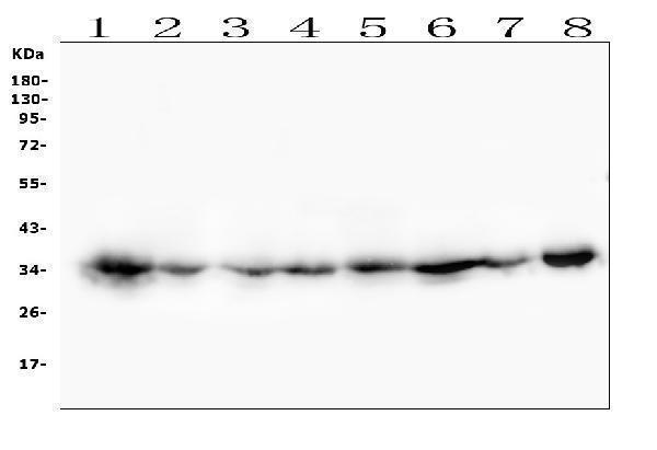
Figure 1. Western blot analysis of CDK1 using anti-CDK1 antibody (M00209-6).
Electrophoresis was performed on a 5-20% SDS-PAGE gel at 70V (Stacking gel) / 90V (Resolving gel) for 2-3 hours. The sample well of each lane was loaded with 50ug of sample under reducing conditions.
Lane 1: human HEK293 whole cell lysates
Lane 2: human A549 whole cell lysates
Lane 3: human HepG2 whole cell lysates
Lane 4: human THP-1 whole cell lysates
Lane 5: human PANC-1 whole cell lysates
Lane 6: human SW620 whole cell lysates
Lane 7: rat RH35 whole cell lysates
Lane 8: mouse NIH/3T3 whole cell lysates
After Electrophoresis, proteins were transferred to a Nitrocellulose membrane at 150mA for 50-90 minutes. Blocked the membrane with 5% Non-fat Milk/ TBS for 1.5 hour at RT. The membrane was incubated with mouse anti-CDK1 antigen affinity purified monoclonal antibody (Catalog # M00209-6) at 0.5 μg/mL overnight at 4°C, then washed with TBS-0.1%Tween 3 times with 5 minutes each and probed with a goat anti-mouse IgG-HRP secondary antibody at a dilution of 1:10000 for 1.5 hour at RT. The signal is developed using an Enhanced Chemiluminescent detection (ECL) kit (Catalog # EK1001) with Tanon 5200 system. A specific band was detected for CDK1 at approximately 34KD. The expected band size for CDK1 is at 34KD.

Click image to see more details
Figure 2. IHC analysis of CDK1 using anti-CDK1 antibody (M00209-6).
CDK1 was detected in paraffin-embedded section of human colon cancer tissues. Heat mediated antigen retrieval was performed in citrate buffer (pH6, epitope retrieval solution) for 20 mins. The tissue section was blocked with 10% goat serum. The tissue section was then incubated with 1μg/ml mouse anti-CDK1 Antibody (M00209-6) overnight at 4°C. Biotinylated goat anti-mouse IgG was used as secondary antibody and incubated for 30 minutes at 37°C. The tissue section was developed using Strepavidin-Biotin-Complex (SABC)(Catalog # SA1021) with DAB as the chromogen.

Click image to see more details
Figure 3. IHC analysis of CDK1 using anti-CDK1 antibody (M00209-6).
CDK1 was detected in paraffin-embedded section of human tonsil tissues. Heat mediated antigen retrieval was performed in citrate buffer (pH6, epitope retrieval solution) for 20 mins. The tissue section was blocked with 10% goat serum. The tissue section was then incubated with 1μg/ml mouse anti-CDK1 Antibody (M00209-6) overnight at 4°C. Biotinylated goat anti-mouse IgG was used as secondary antibody and incubated for 30 minutes at 37°C. The tissue section was developed using Strepavidin-Biotin-Complex (SABC)(Catalog # SA1021) with DAB as the chromogen.

Click image to see more details
Figure 4. IHC analysis of CDK1 using anti-CDK1 antibody (M00209-6).
CDK1 was detected in paraffin-embedded section of human lung cancer tissues. Heat mediated antigen retrieval was performed in citrate buffer (pH6, epitope retrieval solution) for 20 mins. The tissue section was blocked with 10% goat serum. The tissue section was then incubated with 1μg/ml mouse anti-CDK1 Antibody (M00209-6) overnight at 4°C. Biotinylated goat anti-mouse IgG was used as secondary antibody and incubated for 30 minutes at 37°C. The tissue section was developed using Strepavidin-Biotin-Complex (SABC)(Catalog # SA1021) with DAB as the chromogen.

Click image to see more details
Figure 5. IHC analysis of CDK1 using anti-CDK1 antibody (M00209-6).
CDK1 was detected in paraffin-embedded section of mouse testis tissues. Heat mediated antigen retrieval was performed in citrate buffer (pH6, epitope retrieval solution) for 20 mins. The tissue section was blocked with 10% goat serum. The tissue section was then incubated with 1μg/ml mouse anti-CDK1 Antibody (M00209-6) overnight at 4°C. Biotinylated goat anti-mouse IgG was used as secondary antibody and incubated for 30 minutes at 37°C. The tissue section was developed using Strepavidin-Biotin-Complex (SABC)(Catalog # SA1021) with DAB as the chromogen.

Click image to see more details
Figure 6. IHC analysis of CDK1 using anti-CDK1 antibody (M00209-6).
CDK1 was detected in paraffin-embedded section of rat testis tissues. Heat mediated antigen retrieval was performed in citrate buffer (pH6, epitope retrieval solution) for 20 mins. The tissue section was blocked with 10% goat serum. The tissue section was then incubated with 1μg/ml mouse anti-CDK1 Antibody (M00209-6) overnight at 4°C. Biotinylated goat anti-mouse IgG was used as secondary antibody and incubated for 30 minutes at 37°C. The tissue section was developed using Strepavidin-Biotin-Complex (SABC)(Catalog # SA1021) with DAB as the chromogen.

Click image to see more details
Figure 7. Flow Cytometry analysis of PC-3 cells using anti-CDK1 antibody (M00209-6).
Overlay histogram showing PC-3 cells stained with M00209-6 (Blue line). To facilitate intracellular staining, cells were fixed with 4% paraformaldehyde and permeabilized with permeabilization buffer. The cells were blocked with 10% normal goat serum. And then incubated with mouse anti-CDK1 Antibody (M00209-6,1μg/1x106 cells) for 30 min at 20°C. DyLight®488 conjugated goat anti-mouse IgG (BA1126, 5-10μg/1x106 cells) was used as secondary antibody for 30 minutes at 20°C. Isotype control antibody (Green line) was mouse IgG (1μg/1x106) used under the same conditions. Unlabelled sample without incubation with primary antibody and secondary antibody (Red line) was used as a blank control.

Click image to see more details
Figure 8. Flow Cytometry analysis of U20S cells using anti-CDK1 antibody (M00209-6).
Overlay histogram showing U20S cells stained with M00209-6 (Blue line). To facilitate intracellular staining, cells were fixed with 4% paraformaldehyde and permeabilized with permeabilization buffer. The cells were blocked with 10% normal goat serum. And then incubated with mouse anti-CDK1 Antibody (M00209-6,1μg/1x106 cells) for 30 min at 20°C. DyLight®488 conjugated goat anti-mouse IgG (BA1126, 5-10μg/1x106 cells) was used as secondary antibody for 30 minutes at 20°C. Isotype control antibody (Green line) was mouse IgG (1μg/1x106) used under the same conditions. Unlabelled sample without incubation with primary antibody and secondary antibody (Red line) was used as a blank control.
Protein Target Info & Infographic
Gene/Protein Information For CDK1 (Source: Uniprot.org, NCBI)
Gene Name
CDK1
Full Name
Cyclin-dependent kinase 1
Weight
34.095kDa
Superfamily
protein kinase superfamily
Alternative Names
Cyclin-dependent kinase 1; CDK1; Cell division control protein 2 homolog; Cell division protein kinase 1; p34 protein kinase; CDK1; CDC2P; CDC28A; CDKN1; P34CDC2 CDK1 CDC2, CDC28A, P34CDC2 cyclin dependent kinase 1 cyclin-dependent kinase 1|cell cycle controller CDC2|cell division control protein 2 homolog|cell division cycle 2, G1 to S and G2 to M|cell division protein kinase 1|p34 protein kinase
*If product is indicated to react with multiple species, protein info is based on the gene entry specified above in "Species".For more info on CDK1, check out the CDK1 Infographic

We have 30,000+ of these available, one for each gene! Check them out.
In this infographic, you will see the following information for CDK1: database IDs, superfamily, protein function, synonyms, molecular weight, chromosomal locations, tissues of expression, subcellular locations, post-translational modifications, and related diseases, research areas & pathways. If you want to see more information included, or would like to contribute to it and be acknowledged, please contact [email protected].
Specific Publications For Anti-CDK1 Antibody Picoband® (monoclonal, 2G11) (M00209-6)
Hello CJ!
M00209-6 has been cited in 7 publications:
*The publications in this section are manually curated by our staff scientists. They may differ from Bioz's machine gathered results. Both are accurate. If you find a publication citing this product but is missing from this list, please let us know we will issue you a thank-you coupon.
Pseudolaric acid B inhibits neuroglioma cell proliferation through DNA damage response
Suppression of C-myc Expression Associates with Anti-Proliferation of Aloe-Emodin on Gastric Cancer Cells
Blakemore D,Vilaplana-Lopera N,Almaghrabi R,Gonzalez E,Moya M,Ward C,Murphy G,Gambus A,Petermann E,Stewart GS,García P.MYBL2 and ATM suppress replication stress in pluripotent stem cells.EMBO Rep.2021 Mar 28:e51120.doi:10.15252/embr.202051120.Epub ahead of print.PMID:33779025.
Species: Mouse
M00209-6 usage in article: APP:WB, SAMPLE: ESCS, DILUTION:NA
A novel cell cycle blocker extracted from Stellera chamaejasme L. inhibits the proliferation of hepatocarcinoma cells
Human enterovirus 68 interferes with the host cell cycle to facilitate viral production
Coxsackievirus A6 induces cell cycle arrest in G0/G1 phase for viral production
Yang C, Yang J, Sun M, Yan J, Meng X, Ma T. Iubmb Life. 2013 May;65(5):435-44. Doi: 10.1002/Iub.1141. Epub 2013 Feb 26. Alantolactone Inhibits Growth Of K562/Adriamycin Cells By Downregulating Bcr/Abl And P-Glycoprotein Expression.
Recommended Resources
Here are featured tools and databases that you might find useful.
- Boster's Pathways Library
- Protein Databases
- Bioscience Research Protocol Resources
- Data Processing & Analysis Software
- Photo Editing Software
- Scientific Literature Resources
- Research Paper Management Tools
- Molecular Biology Software
- Primer Design Tools
- Bioinformatics Tools
- Phylogenetic Tree Analysis
Customer Reviews
Have you used Anti-CDK1 Antibody Picoband® (monoclonal, 2G11)?
Submit a review and receive an Amazon gift card.
- $30 for a review with an image
0 Reviews For Anti-CDK1 Antibody Picoband® (monoclonal, 2G11)
Customer Q&As
Have a question?
Find answers in Q&As, reviews.
Can't find your answer?
Submit your question
5 Customer Q&As for Anti-CDK1 Antibody Picoband® (monoclonal, 2G11)
Question
Our team were well pleased with the WB result of your anti-CDK1 antibody (monoclonal, 2G11). However we have seen positive staining in leukemic t-cell nucleus. cytoplasm. mitochondrion. using this antibody. Is that expected? Could you tell me where is CDK1 supposed to be expressed?
Verified Customer
Verified customer
Asked: 2019-10-24
Answer
From what I have seen in literature, leukemic t-cell does express CDK1. Generally CDK1 expresses in nucleus. cytoplasm. mitochondrion. Regarding which tissues have CDK1 expression, here are a few articles citing expression in various tissues:
Cervix carcinoma, Pubmed ID: 18220336, 18669648, 18691976, 20068231
Cervix carcinoma, and Erythroleukemia, Pubmed ID: 23186163
Leukemic T-cell, Pubmed ID: 19690332
Lymphoblast, Pubmed ID: 14654843
Mammary cancer, Pubmed ID: 9515786, 14702039
Skin, Pubmed ID: 15489334
Boster Scientific Support
Answered: 2019-10-24
Question
We have observed staining in rat cervix carcinoma. Any tips? Is anti-CDK1 antibody (monoclonal, 2G11) supposed to stain cervix carcinoma positively?
Verified Customer
Verified customer
Asked: 2019-10-03
Answer
Based on literature cervix carcinoma does express CDK1. Based on Uniprot.org, CDK1 is expressed in intestine, mammary cancer, skin, lymphoblast, cervix carcinoma, leukemic t-cell, cervix carcinoma erythroleukemia, among other tissues. Regarding which tissues have CDK1 expression, here are a few articles citing expression in various tissues:
Cervix carcinoma, Pubmed ID: 18220336, 18669648, 18691976, 20068231
Cervix carcinoma, and Erythroleukemia, Pubmed ID: 23186163
Leukemic T-cell, Pubmed ID: 19690332
Lymphoblast, Pubmed ID: 14654843
Mammary cancer, Pubmed ID: 9515786, 14702039
Skin, Pubmed ID: 15489334
Boster Scientific Support
Answered: 2019-10-03
Question
We ordered your anti-CDK1 antibody (monoclonal, 2G11) for Flow Cytometry on skin in the past. I am using human, and We are going to use the antibody for WB next. I would like examining skin as well as cervix carcinoma in our next experiment. Could you please give me some suggestion on which antibody would work the best for WB?
Verified Customer
Verified customer
Asked: 2019-05-23
Answer
I looked at the website and datasheets of our anti-CDK1 antibody (monoclonal, 2G11) and it appears that M00209-6 has been tested on human in both Flow Cytometry and WB. Thus M00209-6 should work for your application. Our Boster satisfaction guarantee will cover this product for WB in human even if the specific tissue type has not been validated. We do have a comprehensive range of products for WB detection and you can check out our website bosterbio.com to find out more information about them.
Boster Scientific Support
Answered: 2019-05-23
Question
We are currently using anti-CDK1 antibody (monoclonal, 2G11) M00209-6 for human tissue, and we are content with the Flow Cytometry results. The species of reactivity given in the datasheet says human, mouse, rat. Is it true that the antibody can work on dog tissues as well?
R. Li
Verified customer
Asked: 2013-07-01
Answer
The anti-CDK1 antibody (monoclonal, 2G11) (M00209-6) has not been tested for cross reactivity specifically with dog tissues, though there is a good chance of cross reactivity. We have an innovator award program that if you test this antibody and show it works in dog you can get your next antibody for free. Please contact me if I can help you with anything.
Boster Scientific Support
Answered: 2013-07-01
Question
Our lab want to know about using your anti-CDK1 antibody (monoclonal, 2G11) for cell division studies. Has this antibody been tested with western blotting on a549 whole cell lysates? We would like to see some validation images before ordering.
B. Parker
Verified customer
Asked: 2013-03-06
Answer
I appreciate your inquiry. This M00209-6 anti-CDK1 antibody (monoclonal, 2G11) is validated on human a549, a549 whole cell lysates, hepg2 whole cell lysates, sw620 whole cell lysates, rat rh35 whole cell lysates, u20s cells. It is guaranteed to work for Flow Cytometry, IHC-P, WB in human, mouse, rat. Our Boster guarantee will cover your intended experiment even if the sample type has not been be directly tested.
Boster Scientific Support
Answered: 2013-03-06
