Click image to see more details
-

-
-
-
-
+2
Product Info Summary
| SKU: | A02423-2 |
|---|---|
| Size: | 100 μg/vial |
| Reactive Species: | Human, Mouse, Rat |
| Host: | Rabbit |
| Application: | IHC, WB |
Customers Who Bought This Also Bought

Product info
Product Name
Anti-Bag1 Antibody Picoband™
SKU/Catalog Number
A02423-2
Size
100 μg/vial
Form
Lyophilized
Description
Boster Bio Anti-Bag1 Antibody Picoband™ catalog # A02423-2. Tested in IHC, WB applications. This antibody reacts with Human, Mouse, Rat.
Storage & Handling
Store at -20˚C for one year from date of receipt. After reconstitution, at 4˚C for one month. It can also be aliquotted and stored frozen at -20˚C for six months. Avoid repeated freeze-thaw cycles.
Cite This Product
Anti-Bag1 Antibody Picoband™ (Boster Biological Technology, Pleasanton CA, USA, Catalog # A02423-2)
Host
Rabbit
Contents
Each vial contains 4mg Trehalose, 0.9mg NaCl and 0.2mg Na2HPO4.
Clonality
Polyclonal
Isotype
Rabbit IgG
Immunogen
A synthetic peptide corresponding to a sequence at the C-terminus of human Bag1, different from the related mouse sequence by two amino acids, and from the related rat sequence by three amino acids.
*Blocking peptide can be purchased. Costs vary based on immunogen length. Contact us for pricing.
Cross-reactivity
No cross-reactivity with other proteins
Reactive Species
A02423-2 is reactive to BAG1 in Human, Mouse, Rat
Applications
A02423-2 is guaranteed for IHC, WB Boster Guarantee
Observed Molecular Weight
33, 46, 56 kDa
Calculated molecular weight
38779 MW
Background of Bag-1
BAG family molecular chaperone regulator 1 (BAG1) is a protein that in humans is encoded by the BAG1 gene. Human BAG1 is mapped to chromosome 9p12, a region associated with hereditary disorders that may involve developmental dysregulation of programmed cell death. The Bag1 protein is rich in glutamic acid residues. Its deduced 274-amino acid protein has a calculated molecular mass of 31 KD. Being the BCL-2-associated athanogene, Bag1 enhances the anti-apoptotic effects of BCL2 and represents a link between growth factor receptors and anti-apoptotic mechanisms.
Antibody Validation
Boster validates all antibodies on WB, IHC, ICC, Immunofluorescence, and ELISA with known positive control and negative samples to ensure specificity and high affinity, including thorough antibody incubations.
Assay dilution & Images
Reconstitution
Add 0.2ml of distilled water will yield a concentration of 500ug/ml.
Assay Dilutions Recommendation
The recommendations below provide a starting point for assay optimization. The actual working concentration varies and should be decided by the user.
Western blot, 0.1-0.5μg/ml, Human
Immunohistochemistry (Paraffin-embedded Section), 2-5μg/ml, Human, Mouse, Rat, By Heat
Validation Images & Assay Conditions
Click image to see more details
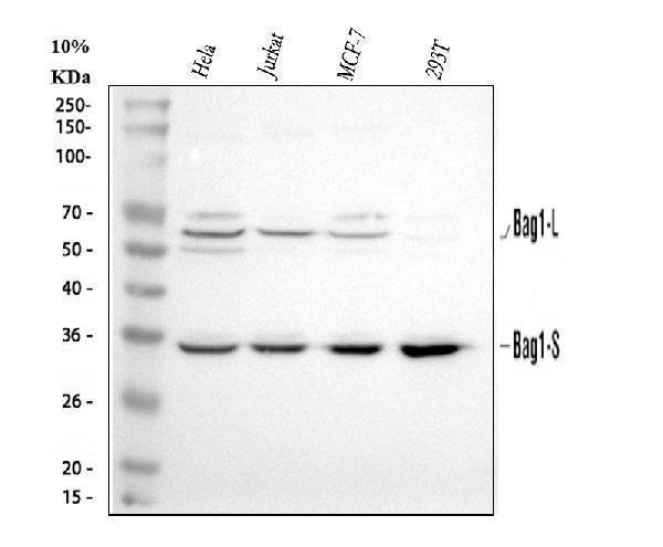
Figure 1. Western blot analysis of Bag1 using anti-Bag1 antibody (A02423-2).
Electrophoresis was performed on a 5-20% SDS-PAGE gel at 70V (Stacking gel) / 90V (Resolving gel) for 2-3 hours. The sample well of each lane was loaded with 30 ug of sample under reducing conditions.
Lane 1: human Hela whole cell lysates,
Lane 2: human Jurkat whole cell lysates,
Lane 3: human MCF-7 whole cell lysates,
Lane 4: human 293T whole cell lysates.
After electrophoresis, proteins were transferred to a nitrocellulose membrane at 150 mA for 50-90 minutes. Blocked the membrane with 5% non-fat milk/TBS for 1.5 hour at RT. The membrane was incubated with rabbit anti-Bag1 antigen affinity purified polyclonal antibody (Catalog # A02423-2) at 0.5 μg/mL overnight at 4°C, then washed with TBS-0.1%Tween 3 times with 5 minutes each and probed with a goat anti-rabbit IgG-HRP secondary antibody at a dilution of 1:5000 for 1.5 hour at RT. The signal is developed using an Enhanced Chemiluminescent detection (ECL) kit (Catalog # EK1002) with Tanon 5200 system. A specific band was detected for Bag1 at approximately 33, 46, 56 kDa. The expected band size for Bag1 is at 39 kDa.

Click image to see more details
Figure 2. IHC analysis of Bag1 using anti-Bag1 antibody (A02423-2).
Bag1 was detected in a paraffin-embedded section of human breast cancer tissue. Heat mediated antigen retrieval was performed in EDTA buffer (pH 8.0, epitope retrieval solution). The tissue section was blocked with 10% goat serum. The tissue section was then incubated with 2 μg/ml rabbit anti-Bag1 Antibody (A02423-2) overnight at 4°C. Peroxidase Conjugated Goat Anti-rabbit IgG was used as secondary antibody and incubated for 30 minutes at 37°C. The tissue section was developed using HRP Conjugated Rabbit IgG Super Vision Assay Kit (Catalog # SV0002) with DAB as the chromogen.

Click image to see more details
Figure 3. IHC analysis of Bag1 using anti-Bag1 antibody (A02423-2).
Bag1 was detected in a paraffin-embedded section of human colorectal adenocarcinoma tissue. Heat mediated antigen retrieval was performed in EDTA buffer (pH 8.0, epitope retrieval solution). The tissue section was blocked with 10% goat serum. The tissue section was then incubated with 2 μg/ml rabbit anti-Bag1 Antibody (A02423-2) overnight at 4°C. Peroxidase Conjugated Goat Anti-rabbit IgG was used as secondary antibody and incubated for 30 minutes at 37°C. The tissue section was developed using HRP Conjugated Rabbit IgG Super Vision Assay Kit (Catalog # SV0002) with DAB as the chromogen.

Click image to see more details
Figure 4. IHC analysis of Bag1 using anti-Bag1 antibody (A02423-2).
Bag1 was detected in a paraffin-embedded section of human tonsil tissue. Heat mediated antigen retrieval was performed in EDTA buffer (pH 8.0, epitope retrieval solution). The tissue section was blocked with 10% goat serum. The tissue section was then incubated with 2 μg/ml rabbit anti-Bag1 Antibody (A02423-2) overnight at 4°C. Peroxidase Conjugated Goat Anti-rabbit IgG was used as secondary antibody and incubated for 30 minutes at 37°C. The tissue section was developed using HRP Conjugated Rabbit IgG Super Vision Assay Kit (Catalog # SV0002) with DAB as the chromogen.

Click image to see more details
Figure 5. IHC analysis of Bag1 using anti-Bag1 antibody (A02423-2).
Bag1 was detected in a paraffin-embedded section of mouse colon tissue. Heat mediated antigen retrieval was performed in EDTA buffer (pH 8.0, epitope retrieval solution). The tissue section was blocked with 10% goat serum. The tissue section was then incubated with 2 μg/ml rabbit anti-Bag1 Antibody (A02423-2) overnight at 4°C. Peroxidase Conjugated Goat Anti-rabbit IgG was used as secondary antibody and incubated for 30 minutes at 37°C. The tissue section was developed using HRP Conjugated Rabbit IgG Super Vision Assay Kit (Catalog # SV0002) with DAB as the chromogen.

Click image to see more details
Figure 6. IHC analysis of Bag1 using anti-Bag1 antibody (A02423-2).
Bag1 was detected in a paraffin-embedded section of rat colon tissue. Heat mediated antigen retrieval was performed in EDTA buffer (pH 8.0, epitope retrieval solution). The tissue section was blocked with 10% goat serum. The tissue section was then incubated with 2 μg/ml rabbit anti-Bag1 Antibody (A02423-2) overnight at 4°C. Peroxidase Conjugated Goat Anti-rabbit IgG was used as secondary antibody and incubated for 30 minutes at 37°C. The tissue section was developed using HRP Conjugated Rabbit IgG Super Vision Assay Kit (Catalog # SV0002) with DAB as the chromogen.
Protein Target Info & Infographic
Gene/Protein Information For BAG1 (Source: Uniprot.org, NCBI)
Gene Name
BAG1
Full Name
BAG family molecular chaperone regulator 1
Weight
38779 MW
Alternative Names
BAG family molecular chaperone regulator 1; Bag1; Bag-1; Bcl-2 associating athanogene-1 protein; Bcl-2-associated athanogene 1; BCL2-associated athanogene; Bcl-2-binding protein; glucocortoid receptor-associated protein RAP46,46-KD; HAP; RAP46 BAG1 BAG-1, HAP, RAP46 BAG cochaperone 1 BAG family molecular chaperone regulator 1|BCL2 associated athanogene 1|BCL2-associated athanogene|Bcl-2 associating athanogene-1 protein|Bcl-2-binding protein|glucocortoid receptor-associated protein RAP46|receptor-associated protein, 46-KD
*If product is indicated to react with multiple species, protein info is based on the gene entry specified above in "Species".For more info on BAG1, check out the BAG1 Infographic

We have 30,000+ of these available, one for each gene! Check them out.
In this infographic, you will see the following information for BAG1: database IDs, superfamily, protein function, synonyms, molecular weight, chromosomal locations, tissues of expression, subcellular locations, post-translational modifications, and related diseases, research areas & pathways. If you want to see more information included, or would like to contribute to it and be acknowledged, please contact [email protected].
Specific Publications For Anti-Bag1 Antibody Picoband™ (A02423-2)
Hello CJ!
A02423-2 has been cited in 2 publications:
*The publications in this section are manually curated by our staff scientists. They may differ from Bioz's machine gathered results. Both are accurate. If you find a publication citing this product but is missing from this list, please let us know we will issue you a thank-you coupon.
Clinicopathologic significance of BAG1 and TIMP3 expression in colon carcinoma
Bai Yx, Yi Jl, Li Jf, Sui H. World J Gastroenterol. 2007 Jul 28;13(28):3883-5. Clinicopathologic Significance Of Bag1 And Timp3 Expression In Colon Carcinoma.
Recommended Resources
Here are featured tools and databases that you might find useful.
- Boster's Pathways Library
- Protein Databases
- Bioscience Research Protocol Resources
- Data Processing & Analysis Software
- Photo Editing Software
- Scientific Literature Resources
- Research Paper Management Tools
- Molecular Biology Software
- Primer Design Tools
- Bioinformatics Tools
- Phylogenetic Tree Analysis
Customer Reviews
Have you used Anti-Bag1 Antibody Picoband™?
Submit a review and receive an Amazon gift card.
- $30 for a review with an image
0 Reviews For Anti-Bag1 Antibody Picoband™
Customer Q&As
Have a question?
Find answers in Q&As, reviews.
Can't find your answer?
Submit your question
1 Customer Q&As for Anti-Bag1 Antibody Picoband™
Question
We are currently using anti-Bag1 antibody A02423-2 for rat tissue, and we are happy with the IHC results. The species of reactivity given in the datasheet says human, mouse, rat. Is it likely that the antibody can work on horse tissues as well?
Verified Customer
Verified customer
Asked: 2019-10-17
Answer
The anti-Bag1 antibody (A02423-2) has not been validated for cross reactivity specifically with horse tissues, but there is a good chance of cross reactivity. We have an innovator award program that if you test this antibody and show it works in horse you can get your next antibody for free. Please contact me if I can help you with anything.
Boster Scientific Support
Answered: 2019-10-17
