This website uses cookies to ensure you get the best experience on our website.
- Table of Contents
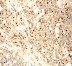
Facts about XIAP-associated factor 1.
Mediates TNF-alpha- induced apoptosis and is involved in apoptosis in trophoblast cells. May inhibit BIRC4 indirectly by activating the mitochondrial apoptosis pathway.
Human | |
---|---|
Gene Name: | XAF1 |
Uniprot: | Q6GPH4 |
Entrez: | 54739 |
Belongs to: |
---|
No superfamily |
BIRC4-binding protein; BIRC4BPBIRC4 binding protein; HSXIAPAF1; XIAP associated factor 1; XIAPAF1XIAP-associated factor 1
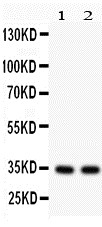
Mass (kDA):
34.626 kDA
Human | |
---|---|
Location: | 17p13.1 |
Sequence: | 17; NC_000017.11 (6755408..6775647) |
Widely expressed. Expression is frequently down-regulated in cancer cell lines. Isoform 5 is widely expressed. Expressed in placenta (at protein level).
Cytoplasm. Nucleus. Mitochondrion. Found in the cytoplasm and nucleus of placental syncytiotrophoblasts. Translocates to mitochondria upon TNF-alpha treatment.; [Isoform 1]: Nucleus.; [Isoform 5]: Nucleus.
XAF1 acts as a negative regulator for members of the IAP protein family (inhibitor apoptosis). It inhibits XIAP through antagonizing anticaspase activity. This marker has been made available by Boster for researchers around the world. Read on to learn more. This article will discuss the many uses of the XAF1 marking.
XAF1 inhibits the caspases-3 to -7 and 9 of the IAP protein family. These caspases trigger apoptosis. Through the BIR3domain, XIAP interacts via XIAP with each of these caspases. It inhibits caspases by binding to their respective BIR3 domains.
XAF1 expression was detected in a variety cancers. Tamm I found that human melanomas express a specific type of ML–IAP. A genetic analysis revealed that two different splicing variants were not functionally comparable. The authors also discussed XIAP’s biological properties and tissue distribution patterns and discussed its potential as a therapeutic target.
The RNA extracted from 293 cell lines was transfected using p3XFLAGRIP1 or HAA-TRADD. Lysate preparation was made by incubating the cells at different concentrations for 24 hrs or incubating with TNFa for one hour. RIP1-associated proteins could be captured by immunoprecipitation using anti-FLAG beads and separated by SDS-PAGE. Transfected proteins were identified with antibodies against actin, XAF1 and other antibodies.
XAF1 is a putative tumor suppressor gene, located on 17p13.2. It has been associated in poor outcome in breast cancer and has been identified in certain types. High XAF1 expression can be associated with activation on the NFkappaB pathway.
IFNG induces proapoptotic responses in immune-deficient mouse models. VSMCs are proapoptotic due to IFNG via STAT1-independent signals. The apoptotic signaling mediated via IFNG is responsible to the proapoptotic effect of XIAP/NOXA.
XAF1 interacts and disrupts TNFR signals by interacting with TRADD1 or RIP1. TNF-a infected cells result in lower levels of TRAF2 or RIP1 protein. XAF1 may influence NF-kB signaling, which is necessary before RIP1 can be activated.
XAF1 is believed to modulate XIAP's ability to inhibit apoptosis. Previously, XIAP had been shown to inhibit apoptosis. Researchers have recently found that XIAP has the ability to influence autophagy. A study by Huang et al. Huang et.al. demonstrated that XIAP inhibits phagy via Mdm2-p53's signaling pathway.
XAF1, a cytoplasmic cytoplasmic proteins, is expressed in low levels in normal tissues and is increased by cytokines. XAF1 has been implicated by many groups in pro-apoptotic response, but there isn’t consensus about its exact role. XAF1 binds XIAP. This is what was suggested in earlier studies. Studies that followed have not confirmed the association between XAF1 & XIAP. Moreover other members of IAP can interact with XAF1, so the implications of these interactions are not clear.
These results suggest that XAF1 might promote pro-apoptotic responses by increasing the sensitivity of cells for pro-apoptotic signals. These observations are important for understanding inflammatory reactions and cytokine interaction in cells. If these findings are replicated in humans, it would indicate that XAF1 is an important negative regulator of TNF-a responsiveness.
Another function that XAF1 has is to regulate the migration of tumor cells. It acts to inhibit tumorigenesis by acting as a coactivator and activator of IRF-1. XAF1 blocks p65/RelA binding with IRFE and can suppress tumor cell movement. XAF1 also inhibits XIAP’s anticaspase action.
XIAP is a group of proteins found in the body. They regulate several cellular processes, including apoptosis and cell growth. Dysregulations in these proteins could cause cancer progression. Although the role XIAP has in tumor biology is not yet clear, XAF1 counteracts XIAP's anticaspase activities by inhibiting Smac.
The role of XAF1 in cancer progression is unknown, but it has been shown to inhibit FAF1-mediated apoptosis in cancer cells. Activation of XAF1's XIAP pathway increases cell survival by inhibiting FAF1 mediated apoptosis. XAF1 promotes apoptosis via multiple pathways including interaction with IRF-1, ubiquitin protasome systems, and more. These effects suggest that XAF1 is a proapoptotic tumour suppressor.
XAF1 is able to associate with TRADD1 or RIP1. Both proteins are induced to degrade after treatment with TNF-a and RIP1 and TRAF2 by transfection. XAF1 expression in combination with TRAF2 decreased TRADD's half life. The anti-XAF1 activity for RIP1 has been decreased.
Moreover, XAF1 is an inhibitor of TNF-a-induced NF-kB activation. It also affects the function and functionality of the TRADD/TRAF2/RIP1 compound. XAF1 might physically block interaction between TRADD/TRAF2 via blocking K63 ubiquitination. Complex formation is dependent on TRADD/TRAF2. Premature K63-ubiquitination may lead to premature proteasomal destruction of TRADD, TRAF2, or RIP1. XAF1 might be more abundant in cytokines-primed inflammatory cells.
The risk of cell suicide is strongly linked to the level of XAF1 expressed in the nucleus. Excessive XAF1 expression can cause cell death and inhibit cell proliferation. Furthermore, overexpression of XAF1 inhibits spindle assembly and induces centrosomal amplification. Moreover, increased XAF1 expression has been associated with cell cycle arrest at G2/M.
XAF1 methylation also affects OS and PFS. However, results from IDh2mut/XAF1M cancers may not be sufficient to draw conclusions about XAF1 status for the general population. Further studies of the disease model are necessary to evaluate the effect of XAF1 survivability. These findings will likely improve cancer treatment for XAF1-mutated patients. They may also have important implications in the treatment of IDh2-mutated people.
This study used a high-resolution mass spectrometry technique to measure the promoter methylation of XAF1 gene. The researchers used a non-parametric Spearman's rank-order correlation to calculate the percent methylation of XAF1. We found a strong association between XAF1's promoter methylation, and cell death using this method. We also tested XAF1 methylation for survival and IDh2 status using a 2-tailed test.
XAF1 knockdowns cells switch from G2 into G1 phase, and accumulate in G1. They don't form colonies. Overexpression of XAF1 can inhibit their growth. The results of the MTT assay were confirmed with viable cell counting. Moreover, XAF1 knockdown cells exhibit enhanced metabolic competence when exposed to TMZ. These cells are more susceptible to cell death.
XAF1 was identified as a novel interferon-stimulating gene that augmented TNF-related apoptosis. XAF1 binds with other IAP proteins like TsIAP and NAIP, and is therefore a nonspecific apoptotic enzyme. XAF1 also inhibits the expression of survivin proteins. XAF1 is therefore a marker of cell death.
The current study reveals XAF1 is an integral part of the cell-cycle and plays a key role in regulating apoptosis. XAF1 knockdown also prevents TMZ-induced g2-arrest. It also suppresses gastric cell proliferation and HCC cell proliferation, as well as inducing cell death in colon cancer patients.
RNA was isolated from cultured cell cultures using a NucleoSpin enzyme. Thermo Fisher Scientific also manufactured a Verso cDNA synthesis kit. Next, total RNA was reverse-transcribed. Final, realtime PCR was performed using the Promega UltraMastermix. Primerdesign (UK) provided ENOX2 and ACTB primers.
XAF1 promoter methylation is closely related to IDh2 mutations. The study found that 18 grade 3 tumors with heterozygous IDh2 deletions had XAF1 methylated transcripts. Six of these tumors had XAF1-methylation. This suggests that IDh2 methylation is a strong prognosticator of tumor grade III. Furthermore, it allows for the detection of rare 2-HG-producing mutations.
PMID: 11175744 by Liston P., et al. Identification of XAF1 as an antagonist of XIAP anti-Caspase activity.
PMID: 17570219 by Chung S.K., et al. Frequent alteration of XAF1 in human colorectal cancers: implication for tumor cell resistance to apoptotic stresses.