This website uses cookies to ensure you get the best experience on our website.
- Table of Contents
Facts about Toll-like receptor 9.
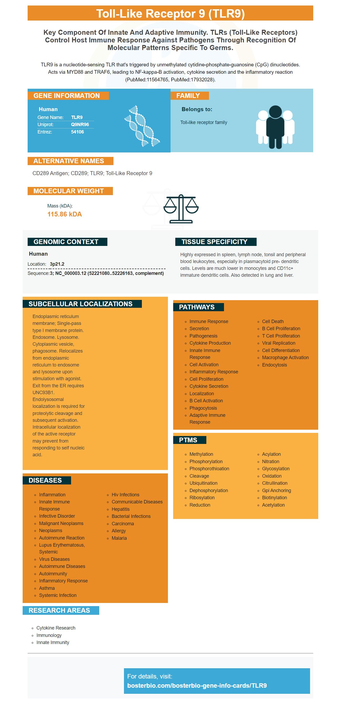
TLR9 is a nucleotide-sensing TLR that's triggered by unmethylated cytidine-phosphate-guanosine (CpG) dinucleotides. Acts via MYD88 and TRAF6, leading to NF-kappa-B activation, cytokine secretion and the inflammatory reaction (PubMed:11564765, PubMed:17932028).
Human | |
---|---|
Gene Name: | TLR9 |
Uniprot: | Q9NR96 |
Entrez: | 54106 |
Belongs to: |
---|
Toll-like receptor family |
CD289 antigen; CD289; TLR9; toll-like receptor 9
Mass (kDA):
115.86 kDA
Human | |
---|---|
Location: | 3p21.2 |
Sequence: | 3; NC_000003.12 (52221080..52226163, complement) |
Highly expressed in spleen, lymph node, tonsil and peripheral blood leukocytes, especially in plasmacytoid pre- dendritic cells. Levels are much lower in monocytes and CD11c+ immature dendritic cells. Also detected in lung and liver.
Endoplasmic reticulum membrane; Single-pass type I membrane protein. Endosome. Lysosome. Cytoplasmic vesicle, phagosome. Relocalizes from endoplasmic reticulum to endosome and lysosome upon stimulation with agonist. Exit from the ER requires UNC93B1. Endolysosomal localization is required for proteolytic cleavage and subsequent activation. Intracellular localization of the active receptor may prevent from responding to self nucleic acid.
This article will discuss the potential uses for the TLR9 marker, which is a DNA receptor. This receptor can detect DNA in viruses and trigger innate immune reactions. This helps the virus cloak their genome from host's immune system. In addition, this receptor is useful for cloaking the AAV genome, which can be harmful to a host's immune system.
TLR9 receptor ligands are known to activate MAPKs. An agonist for TLR9 can be used to treat inflammation or autoimmune conditions. TLR9 agonists or antagonists can be helpful in treating many types of cancer. Activation of TLR9 is correlated with higher expression of the nuclear proteins HMGB1 and RAGE. The latter are released from damaged cells and bind to TLR9. The former activates TLR9 and causes it to send signals through MAPKs. Inflammatory response and myocardial ischemia are associated with innate immunity.
The cytoplasmic sensors recognize a variety of DNA types, including bacterial DNA. TLR9 recognizes specific immunostimulatory sequences that are common in bacterial DNA but not in vertebrate DNA. These sequences are common in antibiotic resistance genes. TLR9 activation results in cells releasing inflammatory cytokines as well type I interferons, which induce cytotoxic T cell and humoral immunity.
TLR9, which can be expressed in HEK–Blue cell cultures, was expressed by either mouse or human. These cells were cultured in DMEM with antibiotics and maintained in selection medium. The cells were then seeded in 96-well plates that contained medium that contains an embryonic alkalinephosphatase reporter. The cells then expressed DNA. The DNA-based biosensors can help us understand the mechanism of aging and to detect the effects of abiotic chemicals on our bodies.
Boster Bio's TLR9 markers may be the missing piece in the investigation into COVID-19. It may be responsible, in large part, for multi-organ complications of COVID-19. It may also contribute to the pathology or the virus in more susceptible individuals. The TLR9 marker is designed to detect DNA in viruses. It combines a broad spectrum of viralRNA with measures of viral load, susceptibility, and susceptibility.
In addition to detecting DNA from viruses, TLR9 also induces regulatory T cells called Tregs, which are involved in allergic asthma. This cell type is important because it contributes in the immunosuppression and immune escape opportunities for SARS-2. Patients with COVID-19 infection have low eosinophils, which can lead to severe disease. Interestingly, the presence of TLR9 in allergic asthma is indicative of a Th2-skewed immune response.
In addition to being expressed intracellularly, TLR9 is also found in the endo-lysosomal compartments of specialized immune cells. TLR9 is activated when DNA recognizes RNA motifs that have unmethylated CpG sequencings. It is expressed more in microbial than in viral genomes. These DAMPs play roles in the regulation and homeostasis of the immune system.
TLR9 receptor is a key component of the innate immunity system. It recognizes microbial origin conserved determinants and initiates signaling events that trigger an immune response. Innate signaling occurs within a cell's complex cellular architecture and involves several cellular processes. Innate signaling triggers signals that regulate cell behavior and protect cells. They can also promote immunological memory.
ECs are the first cells to interact with microbial components in the circulation, and their recognition is a critical component of early innate immunity. ECs also have been shown to express TLRs as well as NLRs. These cytokines are crucial in regulating innate immunity responses. ECs are important antigen-presenting cell and trigger inflammation.
TLRs are the first line of defense against pathogens and are essential for the clearance of infected cells. Toll-like receptacles function as receptors for multiple protein families, including the Nod/IL-1 (Toll/IL-1) and Nod/IL-1 (Nod-like). TLRs trigger signaling pathways by binding specific molecular patterns. This is the first line of defense against invaders for a host.
Activated TLR9 expression enhances cellular defense against parasites. Mice that lack TLR9 expression do not have significant parasite burdens after the experiment. TLR9 deficient mice also have significantly lower numbers of macrophages than WT mice. These results indicate that Boster Bio's TLR9 mark can induce innate immune response.
The TLR9 was originally designed to detect pathogenic virus infections. However, the immune system may not be able to distinguish between a virus and friendly cells if it infects the body. Boster Bio's latest discovery could help scientists overcome this problem. The new gene that targets TLR9, a protein found on the surface cells of cells called Cytotoxic T Lymphocytes (CTLs), may be able to cloak the AAV genome and suppress the immune response.
Gene therapies have historically been hindered by the immune system. Boster Bio developed a new gene treatment that uses a TLR9 mark to protect the AAV genome from immune reactions. This would make it easier and more efficient to deliver the genetic package. The researchers also showed that the masked AAV induced the same level and degree of inflammation as the uncloaked variant, but with a delayed response.
The TLR9–MyD88 pathway is crucial in activating human pDCs that generate AAV-neutralizing antibody. The TLR9/MyD88 pathway is also crucial in the development transgenes. Transgene-specific CD8+ T cell activation can be achieved by activating the TLR9-MyD88 pathway.
The use of e-cigarettes has increased the prevalence of the TLR9 gene, which may play a role in e-cigarette-associated atherosclerosis. TLR9 and cancer invasion have been studied by researchers through the study of the ligand CPG ODN2216. It was found that the ligand weakened Panc-1 cell migration, membrane anchoring, and clone proliferation. The effect of TLR9 on the invasion capability of human pancreatic cancer cells was not fully understood, but the discovery of this gene was an important step forward for understanding the role of TLR9 in this disease.
The TLR9 protein mediates trans-repression of cytokine expression. TLR9 plays a protective role in intestinal inflammation. It has been shown that it inhibits the activation pro-inflammatory NFkappa-B signaling. TLR9 inhibits proinflammatory NFkappa B signaling, which results in suppression and inhibition of inflammatory responses.
A study with humans found that e-cigarette extract caused elevated levels of TLR9 and decreased plaque formation. It also suppressed the production of inflammatory cytokines. It may be able to reduce the symptoms of atherosclerosis in people. It is possible to pursue this drug even though it is still in its early stages.
TLR9 inhibitors can reduce the upregulation NFKB and TLR9 in human arterial plaques. These two receptors have a close relationship and may be linked to autoimmune diseases. Boster Bio's study is the first to examine the interaction of TLR9/BCR. However, the study also revealed a link between these receptors.
Research is a must-have tool that uses high-affinity prima antibodies. Boster Bio TLR9Marker is an excellent choice because it binds with TLR9 receptors, induces affinity maturation, and somatic mutation of the B cells. This antibody is well cited by researchers, and has been validated for use in Western Blotting, Immunohistochemistry, and ELISA.
Within one week of vaccination or infection, B cells form germinal centers and gradually fade away as the immune response decreases. These centres are divided into two anatomical compartments, a dark zone and a light zone, and have two contrasting functions: antigen-driven selection and affinity maturation. The germinal centre contains B cells that either become plasma cells or long-lived, memory B cells.
TLR9 is high in memory B cells. They are directly triggered via CpG. This natural process has been used for antigen-specific B cell growth and is believed to be an important mechanism to preserve serological memory. CpG stimulation was also used to increase antigen-specific cells. The Batista lab has studied the role TLRs have in specific cell differentiation and proliferation.
PMID: 11022119 by Du X., et al. Three novel mammalian Toll-like receptors: gene structure, expression, and evolution.
PMID: 11022120 by Chuang T.-H., et al. Cloning and characterization of a sub-family of human Toll-like receptors: hTLR7, hTLR8 and hTLR9.