This website uses cookies to ensure you get the best experience on our website.
- Table of Contents


1 Citations 9 Q&As


Facts about Trefoil factor 1.

.
| Human | |
|---|---|
| Gene Name: | TFF1 |
| Uniprot: | P04155 |
| Entrez: | 7031 |

| Belongs to: |
|---|
| No superfamily |

BCEI; BCEIbreast cancer, estrogen-inducible sequence expressed in; Breast cancer estrogen-inducible protein; breast cancer estrogen-inducible sequence; D21S21; gastrointestinal trefoil protein pS2; hP1.A; HPS2; PNR-2; Polypeptide P1.A; Protein pS2; PS2; TFF1; trefoil factor 1; trefoil factor, BCE1, human pS2 induced by estrogen from human breast cancercell line M10HP1.A
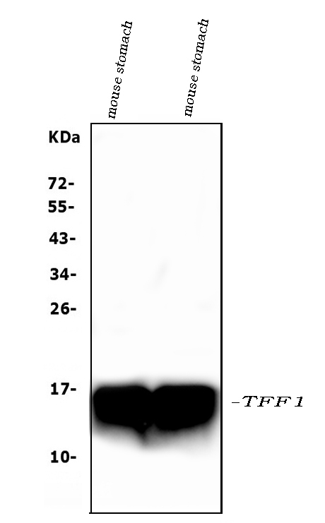
Mass (kDA):
9.15 kDA

| Human | |
|---|---|
| Location: | 21q22.3 |
| Sequence: | 21; NC_000021.9 (42362282..42366535, complement) |
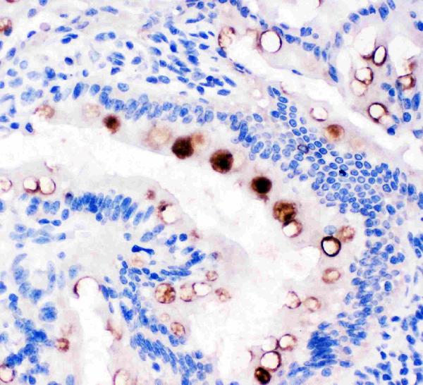
Found in stomach, with highest levels in the upper gastric mucosal cells (at protein level). Detected in goblet cells of the small and large intestine and rectum, small submucosal glands in the esophagus, mucous acini of the sublingual gland, submucosal glands of the trachea, and epithelial cells lining the exocrine pancreatic ducts but not in the remainder of the pancreas (at protein level). Scattered expression is detected in the epithelial cells of the gallbladder and submucosal glands of the vagina, and weak expression is observed in the bronchial goblet cells of the pseudostratified epithelia in the respiratory system (at protein level). Detected in urine (at protein level). Strongly expressed in breast cancer but at low levels in normal mammary tissue. It is regulated by estrogen in MCF-7 cells. Strong expression found in normal gastric mucosa and in the regenerative tissues surrounding ulcerous lesions of gastrointestinal tract, but lower expression found in gastric cancer (at protein level).
Secreted.


These are the factors to take into consideration when evaluating the Boster bio Anti-pS2/pNR-2Trefoil Factor 1 (Estrogen Regulated Protein) Monoclonal Antibody. Ensure the antibody has been validated on Immunofluorescence, immunohistochemistry, and Western blot. Boster's antibody is validated for all of these platforms.
Boster Bio has given this monoclonal antibody its catalog number M01391. It reacts with Humans and is available at 1.0mg/l. It is composed of 28 amino acid residues. It is also available as a monoclonal antibody a lyophilized format. It is perfect for a variety of applications, as well as in various other tests.
This monoclonal antibody was developed in line with the Human Protein Atlas project. This project aims at characterizing the human proteome using antibodies derived from a variety of human tissues. The antibodies were developed and validated using immunofluorescence on hundreds of disease and normal tissues. Each Prestige Antibody is also provided with characterization information. The portal also has protocols for the use this antibody.
Boster Bio offers a wide selection of boster-validated antibodies that include ELISA kits and picogram sensitive polyclonal antibodies. To ensure high affinity, all boster-validated antibodies have been tested against more than 250 tissue samples. They also have been quantitatively validated by testing the antibodies against known quantities of recombinant proteins and untransfected cell lines.
Western Blotting is a great tool for antibody validation. Boster tests all of its products using WB as this method requires the antibodies be validated using identical samples in a controlled setting. Fortunately, Boster offers customized services, BeNeLux deliveries, and other benefits that make them an invaluable source. If you're looking to purchase boster bio-antibodies, contact Sanbio to find out more about their products and services.
Reproducibility requires collaboration between the vendors, the user and the publisher. The user has to verify the effectiveness of an antibody and conduct well-designed tests. The seller should offer high-quality antibodies as well as a detailed description of its procedures. In addition, the publisher must establish guidelines for validation and implement them. Researchers should also work with publishers and antibody manufacturers to create validation-enabling technologies that increase the reproducibility and quality of research. All of this will help the scientific community, improve the quality of antibodies, and make their research easier and more efficient.
Boster tests primary antibodies as well as validating WB. While primary antibodies have been evaluated for their ability to recognize their target protein in a particular test There could be additional bands present. These could indicate that the assay must be improved. A new primary antibody is needed. Boster's expertise facilitates this process. The company has more than 30 years of experience in the validation of immunoassays.
In addition to their array of antibodies, Boster also manufactures ELISA kits and picogram sensitivity polyclonal antibodies. Boster provides over 12,000 antibodies, all approved for use in IHC and WB. Every antibody is tested against a range of tissues to ensure high specificity and affinity. In addition, each antibody is tested against the known quantities of Recombinant proteins.
The process of validation of antibodies involves the preparation of TMAs for IHC. Because the technical parameters of TMAs are uniform, they are the best method to collect IHC data. This method is particularly important for antigens that tend to degrade in cut sections because of water. This makes it difficult to validate antibodies. In addition, the creation of TMAs can require the use of a positive and negative control cell line.
Boster validation ensures that every antibody performs well in IHC. The validation process includes obtaining key information from published references. This information is crucial for the publication of IHC results. All antibodies identified by IHC are checked by the Boster team before being released for clinical use. Although the process of validation may be lengthy, it's worthwhile in the long term. It is important to determine specific antibodies that can be used for specific purposes when establishing biomarkers. For instance, a patient can be diagnosed with a single antibody or with a variety of antibodies.
For instance, p21 is an antibody found in the human colon. Doctors can make use of the Boster validation process to assist them in selecting a drug that is based on this mutation. It could even replace DNA sequencing as an economical alternative to this method. It is also important for researchers working in the clinical setting where antibodies are used to determine the health of patients. When used correctly these antibodies can be used to determine the effectiveness of treatment for patients.
The Boster validation process involves using the antibody on tissues to test its specificity and its sensitivity. The antibody dilution must be adjusted to guarantee a signal-to-noise ratio. A consistent pattern in the same tissue indicates that the antibody is valid for the purpose. If it's not the next step, it is to move on to Level 3 validation. For the gastric cancer case such as HER2, for instance, would be considered to be Level 2 validation.
When you are choosing an antibody to use in your research, it's essential to know if it has been approved by the manufacturer. Boster, an antibody manufacturer has strict protocols to test all of its antibodies. This includes rigorous validation of the antigens used and the antibodies themselves. A high-quality antibody will help you save time and money. Companies that test their antibodies are ones that are more consistent than the standard.
Many companies claim to have validated their products, but this information may vary. Certain companies provide the most thorough validation, while others don't. Each vendor tries to strike an appropriate balance between quality goods and profits. The companies selected included Cell Signaling Technology, Boster, and GeneTex. They claim that their products have been thoroughly tested to ensure they are free of contamination and cross-reactivity.
Reproducibility is a different criterion to consider to be considered for validation. Reproducibility refers to the degree to the extent an antigen has been tested against the exact same target in two different tests. In practice, the use of a single antibody by different researchers in different laboratories can result in differences in the results obtained with different antibodies. Additionally, these methods are costly and labor-intensive. It can be difficult to compare results from different laboratories and often the results are discarded early.
Blocking peptides can be used to further verify the specificity of antibodies. These peptides are derived form sequences that make antibodies. These peptides can be used to stain tissue that contains the protein of interest when the antibodies are infused in large quantities. If the antibody is unable to recognize a particular protein, the blocking peptides won't stain the tissue. These antibodies are the main reason for the rise in immunoglobulin concentrations in cells.
Boster employs multiple methods to verify antibodies. Validation involves testing the specificity of an antibody against various cell lines and tissue microarrays. This confirms the antibody's ability to recognize the target and its ability to reproduce across cell lines. Furthermore, it includes positive and negative control lines. Visit Boster's site for more details. Boster has tested a wide range of antibodies to Immunofluorescence.
PMID: 6324130 by Jakowlew S.B., et al. Sequence of the pS2 mRNA induced by estrogen in the human breast cancer cell line MCF-7.
PMID: 3838275 by Prud'Homme J.-F., et al. Cloning of a gene expressed in human breast cancer and regulated by estrogen in MCF-7 cells.
*More publications can be found for each product on its corresponding product page