This website uses cookies to ensure you get the best experience on our website.
- Table of Contents
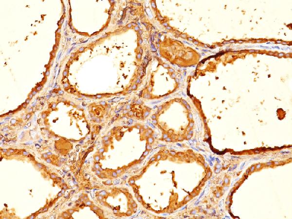
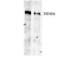
Facts about Thyroglobulin.
Human | |
---|---|
Gene Name: | TG |
Uniprot: | P01266 |
Entrez: | 7038 |
Belongs to: |
---|
type-B carboxylesterase/lipase family |
AITD3; AITD3TGN; cog; TDH3; Tg; TGN; Thyroglobulin
Mass (kDA):
304.79 kDA
Human | |
---|---|
Location: | 8q24.22 |
Sequence: | 8; NC_000008.11 (132866943..133134902) |
Thyroid gland specific.
Secreted. Secreted into the thyroid follicle lumen (PubMed:19509106). Localizes to colloid globules, a structure formed in the thyroid follicle lumen consisting of cross-linked TG arranged in concentric layers (PubMed:8626858, PubMed:11082042).
The TG marker is used in many ways. It can be used to screen and diagnose differentiated thyroid carcinoma or monitor the recurrence following a thyroidectomy. Keep reading to find out the best uses for TG. In this article, we'll discuss the most common applications for TG and explain why it might not be expressed in all patients. It can also help you determine if your hypothyroidism is present.
Serum thyroglobulin (TG) is a tissue-specific protein that plays a role in biosynthesis of thyroid hormones. It is produced by thyroid cells and differentiated cancer cells. TG is commonly measured by a neck ultrasound in patients suffering from differentiated thyroid cancer. Patients with differentiated thyroid carcinoma may also benefit from TG for postoperative follow up.
Tg is a tissue-specific antigen produced by thyroid follicular cells and represents the most sensitive marker of DTC. Tg cannot detect recurrent DTC, but imaging technologies are available to detect the presence of distant metastases. Tg, a sensitive marker for thyroid cancer, can be detected in detectable amounts.
The most commonly accepted follow-up tool for patients with differentiated thyroid cancer is the serum Tg level. It is typically measured while the patient is receiving thyroid hormonal treatment. One study examined the accuracy of Tg measurement on treatment in detecting remnant or metastatic disease. This retrospective study involved 26 high risk DTC patients. The patients did not have any history of thyroid cancer.
Serum TG is a marker for recurrence after thyroidectomy. It must be checked every six to 12 month because the residual thyroid gland can grow and harbour malignant cell. Iodine scintigraphy should be performed on patients with low serum TG levels to detect recurrence. It can also be used for the detection of cancer cells in the thyroid in patients at high risk.
Radioactive Iodine Therapy is a common treatment option for thyroid cancer. It suppresses thyroid-stimulating hormonal (TSH) levels and is highly effective. Recurrence is less likely if the TSH level is low, but sustained high levels increase the risk. To ensure that treatment is effective, survivors should monitor their thyroid function. This procedure could be repeated many years later.
According to the American Cancer Society, 52,070 cases will be diagnosed with thyroid cancer in 2019. The disease is the fastest growing cancer in the United States. There are 2,170 expected deaths from the disease in 2019. Increasing numbers of imaging and diagnostic tests are contributing to the rapid growth of the disease. The survival rate of patients with thyroid cancer depends on a number of factors, including whether or not they develop other forms of the disease.
Hypothyroid goitre patients may not express the TG marker in all cases, especially those with a TG gene variant. The serum TG of patients with hypothyroid goitre may not increase despite an elevated TSH. Therefore, it is important to carefully interpret a rising serum TG as a sign of hypothyroid goitre.
The TG marker does not appear in all patients with hypothyroid disease. However, there are some overlaps between patients with the condition and those without it. Tg may not be expressed in hypothyroid patients. This is due to a lack of TSH hormone which regulates T4 synthesis. Patients with the TGW2346R gene may not be able to express the TG marker.
In a study using a TGcog/cog mouse model, a Tg protein of 330 kDa was purified from thyroid tissue. It was the T4-containing protein with the highest concentration and migrated along with the WT Tg. However, the mutant Tg had a lower efficiency at producing T4 than the WT Tg and was loaded on Tg protein. This indicated the presence T4 inside the patient's body.
Tg levels in healthy people are low. However Tg levels rise in those with tissue damage or elevated TSH. In a further study, I2 was administered to hypothyroid-goitre patients. It caused antibody production in eight to twenty percent of patients. I2 administration caused intra-thyroidal lymphocyte formation in some patients. These mechanisms include the mass release antigens following the destruction of thyrocytes, and the generation new epitopes via high I2 content.
Another study of elderly patients found that patients with hyperthyroid goitre had a higher likelihood of having a lower TG marker. The pro-inflammatory chemicals in the blood of patients who received the treatment were lower, which could have clinical implications. Inflammation is also known as a risk factor for cardiovascular disease and atherosclerosis. Although this study was limited in scope to a small number of patients, it did identify potential causes of TG.
PMID: 3595599 by Malthiery Y., et al. Primary structure of human thyroglobulin deduced from the sequence of its 8448-base complementary DNA.
PMID: 9186272 by van de Graaf S.A.R., et al. The revised 8307 base pair coding sequence of human thyroglobulin transiently expressed in eukaryotic cells.