This website uses cookies to ensure you get the best experience on our website.
- Table of Contents

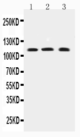
Facts about Short transient receptor potential channel 4.

Probably operated by a phosphatidylinositol second messenger system activated by receptor tyrosine kinases or G-protein coupled receptors. Mediates cation entry, with an enhanced permeability to barium over calcium.
| Human | |
|---|---|
| Gene Name: | TRPC4 |
| Uniprot: | Q9UBN4 |
| Entrez: | 7223 |

| Belongs to: |
|---|
| transient receptor (TC 1.A.4) family |

hTrp4; hTrp-4; MGC119570; MGC119571; MGC119572; MGC119573; short transient receptor potential channel 4; transient receptor potential 4; transient receptor potential cation channel, subfamily C, member 4; TRP4HTRP4; TrpC4; Trp-related protein 4
Mass (kDA):
112.101 kDA

| Human | |
|---|---|
| Location: | 13q13.3 |
| Sequence: | 13; NC_000013.11 (37632063..37869772, complement) |
Strongly expressed in placenta. Expressed at lower levels in heart, pancreas, kidney and brain. Expressed in endothelial cells. Isoform alpha was found to be the predominant isoform. Isoform beta was not found in pancreas and brain.
Membrane; Multi-pass membrane protein. Cell membrane; Multi-pass membrane protein. Enhanced insertion into the cell membrane after activation of the EGF receptor.


If you're interested in finding the right antibody for your research project, you've come to the right place. Boster produces high-affinity primary antibodies that have been widely cited in the scientific community and validated by Western Blotting, Immunohistochemistry, and ELISA. Read on to learn more about these antibodies and their uses in biomedical research. Here, you'll learn about Boster and what sets them apart from their competition.
While commercially available antibodies can be useful tools for your research, there is still a risk of poor quality and high costs. A study by Berglund revealed that failure rates for 51 represented vendors ranged from zero to 100%. As a result, commercial antibodies are often a waste of money and time. In fact, US$800 million a year is wasted on poorly performing antibodies in biomedical research. Many experiments fail because of a bad antibody.
The validation of His primary antibodies must be based on the specificity and selectivity of the antigen. Validation recommendations vary for each immunoassay. Even small differences in assay conditions can affect the performance of antibodies. Consequently, it is important to evaluate and validate antibodies for reproducibility and to ensure the best quality data. Performing validation on Western Blotting, Immunohistochemistry, and ImmunoELISA should be the first step for a reproducible experiment.
The validation of His primary antibodies on Western Blotting, Immunohistochemical, and ImmunoELISA methods is based on the fact that the antibodies detect the target molecule in its native conformation. Different organs, cell types, and subcellular fractions may display different expression patterns. Co-staining with established antibodies helps ensure accuracy.
The validation of His primary antibodies on Western blotting requires appropriate positive and negative controls. This helps to identify potential sources of error and to intervene before results are compromised. Positive controls are important because they provide information on the immunoblotting protocol used. Positive control results in the positive lane mean that the immunodetection protocol worked and lends validity to other results. Conversely, negative results on positive controls indicate that a certain antibody was responsible for a negative result.
Abcam manufactures primary antibodies. These antibodies are HRP-conjugated mouse and rabbit monoclonal IgG1 isotype controls. Using these as controls, you can see the specificity of each primary antibody. When using these primary antibodies, always remember to check the manufacturer's label for authenticity. The majority of antibodies are validated on Western Blotting before you use them.
To confirm that His primary antibodies are compatible with a target protein, you can use an affinity tag to modify the protein and apply a fluorescent tag. Then, apply the antibody on the membrane, followed by a well-characterized immunoreagent that binds the tag. The results should correlate on two blots to validate antibody specificity. This technique is known as multiplex fluorescence Western blotting.
The concentration of His primary antibodies should be confirmed on the same kind of sample. If the primary antibody is not purified, the concentration of the secondary antibody should be checked with the antigen itself to make sure that it is a specific antibody. If the concentration of the secondary antibody is too high or too low, it may lead to off-target binding and faint signals. The recommended dilution range depends on the type of secondary antibody conjugate used and the detection method.
The validation of His primary antibodies on Western Blotting, Immunohistochemical, and ImmunoELISA is critical for the success of pan/PTM analysis. Since the modified and unmodified forms of the target protein will co-migrate closely, validation on Western Blotting and Immunohistochemistry is critical. For optimal results, His primary antibodies should be specific against both forms of the antigen.
PMID: 11042129 by McKay R.R., et al. Cloning and expression of the human transient receptor potential 4 (TRP4) gene: localization and functional expression of human TRP4 and TRP3.
PMID: 11163362 by Mery L., et al. Alternative splice variants of hTrp4 differentially interact with the C-terminal portion of the inositol 1,4,5-trisphosphate receptors.