This website uses cookies to ensure you get the best experience on our website.
- Table of Contents
4 Citations 9 Q&As
2 Citations 9 Q&As
2 Citations
7 Citations
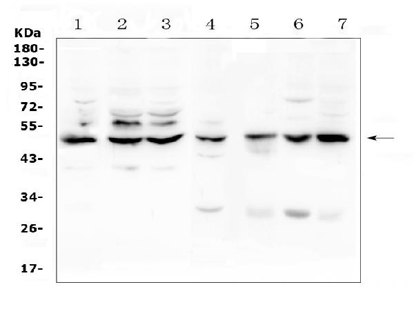
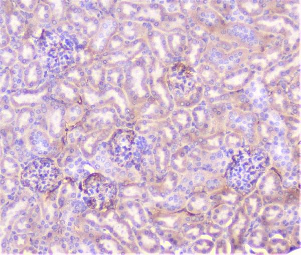
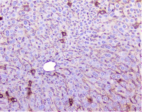
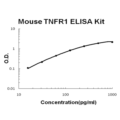
Facts about Tumor necrosis factor receptor superfamily member 1A.
Receptor for TNFSF2/TNF-alpha and homotrimeric TNFSF1/lymphotoxin-alpha.
The adapter molecule FADD recruits caspase-8 to the activated receptor.The resulting death-inducing signaling complex (DISC) performs caspase-8 proteolytic activation which initiates the subsequent cascade of caspases (aspartate- specific cysteine proteases) mediating apoptosis. Contributes to the induction of non-cytocidal TNF effects including anti-viral state and activation of the acid sphingomyelinase.
Human | |
---|---|
Gene Name: | TNFRSF1A |
Uniprot: | P19438 |
Entrez: | 7132 |
Belongs to: |
---|
No superfamily |
CD120a antigen; CD120a; FPF; p55; p55-R; p60; TNF RI; TNFARMGC19588; TNF-R; TNF-R1; TNFR1TBP1; TNFR55; TNF-R55; TNFR60; TNFRI; TNF-RI; TNF-R-I; TNFR-I; TNFRSF1A; tumor necrosis factor binding protein 1; Tumor necrosis factor receptor 1; tumor necrosis factor receptor 1A isoform beta; tumor necrosis factor receptor superfamily member 1A; tumor necrosis factor receptor superfamily, member 1A; tumor necrosis factor receptor type 1; Tumor necrosis factor receptor type I; tumor necrosis factor-alpha receptor
Mass (kDA):
50.495 kDA
Human | |
---|---|
Location: | 12p13.31 |
Sequence: | 12; NC_000012.12 (6328771..6342076, complement) |
Cell membrane; Single-pass type I membrane protein. Golgi apparatus membrane; Single-pass type I membrane protein. Secreted. A secreted form is produced through proteolytic processing.; [Isoform 4]: Secreted. Lacks a Golgi-retention motif, is not membrane bound and therefore is secreted.
The TNFRSF1A Marker has been confirmed by Boster Bio. This antibody is derived from human TNF alpha an cytokine that has multiple functions within the superfamily of tumor necrosis factors. It is produced by macrophages. It binds to several receptors to regulate many biological processes. It is involved in an autoimmune disease and has neuroprotective properties. Boster Bio is dedicated to verifying all antibodies.
The Boster Bio Anti-TNF Alpha Monoclonal Antibody is designed for use in Boster Bio. It reacts with Human, Mouse, and Rat TNF alpha. The antibody contains 5 mg of bovine serum albumin and peptides. Based on the length of the immunogen, blocking peptides can be purchased separately. The price of blocking peptides can vary, but is usually between $80 and $120.
TNF alpha, a multifunctional proinflammatory cell cytokine, has been classified as a member the tumor necrosis factors (TNF) superfamily. It is mainly produced and released by macrophages. This protein binds with various receptors and controls a myriad of biological processes. It is believed to be linked with various autoimmune diseases. However studies of knockouts in mice suggest that it may have neuroprotective properties. All Boster antibodies have been validated against positive samples. This allows them to be used in a variety situations.
Several studies have examined the genetic basis of TNFRSF1A mutations and have concluded that this gene rarely mutated in nonfamilial recurrent fevers of unknown etiology. However, Aganna et al. Aganna and colleagues. found no TNFRSF1A mutations in two families with dominantly inherited TRAPS despite the fact their clinical histories were similar. These results suggest that there is an inheritance heterogeneity that is present in periodic fever syndromes.
RefSeq provides genome, transcript and protein data. It includes records from more than 16000 organisms and two 10(6) x 10(6) RNA. It also includes cross-links to related sources of information. The RefSeq record TNFRSF1A includes annotations and gene predictions of more than 16.000 organisms. The UCSC RefSeq database uses an established format for organizing its annotation tracks.
RefSeq database contains a large number of homologous protein-coding gene. About 50 percent of orthologous proteins-coding genes are backed by splicing conservation between three reference genomes. Approximately 7,500 ortholog sets are identified in more than half of analyzed organisms. Most of these have highly conserved sequences and splicing and could function as the minimum core proteins. Most proteins pass tests to detect lack of splicing conservation or domain annotation.
The TNFRSF1A gene is linked to susceptibility to multiple sclerosis (MS). This gene is closely connected to MS according to genetic research. Researchers have discovered that rs1800693 is closely associated with MS but not with any other autoimmune disorders. In addition, Gregory et al. have established that rs1800693 represents a causal variant within the TNFRSF1A gene.
The TNFRSF1A marker is a receptor for the TNF-alpha and homotrimeric TNFSF1/lymphotoxin-alpha proteins. After activation, FADD recruits caspase-8 into the activated receptor. When activated, caspase-8 initiates a cascade that involves a variety of caspases. Activated TNFRSF1A plays important roles in the immune system, antiviral state and activation acid-sphingomyelinase.
The inhibition of necroptosis and caspases offers additional protection against TNF-induced inflammation. TNF-induced inflammation may also be prevented by blocking RIPK1. Thus, the TNFRSF1A marker could be useful in the field of research and treatment of human inflammation-related disorders. There are many applications for TNFRSF1A markers, but there is a lot of research to be done.
TNFa is a co-stimulator with TNFa which may enhance the efficiency of your immune system. TNFRa is a signaling molecule and increases the number of monocytes, neutrophils and lymphocytes. A functioning TNFRF1A results in an enhanced immune system's ability to recognize microbes.
TNF-a signaling is a part of cell responses such as proliferation and apoptosis. Following activation of the TNFR, cellular responses reflect the existence of a complicated regulatory network. We will look at TNF-a's signaling in greater detail later. Its role in the immune response and organ regeneration is considered within the context of disease and treatment. Its role in cancer remains unknown, but its significance is becoming more widely acknowledged by researchers in the field.
In breast cancer, TNFRSF1A is a direct STAT3 target gene. It regulates the activity of NFkB in TNBC cell lines. The presence of it in breast cancers could be a biomarker that can discover new STAT3 inhibitors. Furthermore, TNFRSF1A has been reported in primary breast cancers. We provide evidence of the TNFRSF1A mark.
In this study, a family with a son who is deficient in TNFRSF1A was screened for mutations in TNFRSF1A. High incidence of RTH was seen in the TNFRSF1A mutant cases. One of the daughters was diagnosed with an extremely low-grade fever (37.5 degC). The unusual inflammatory markers detected in the mother were not normal. The symptoms lasted for three weeks, and then immediately remitted. The acute inflammatory reactions were within the normal range.
TNF-receptor-associated periodic syndrome (TRAPS) is one of the systemic autoinflammatory disorders. Most AIDs can be genetically or genetically affected. There are signs that could indicate but genetic testing is essential to confirm the diagnosis. One of the complications of TRAPS is AA amyloidosis. To determine the reason for TRAPS A novel in-frame insert of TNFRSF1A was discovered. The structural changes caused by this insertion were analyzed through three-dimensional modeling.
A new method to determine TNFa receptor shedding is sensitiver than other tests. It also distinguishes between LOR (Long Time Response) and antibody levels. This test could be used in clinical practice for optimizing the process of making decisions and reducing complications. These studies have shown positive results.
The TNFRSF1A genes provide instructions for the production of the tumor-necrosis factor receptor 1 protein. This protein spans a cell’s membrane. The receptor binds with tumor necrosis factors (TNF) and creates the trimer. This trimer is crucial for TNFR1 protein function. The protein is also found in tumors, and it is a marker for tumor-related NFKB Kinase.
MIF is an important chemical, playing an important role in monocyte recruitment and the arrest of monocytes. MIF is linked to various receptors such as CXCL2, CXCL4, CD74 and CXCL4. The marker was discovered by Tillmann et al. in 1996. Since 1996, the Boster bio database has added the TNFRSF1A mark.
PMID: 2158862 by Loetscher H., et al. Molecular cloning and expression of the human 55 kd tumor necrosis factor receptor.
PMID: 2158863 by Schall T.J., et al. Molecular cloning and expression of a receptor for human tumor necrosis factor.
*More publications can be found for each product on its corresponding product page