This website uses cookies to ensure you get the best experience on our website.
- Table of Contents
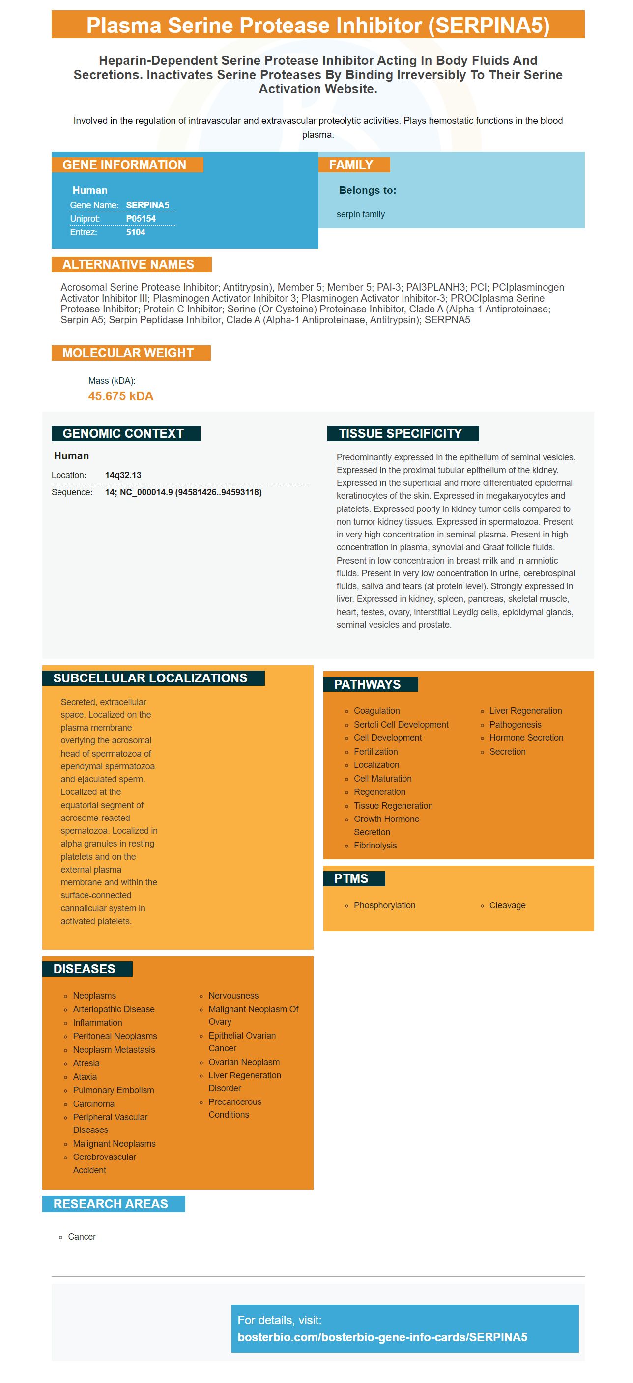


Facts about Plasma serine protease inhibitor.

Involved in the regulation of intravascular and extravascular proteolytic activities. Plays hemostatic functions in the blood plasma.
| Human | |
|---|---|
| Gene Name: | SERPINA5 |
| Uniprot: | P05154 |
| Entrez: | 5104 |

| Belongs to: |
|---|
| serpin family |

Acrosomal serine protease inhibitor; antitrypsin), member 5; member 5; PAI-3; PAI3PLANH3; PCI; PCIplasminogen activator inhibitor III; Plasminogen activator inhibitor 3; plasminogen activator inhibitor-3; PROCIplasma serine protease inhibitor; Protein C inhibitor; serine (or cysteine) proteinase inhibitor, clade A (alpha-1 antiproteinase; Serpin A5; serpin peptidase inhibitor, clade A (alpha-1 antiproteinase, antitrypsin); SERPNA5
Mass (kDA):
45.675 kDA

| Human | |
|---|---|
| Location: | 14q32.13 |
| Sequence: | 14; NC_000014.9 (94581426..94593118) |
Predominantly expressed in the epithelium of seminal vesicles. Expressed in the proximal tubular epithelium of the kidney. Expressed in the superficial and more differentiated epidermal keratinocytes of the skin. Expressed in megakaryocytes and platelets. Expressed poorly in kidney tumor cells compared to non tumor kidney tissues. Expressed in spermatozoa. Present in very high concentration in seminal plasma. Present in high concentration in plasma, synovial and Graaf follicle fluids. Present in low concentration in breast milk and in amniotic fluids. Present in very low concentration in urine, cerebrospinal fluids, saliva and tears (at protein level). Strongly expressed in liver. Expressed in kidney, spleen, pancreas, skeletal muscle, heart, testes, ovary, interstitial Leydig cells, epididymal glands, seminal vesicles and prostate.
Secreted, extracellular space. Localized on the plasma membrane overlying the acrosomal head of spermatozoa of ependymal spermatozoa and ejaculated sperm. Localized at the equatorial segment of acrosome-reacted spematozoa. Localized in alpha granules in resting platelets and on the external plasma membrane and within the surface-connected cannalicular system in activated platelets.


SERPINA5 is a protein that is located on many cells in the body. This protein has been identified in human samples and has multiple uses, including protein transfer. Here are some examples of its applications. To learn more about it, read on! Boster Bio: Best Uses Of The SERPINA5 Marker
Biological assays use antibodies to detect SERPINA5 (or SERPIN) in a variety of animal samples. Boster Bio uses rabbit and mouse to develop antibodies to this marker. SERPINA5 is an inhibitor of serine proteases that regulate intravascular and extravascular proteolytic activity. It also plays hemostatic functions in blood plasma. Boster Bio antibodies have been extensively validated in ELISA, Western Blotting, and immunohistochemistry.
The enhanced chemiluminescence (ECL) chemistry of Boster Bio's ECL cheiluminescent detection system allows researchers to detect protein levels in medium and high-expression samples in a matter of hours. This technique is superior to colorimetric assays, which require a long incubation period for protein detection. The system's sensitivity makes it the ideal choice for research.
The chemistry behind the process is based on immunoblotting, a simple analytical technique that involves electrophoretic separation of proteins, immobilization on a membrane, and detection by standard methods. While most laboratories use commercial chemiluminescence kits to perform immunoblotting experiments, self-prepared reagents are superior for several reasons. These include better sensitivity, shorter signal duration, ease of use, and a longer shelf life.
The Hybond-ECL reagent offers excellent sensitivity and low background, making it an ideal choice for multiple blottings. Hybond ECL membranes feature a pore size of 0.45, resulting in excellent sensitivity and low background. Hybond ECL membranes also feature a reinforced nitrocellulose membrane, which provides superior binding capacity. This system is compatible with WesternBright substrates and systems.
The reagents used for ECL chemistry include luminol-based, acridan-based, and 1,2-dioxetane-based reagents. Most commonly used chemiluminescent reagent is luminol, which decays to a lower energy state and releases photons. These reagents can be used to detect proteins in a wide range of samples.
The Clarity Max reagents are compatible with all horseradish peroxidase conjugates. This means they can be used with any western blots. In addition, Clarity Max are ideal for a wide range of applications. If you're interested in testing for protein levels in Western blots, you should consider this reagent.
The SERPINA5 chromogenic detection kit from Boster offers a variety of solutions for your IHC and chromogenic blot experiments. The system utilizes nitro-blue tetrazolium and an alkaline phosphatase reaction to produce a black-purple precipitate. By adding an ELISA kit to your workflow, you'll save up to 55% on your research.
Membrane staining is an important part of blot analyses. Proteins are transferred to the membrane by electroblotting, direct spotting of protein solutions, or contact blots. Membrane staining allows researchers to monitor the extent of transfer. In this unit, we describe the different methods for staining proteins after electroblotting. We use seven general stains and three fluorescent stains to analyze protein transfer.
Protein transfer efficiency was determined by measuring the amount of protein remaining after wet and rapid semi-dry membrane staining. We detected three HMW protein targets using these two methods. After the transfer process, gels were stained to determine how much protein was left on the membrane. Membrane staining was performed using a reversible dye. Regardless of the method used, the amount of protein left on the membranes was equal in both cases.
MEmCode Reversible Protein Staining is another option for improving protein transfer efficiency by membrane staining. It is compatible with different types of membranes and is a more reliable method to measure transfer efficiency. The MemCode stain also completely removes itself from the membrane at the end of the transfer procedure, allowing for reuse of the blots.
To use this method, you should clean the nitrocellulose or PVDF membranes with phosphate-buffered saline and apply the MEmCode Reversible Stain. The staining procedure can take up to 20 minutes, depending on the target protein and the membrane type. It is important to wash the membrane thoroughly after the staining process, since the protein bands can easily be over-stained and cannot be detected.
Progen's quality initiative was launched in response to the growing demand for reliable, reproducible antibodies in the research community. CiteAb data is based on analysis of hundreds of thousands of antibody citations to provide a quantitative view of the research antibody market. Progen's antibodies are praised in many scientific publications, but some of them are less cited than others. This negative coverage may also give other suppliers an opportunity to catch up.
Boster Bio is a leader in producing high-affinity primary antibodies that are highly specialized for specific applications. Their products have been validated for ELISA, FC, Western Blotting, and Immunohistochemistry, and are recommended for use in multiple fields. Boster also offers rabbit polyclonal antibodies, a free secondary antibody with every primary antibody purchase, and validated for both mouse and human samples.
Prominent in the research community, Agrisera manufactures primary and secondary antibodies for plant-based applications. Its antibodies are reactive to thousands of plant species, and have been cited in dozens of scientific journals. They were recently recognized as a Plant Science Antibody Supplier of the Year, and their products were cited in thousands of scientific articles. They have also been awarded the highest number of antibody citations in the field during 2018.
Prominent in the field of immunology, Rockland provides high-affinity and high-specific antibodies for research applications. The high affinity and specificity of Rockland antibodies make them a valuable choice for the research community. Rockland Primary Antibodies are available for use in immunoblotting, flow cytometry, and immunohistochemistry. With their high specificity, they can be used in virtually all immunological applications requiring high-titer and sensitivity.
PMID: 3027058 by Suzuki K., et al. Characterization of a cDNA for human protein C inhibitor. A new member of the plasma serine protease inhibitor superfamily.
PMID: 2173165 by Meijers J.C.M., et al. Evidence for a glycine residue at position 316 in human protein C inhibitor.


