This website uses cookies to ensure you get the best experience on our website.
- Table of Contents
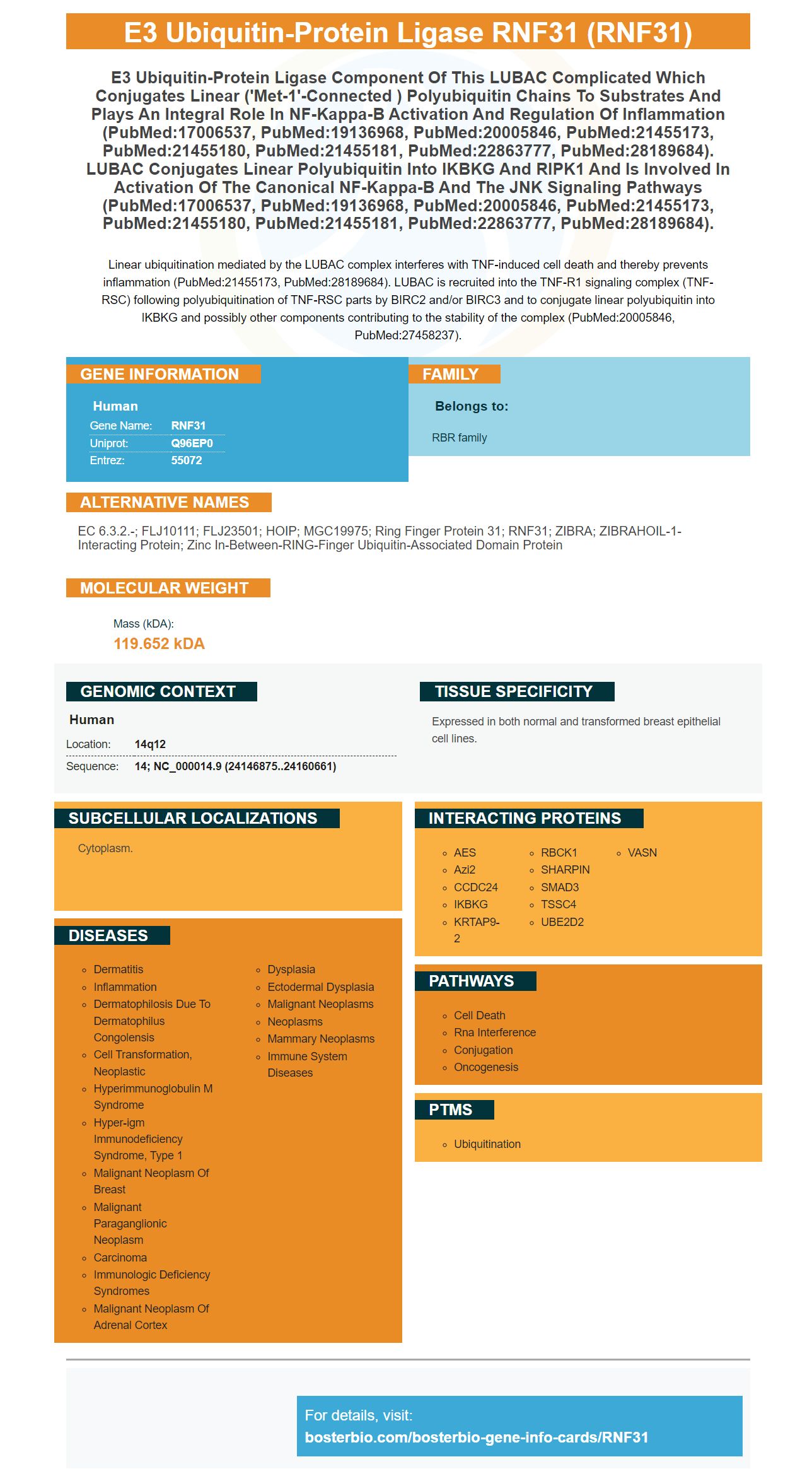
Facts about E3 ubiquitin-protein ligase RNF31.
Linear ubiquitination mediated by the LUBAC complex interferes with TNF-induced cell death and thereby prevents inflammation (PubMed:21455173, PubMed:28189684). LUBAC is recruited into the TNF-R1 signaling complex (TNF-RSC) following polyubiquitination of TNF-RSC parts by BIRC2 and/or BIRC3 and to conjugate linear polyubiquitin into IKBKG and possibly other components contributing to the stability of the complex (PubMed:20005846, PubMed:27458237).
Human | |
---|---|
Gene Name: | RNF31 |
Uniprot: | Q96EP0 |
Entrez: | 55072 |
Belongs to: |
---|
RBR family |
EC 6.3.2.-; FLJ10111; FLJ23501; HOIP; MGC19975; ring finger protein 31; RNF31; ZIBRA; ZIBRAHOIL-1-interacting protein; Zinc in-between-RING-finger ubiquitin-associated domain protein
Mass (kDA):
119.652 kDA
Human | |
---|---|
Location: | 14q12 |
Sequence: | 14; NC_000014.9 (24146875..24160661) |
Expressed in both normal and transformed breast epithelial cell lines.
Cytoplasm.
As the inventor of the RNF31 marker, Steven Boster has a unique insight into what it takes to make a great antibody. In his quest to improve the human immune system, Boster created several products including high-affinity primary antibodies and IHC assays. By the late 90s, he had grown his business to become the largest catalog antibody company in China. In the early 2000s, he developed a proprietary ELISA platform, PicoKine(tm) - which utilizes the company's own trade secrets to deliver a high-sensitivity ELISA kit.
High-affinity primary antibodies against the RNF32 marker from Boster Bio are designed to target the protein in cancer immunotherapy applications. This marker is present on the surface of a variety of cancer cells, allowing the researchers to target these antigens to control the immune response. High-affinity antibodies have high specificity, and their use is ideal for tumor immunotherapy research.
The RNF31 gene codes for a protein with a RING finger motif. This protein is known to be involved in both protein-protein and DNA-protein interactions. Two transcript variants encoding two different isoforms have been identified. Boster Bio antibodies recognize RNF31 in a wide variety of cell types, including eukaryotic cells and human lymphocytes.
In addition to the RNF31 antibody, the Boster Bio Western blot antibody detection methods also feature a load control, which is an anti-antibody to a housekeeping protein that is expressed at equivalent levels in all cells and tissues. While the RNF31 antibody can be detected in many cell types, it must be extracted correctly. The sample preparation and protein extraction methods determine the quality of the blot. The Boster Bio RNF31 Western blot antibody detection methods include detailed instructions for both sample preparation and protein extraction. Moreover, they come with comprehensive troubleshooting and support.
To perform a Western blot analysis, the antibodies must be incubated in a blocking buffer. The blocking reagents reduce the non-specific binding of the antibodies to reduce background. Nonfat dried milk is a widely available and inexpensive solution for blocking, but it is important to determine the appropriate reagent. You may want to experiment with various methods to see which one yields the best results.
Polyacrylamide gel electrophoresis is an important part of the western blot analysis, since it allows you to detect a target protein as low as one nanogram. This high-resolution method is ideal for detection of low levels of proteins in a variety of molecular biology disciplines. While polyacrylamide gel electrophoresis is the most commonly used western blot antibody detection method, it can be problematic in cases where the target protein is a large one.
The Boster Bio ECL chemiluminescent detection systems have the potential to significantly increase the sensitivity of your protein-based assays. Compared with colorimetric assays, ECL offers superior sensitivity. Boster Bio's ECL reagents detect both low and medium-expression proteins. This system allows you to detect multiple targets simultaneously.
This chemistry is commonly used in Western blot applications, such as immunoblots. The Boster ECL chemiluminescent detection system detects antibodies to the G-protein-coupled receptor family. The ECL chemiluminescent substrate produces a signal that is proportional to the amount of HRP-labeled antibodies in your sample. The signal's intensity is used for indirect protein quantification. Images of antigens are readily available using photographic film or imaging instruments.
The Boster Bio ECL chemiluminescent detection systems can detect a wide variety of proteins in a single sample. Its sensitivity and accuracy make it a valuable addition to your laboratory workflow. A good ECL detection system should provide reliable results every time. And since Boster Bio ECL reagents are optimized for western blotting, your results will always be consistent.
The ECL chemiluminescent detection systems eliminate the need for expensive darkroom space and developing reagents. The CCD camera-based imagers have a higher dynamic range than traditional film, minimizing the need for multiple exposures. Additionally, you can save on costs by using a Boster Bio ECL chemiluminescent detection system. You'll save time, money, and effort using this system.
The best Western blotting conditions last anywhere from 6 to 24 hours. The length of time light is generated depends on the enzyme-to-substrate ratio and the amount of substrate on the blot. Too much of an enzyme conjugate will cause poor signal quality, dark background, and short signal duration. Optimizing the enzyme-to-substrate ratio is essential for ensuring reproducible results.
Chromatin detection uses the DAB chromogenic substrate. This dye combines with the fluorescent agent hexahodamine, resulting in a color shift from pink to blue. In addition to this color change, NF-kB activation also drives the expression of the alkaline phosphatase enzyme, resulting in a blue precipitate. The DAB chromogenic detection system includes an enhanced HRP substrate kit from Boster Bio.
MemCode Reversible Stain is a powerful solution for membrane staining. This reversible stain provides direct visualization of protein bands. MemCode Reversible Stain is suitable for both nitrocellulose and PVDF membranes. The solution can be removed from the membrane in as little as 20 minutes, although the stain may remain on the membrane for longer depending on the protein and staining procedures used.
The sensitivity of covalent fluorescent stains depends on the amino acid composition of the proteins. The sensitivity of Sypro Ruby is comparable to that of colloidal gold, but is compatible with both nitrocellulose and PVDF membranes. The minimum amount detected by Sypro Ruby depends on the amount of protein loaded onto the gel, but the amount on the blot may be slightly less. The sensitivity of this stain is greatly increased when using minigels (each 8 cm x 10 cm x 1.0 mm).
The RNF31 marker from Boster Bio is compatible with a variety of downstream applications, such as protein purification. Its unique property is that it improves membrane staining efficiency by approximately three-fold. Since RNF31 is a molecular weight protein, it is easy to use, which allows researchers to increase the efficiency of their protein transfer experiments.
Compared to Bis-Tris gel, RNF31 significantly improves membrane staining. The transfer efficiency of 190 kDa EGFR is improved with this marker. With this protein, the transfer time is reduced to approximately eight to 10 minutes at 25 V. In addition, it is easier to transfer larger proteins with the RNF31 marker, as it is soluble in ethanol.
PMID: 15093743 by Thompson H.G.R., et al. Identification of the protein Zibra, its genomic organization, regulation, and expression in breast cancer cells.
PMID: 17006537 by Kirisako T., et al. A ubiquitin ligase complex assembles linear polyubiquitin chains.