This website uses cookies to ensure you get the best experience on our website.
- Table of Contents
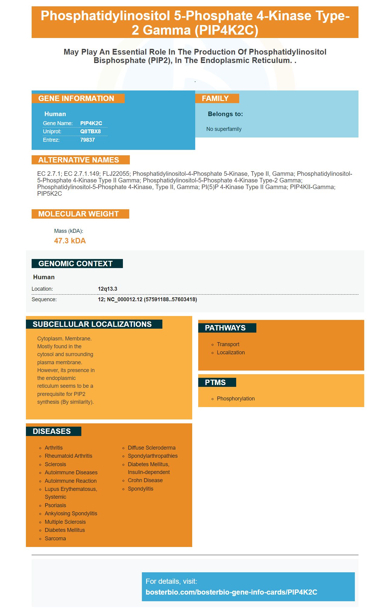
Facts about Phosphatidylinositol 5-phosphate 4-kinase type-2 gamma.

.
| Human | |
|---|---|
| Gene Name: | PIP4K2C |
| Uniprot: | Q8TBX8 |
| Entrez: | 79837 |

| Belongs to: |
|---|
| No superfamily |

EC 2.7.1; EC 2.7.1.149; FLJ22055; phosphatidylinositol-4-phosphate 5-kinase, type II, gamma; Phosphatidylinositol-5-phosphate 4-kinase type II gamma; phosphatidylinositol-5-phosphate 4-kinase type-2 gamma; phosphatidylinositol-5-phosphate 4-kinase, type II, gamma; PI(5)P 4-kinase type II gamma; PIP4KII-gamma; PIP5K2C
Mass (kDA):
47.3 kDA

| Human | |
|---|---|
| Location: | 12q13.3 |
| Sequence: | 12; NC_000012.12 (57591188..57603418) |
Cytoplasm. Membrane. Mostly found in the cytosol and surrounding plasma membrane. However, its presence in the endoplasmic reticulum seems to be a prerequisite for PIP2 synthesis (By similarity).


This article will discuss the history and current projects of Steven Boster's high-affinity primary antibodies and PIP4K2C marker. This will allow you to decide which antibody is best for your research. Steven Boster, a pioneer in biomarker research, is worth a look. His first product was developed in 1993, making him a pioneer in the field. His work earned him the nickname, "The guy who converts Science in the Lavatory".
We used a PIP4K2C -specific antibody in this study to examine the effects of Treg cells upon the PI3K/mTORC1 pathway. Tregs play a fundamental role in peripheral tolerance and are important for limiting the effects of legitimate immune responses. Additionally, recruiting Tregs may suppress T-effector and tumor cell signaling. A variety of human diseases can be treated by pharmacological tuning Treg activity.
The PIP4K2C-specific antibody was generated by combining the antibody against PIP4K2C, a distinct PIP4K2C marker. PIP4Kg is highly expressed in mouse tissues, which is consistent with its subcellular compartmentalization. By combining these two proteins, high-affinity primary antibodies against PIP4K2C can recognize the protein in different tissues, thereby detecting a variety of pathogens.
Researchers first created a polyclonal antibody against PIP4Kg to generate PIP4Kg-specific antibodies. This polyclonal antibody was indistinguishable from PIP4Ka or PIP4Kb. Afterward, they used purified recombinant PIP4Ks and analyzed the resulting binding using surface plasmon resonance.
After the preparation of the PIP4K2C-specific antibody, it was necessary to carry out immunohistochemical staining with various primers for this application. We first prepared PBMC samples using 20% methanol, 5% hydrogen oxide. Next, we used the Alexa dye-conjugated primal antibody and RNA to perform immunofluorescence microscopy. We then performed FACS analysis using both the PIP4K2C and non-mouse mRNA samples.
Second, we generated a heat map based on PIP4K signaling. Treg cells can use PIP4K to either induce or suppress CD3 mediated apoptosis. PIP4Ks regulate Treg cell proliferation, apoptosis, and other functions independently of FOXP3-mediated signaling. These are all essential components of the PIP4K2C derived mAbs.
PCA was also performed for the Treg-shPIP4K–DEG genetic set. The Treg-shPIP4K-DEG gene set showed downregulation, indicating that PIP4K is down-regulated in Tregs. RNA-seq data revealed a similar pattern for both genes. It appears that the PIP4K gene sets regulates the expressions of both cluster 1 and 2 genes.
PIKfyve, or phosphatidylinositol-3-phosphate 5-kinase type III, is an evolutionarily conserved PIK. It is found in animals as well plants and fungi. PIKfyve includes a zinc-finger domain, or FYVE. It is named after the Fab1p yeast orthologue. Its zinc finger domain is composed of a small, cysteine-rich Zn2+ binding area. This PIP4K2C marker also has a basic motif in the first b-strand of the HHCR, allowing it to bind phosphatidylinositol.
The PIP4K2A, 2B and 2B isoforms share an 80% homology and share the same Ser326 amino acid. The 2B form is phosphorylated on Th322, and Pin1 bounds the protein through its WW-domain. Experiments with mutant PIP4K2B have shown that the two isoforms interact via their WW-domains.
PI-4,5,P2 is necessary for synaptic pod docking and fusion. The PIP4K isozymes can generate PI-4,5-P2 by two distinct enzyme families, type 1 phosphatidylinositol-4,5-phosphate 5-kinase and type 2 phosphatidylinosidase. This study characterized PI-5-P2 distribution using genetic deletions from the PIP4K2B gene.
PI5P binds specifically to the SPOP Protein, which inhibits PIP4K2B Activity. In addition, PI5P negatively modulates Cul3-SPOP complex activation, promoting degradation of PIP4K2B. The PIP4K2B/p38 MAPK signaling is mediated by the PIP4K2B (complex). PI5P not only has negative effects on nuclear PI5P, but also promotes the growth and survival in cancer cells through several pathways.
Recent studies have shown that PIP4K2C is present in the Golgi, a part of kidney cells. This result suggests that the gene regulates vesicular transport. But what is the gene's function? Researchers have yet to discover exactly what it does, but it is a fascinating project. We can't ignore the implications these findings could have. The future of the marker lies in a wide variety of research fields.
The WW-domain shares a common ser/threonine residue. This is a common feature between both enzymes. Pin1 binds to Ser/Thr phosphorylated protein through the WW-domain. This interaction between PIP4Ks has important consequences for the enzymes that generate PI5P upon exposure to H2O2. Pin1 -/ cells produce high amounts H2O2 PI5P. They also have a higher resistance to oxidative stresses.
PIP4K2C is a cellular protein that encodes a subunit of a type-2 phosphatidylinositol-4,5-phosphate kinase. This enzyme is responsible for converting phosphatidylinositol-5-phosphate (PI5P) into phosphatidylinositone-4,5-bisphosphate. It serves multiple functions and has many activities.
Mutants in the PHD finger sequence bind only PI5P(DW) and H3K4me3(KK) in a specific manner. These mutants differentially regulate genes involved for myogenesis and MYOG. Despite MYOG expression levels being unaffected in these mutants cells, transcriptomic tests indicate that changes at the nucleus in PI levels modulate gene expression.
PMID: 14702039 by Ota T., et al. Complete sequencing and characterization of 21,243 full-length human cDNAs.
PMID: 16541075 by Scherer S.E., et al. The finished DNA sequence of human chromosome 12.


