This website uses cookies to ensure you get the best experience on our website.
- Table of Contents
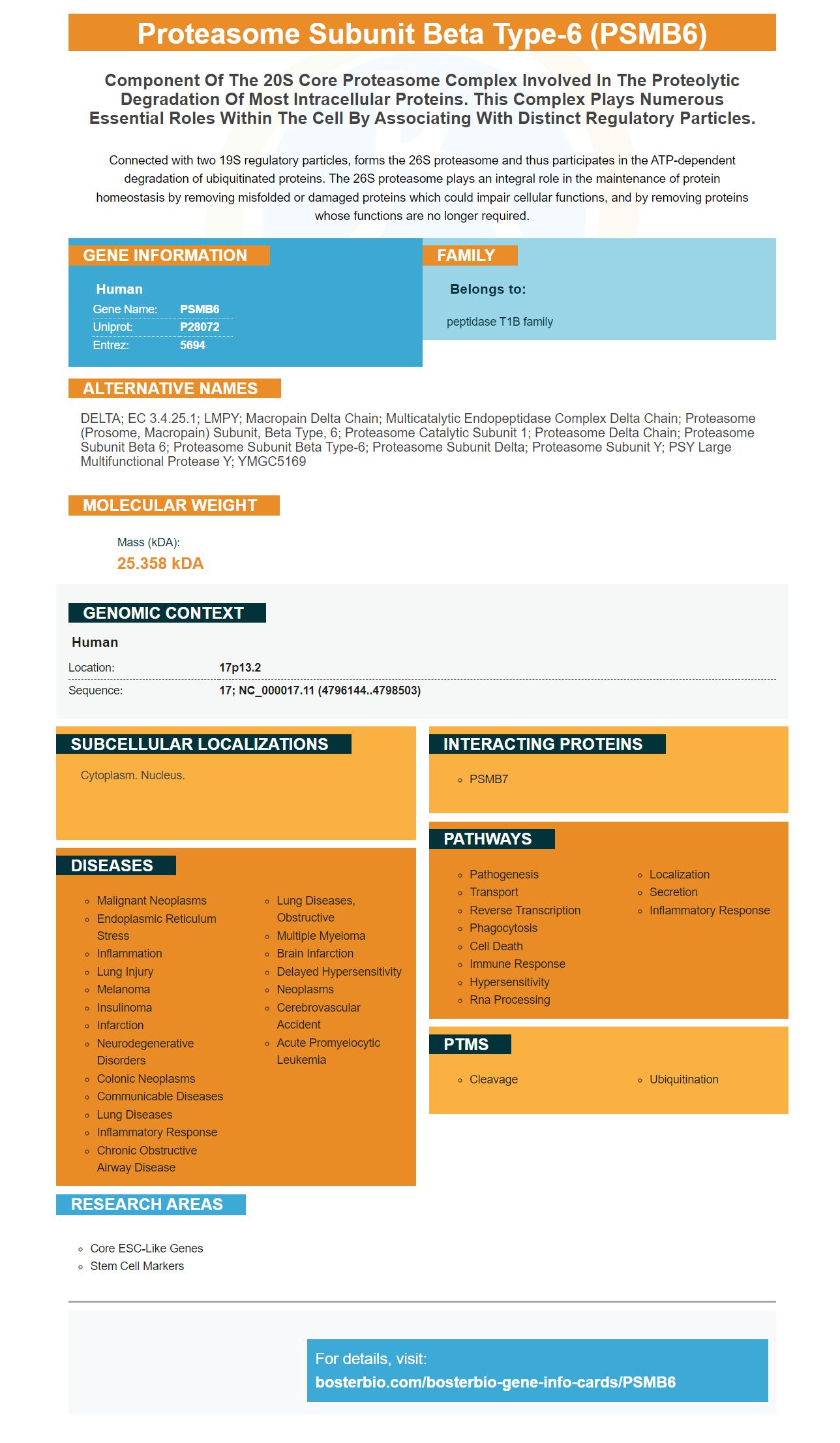


Facts about Proteasome subunit beta type-6.

Connected with two 19S regulatory particles, forms the 26S proteasome and thus participates in the ATP-dependent degradation of ubiquitinated proteins. The 26S proteasome plays an integral role in the maintenance of protein homeostasis by removing misfolded or damaged proteins which could impair cellular functions, and by removing proteins whose functions are no longer required.
| Human | |
|---|---|
| Gene Name: | PSMB6 |
| Uniprot: | P28072 |
| Entrez: | 5694 |

| Belongs to: |
|---|
| peptidase T1B family |

DELTA; EC 3.4.25.1; LMPY; Macropain delta chain; Multicatalytic endopeptidase complex delta chain; proteasome (prosome, macropain) subunit, beta type, 6; proteasome catalytic subunit 1; Proteasome delta chain; proteasome subunit beta 6; proteasome subunit beta type-6; proteasome subunit delta; Proteasome subunit Y; PSY large multifunctional protease Y; YMGC5169
Mass (kDA):
25.358 kDA

| Human | |
|---|---|
| Location: | 17p13.2 |
| Sequence: | 17; NC_000017.11 (4796144..4798503) |
Cytoplasm. Nucleus.


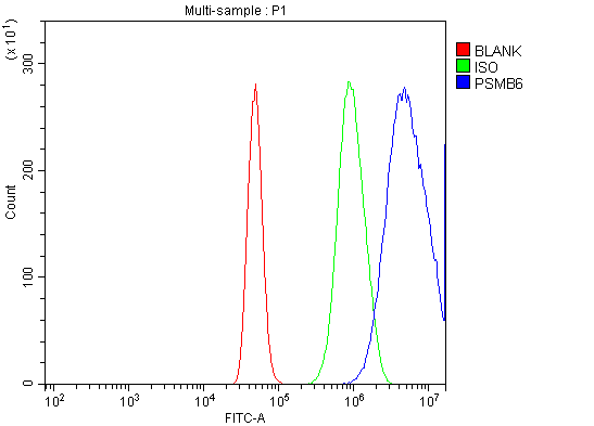
Methods for detecting the Boster Bio Anti Proteasome subunit Beta-type-6 PSMB6 marker. This article will explain how to test the antibody and detect the protein using an autoradiography film, DAB chromogenic detection device, or other methods. Below are the results. You can also find out about other uses for this marker.
Boster Bio Anti-Proteasomal Subunit beta type-6 PSMB6 markers react with proteins expressed in cells and tissues. PSMB6 is a component of the 20S–PA200/28 Complex and exhibits peptidylglutamyl Hydroolizing Activity. This activity is caused by the reduction of acidic residues.
The plasmid is composed of four expression cassettes. To generate the pBacDD-2Cap (d41) vector, the construct was digested using HindIIII, MluI and BsiWI. The fragment was then subcloned at multiple cloning stations of the vector pGem–T–easy-DD.
PCR was used in order to amplify the PCV2Cap Gene. A 579 bp PCR product was then generated. This was subcloned into the XhoI and PstI restriction sites of the pBacSC vector. Sequencing the resulting plasmid confirms that it is authentic.
MG132 treatment boosted Ump1 activity by 2.5-6fold. MG132 treatment also increased mRNA levels. ZPAC and Ump1 both co-localized in nuclear dot structures in two-cell embryos. These co-localized protein were also found in the cytoplasm, nucleus and cytoplasm of 2-cell embryos.
Two methods were used to detect antibodies. We first blotted the PSMB8 and PSMB9 on different blots, due to their close proximity in size. The anti-PSMB8 antibody was used to detect antibodies against PSMB9, and the PSMB9 marker on a single blot. We then analysed the data and used Image Lab (4.1) to quantify immunoblots.
The PSMB6 subunit mark was also used for assessing the expression levels the various proteasome proteins. PSMB6 was significantly up-regulated by the D2, while PSMC2 (and PSME1) were largely cytosol-bound. These antibodies were specific for PSME1 (but not PSMC2), but were very specific for PSME1 (but not PSMB6).
These markers can also be used to measure the expression of immunoproteasomes in cell cultures. These markers are produced when the fusion protein (PSMB9) of immunoproteasome is present in an intact cells. Cells were harvested after transfection and analyzed. We first used lentiviruses that contained the PSMB8 or PSMB9 genes to detect antibodies. Next, we tested the cells with these proteins.
We have also examined the PSMB6 gene to assess the effects of PD-1 therapy in myogenic defects. PSMB6 expression suppression results in myogenic defects, and the immune response is inhibited by the presence of PSMB6 in cells. PSMB6 is not a predictive factor of survival in PD-1. However this may be a possibility for predicting survival in patients with aggressive disease.
This marker is also useful for PSMB8 (PSMB9). PSMB8 is not the only biomarker for melanoma. PSMB9, PSMB8, and PSMB8 are also important. They can also be used to detect antibodies that are active against melanoma. They will decrease activity in the 26S proteasome if they are induced. It is therefore suggested that PSMB9 and PSMB8 are both useful biomarkers in detecting melanoma.
The PSMB6 marker has been identified as an alternative marker for the detection of antibodies against proteasome 20S Y. It was previously discovered in serum derived excosomes from patients suffering from mGC. It has been used in ICC/IF and Western blot procedures. PSMB6 can be used as a marker for the microgranule cells' mGC and in immunoproteasome destruction experiments.
Autoradiography film tests were previously performed using whole body sections that had been fixed in buffered formaldehyde solution for 48 hours. This film is 0.2mm thick and is embedded into the sample with a photographic emulsion. This emulsion creates a latent picture, and then the tissue sample is exposed to a developing reagent. The image is then taken off-line and an optional counterstaining step is done. To identify specific cells or tissues, counterstaining steps can also be added to histological sections. To determine the best time to expose each film, several exposures were made. The one with the highest contrast range was chosen for analysis.
Digital autoradiography uses phosphor imaging plates. These plates allow for increased linear range, sensitivity, and reduced exposure time. They also simplify the development process, leading to a higher throughput and less time for data analysis. Despite its many advantages, film autoradiography remains the most common method for imaging the human body. However, scanning technology has allowed digital autoradiography to be a great alternative to traditional film.
An autoradiography film, which is used to analyze radioactive substances' tissue-specific binding, is widely used. It can be used in order to determine the tissue location for radioactive substances that have been bound by enzymes, receptors, nucleic acid, or other cells. It has many uses, from biomedical research to environmental studies to industry. It is also useful in imaging the distributions of radioactive materials within the body. A sample prepared for autoradiography must be preserved in order to get accurate results.
Autoradiography, a powerful imaging technique, provides high resolution images and quantitative images. It can be used for many analytical assays. In the past, photographic emulsions were used for acquiring radioactivity distributions. These films have a high resolution but are time-consuming and fickle. Today, digital autoradiography has completely replaced the use of film for the same purposes. It is non-destructive, does not use chemical processing and has a wide dynamic range.
Chromogenic detection systems are used to analyze biomolecules and proteins. They can be used to assess food safety and environmental pollutants in resource-constrained environments. Organic or inorganic chemical reactions are used to detect chromogenic detection methods. We will be discussing the benefits of chromogenic methods and their applications in new fields. Continue reading to learn more.
Chromogenic detection systems work by using a dye that is tagged with an enzyme that reacts with a substrate to form an insoluble colored product. The presence of alkaline phosphatase, a chromogenic enzyme that reacts with a substrate to form a dye will affect the color. Horseradish peroxidase is used as the substrate, but alkalinephosphatase may also be used. Either enzyme can either be detected using chromogenic procedures.
There are many advantages of chromogenic detection compared to immunofluorescence. First, chromogenic detection has a higher sensitivity than immunofluorescence. DAB's colored precipitates are photo-stable and make it easier to see multiple antigens. However, overlapping colors can obscure the results. The fluorescent detection techniques produce a color precipitate that is sensitive to light. The chromogenic screens can be stored for many years, and are often used as diagnostics and clinical research tools.
The versatility of chromogenic detection is another advantage. It is more popular than fluorescent detection systems. Both methods have their advantages and disadvantages. It will depend on the results of your experiments. Both types can be used for the detection of a variety of compounds. There are many uses of chromogenic identification. It will not take long to find the right one for you.
PMID: 8066462 by Akiyama K.-Y., et al. cDNA cloning and interferon gamma down-regulation of proteasomal subunits X and Y.
PMID: 1888762 by DeMartino G.N., et al. The primary structures of four subunits of the human, high-molecular- weight proteinase, macropain (proteasome), are distinct but homologous.
