This website uses cookies to ensure you get the best experience on our website.
- Table of Contents
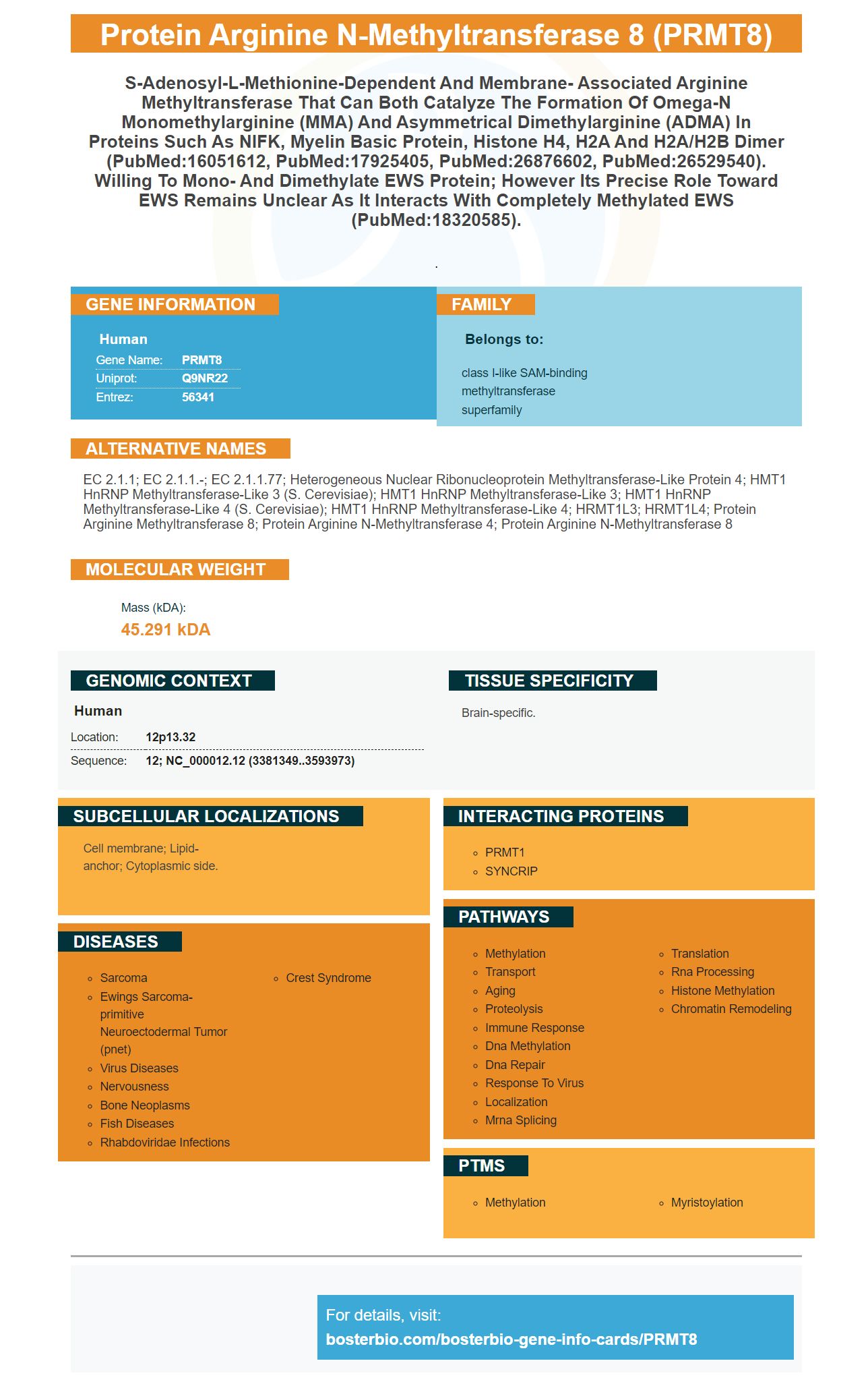
Facts about Protein arginine N-methyltransferase 8.
.
Human | |
---|---|
Gene Name: | PRMT8 |
Uniprot: | Q9NR22 |
Entrez: | 56341 |
Belongs to: |
---|
class I-like SAM-binding methyltransferase superfamily |
EC 2.1.1; EC 2.1.1.-; EC 2.1.1.77; Heterogeneous nuclear ribonucleoprotein methyltransferase-like protein 4; HMT1 hnRNP methyltransferase-like 3 (S. cerevisiae); HMT1 hnRNP methyltransferase-like 3; HMT1 hnRNP methyltransferase-like 4 (S. cerevisiae); HMT1 hnRNP methyltransferase-like 4; HRMT1L3; HRMT1L4; protein arginine methyltransferase 8; protein arginine N-methyltransferase 4; protein arginine N-methyltransferase 8
Mass (kDA):
45.291 kDA
Human | |
---|---|
Location: | 12p13.32 |
Sequence: | 12; NC_000012.12 (3381349..3593973) |
Brain-specific.
Cell membrane; Lipid-anchor; Cytoplasmic side.
If you are interested in studying the protein PRMT8, you can use the PRMT8 antibody. The antibody has good affinity and has been validated on Western Blotting, Immunohistochemistry, and ELISA. This article outlines the best ways to use PRMT8 antibodies and the techniques that you can use to detect its plasma membrane localization. You will also learn about PRMT8's localization in the Plasma Membrane and Mitochondrial.
The N-terminal extended region of the PRMT8 gene plays a key role in promoting its localization to the plasma membrane. PRMT8(DN15), a mutant PRMT8, was found to have a high level of cytosol enrichment in HEK293T cells as well as cultured cortical neurons. As a result, PRMT8-GFP largely occupied the plasma membrane in both cell types.
Northern analysis was used to determine the molecular location of PRMT8. Northern blotting revealed that PRMT8 is a brain-specific transcript. To ensure equal loading, b-actin probed the same membrane. Further analysis of PRMT8 sequence revealed a myristoylation motif in its unique N-terminal area.
Lee et.al. cloned PRMT8 successfully. The homology between the two genes was confirmed and the resulting protein was named the human PRMT8 gene. PRMT8 shares an amino acid identity of 80% with PRMT1 (602950). Although the gene was found in brain cells only, database analysis revealed that it is also expressed in other tissues. Further research is needed in order to determine the exact substrates PRMT8.
To pinpoint the exact location PRMT8 on the chromosomes we created two fluorescent proteins (FLAG–PRMT8 or GFP–PRMT8G-A) using human cDNA. We then digested the DNA with EcoRI/SalI and created GST-PRMT8. The homology was tested afterward. The analysis revealed PRMT8 is located on the chromosomes 12, 19 & 6.
MitoTracker-labeled with a fluorescent protein, PRMT8(N20/G2A/N20), and K4,15A,R6,13,14A/N20-GFP-labeled cells was detected. Glucose-PRMT8-GFP labeled cells showed that PRMT8 (K3,15A,R7,13,14A, and N20)-GFP localized to the cytoplasm.
PRMT8 is well-known to play multiple roles in the nucleus. However very few studies have looked at PRMT's role in brain neurons outside of the nucleus. PRMT8 has been found in the hippocampal neural neuropil of the mouse's brain. This supports local synthesis. PRMT8 expression was also found to correlate with dendritic spinal maturation, which is the location of most excitatory synapses.
RAGE is a unique protein with intrinsicoligomerization tendencies. It has limited the ability of classical methods to study its structures. RAGE is composed of three extracellular domains, a TM region, and an intracellular tail, and four truncated variants. The first two were used for molecular modeling. Native MS was used to examine the oligomeric subdomains C2 & C3. They were found to have oligomeric forms, and their signals vanished when FL-RAGE was supplemented with 0.1% formic acid.
The interaction between the Rossman folding and the dimerization arm of PRMT8 protein increased plasma membrane localization by PRMT8's protein oligomerization. Oligomerization/dimerization of PRMT8 enhanced its plasma membrane localization in both human and mouse cells. The mechanisms underlying this enhanced localization, however, are still being explored. We have here described the mechanism behind PRMT8 oligomerization.
The mechanism by which oligomerization/dimerization is accomplished depends on the type of oligomerization. For the formation oligomers large in size and distribution, both domains must exist. This mechanism enhances the localization in membrane-less cell bodies. The study suggests that this is a general mechanism for the concentration of functional components within membrane-less cellular bodies.
PECAM-1 oligomerization/dimerization improves integrin function. AP1510 (a bivalent membrane-permeable FKBP dimerizer) has been shown that it enhances PECAM-1/PECAM-1 relationships within the plasma membrane. These results suggest that PECAM-1 dimers can be used in integrin-like drugs.
Although all C1 variants contain cysteine residues, the AUCs of each showed that dimers are predominant in C2's spectra. In these conditions, the intensity and frequency of peaks associated with the highly charged monomers increased. A small percentage of dimeric species also appeared in the peak values of C2. So, the dimerization/dimerization of C2 improves its plasma membrane localization.
Although the BACK domain in a SPOP mutBTB oligomerization-induced ubiquitination efficiency is weaker than that of FL-RAGE monomers, it still forms a dimer of itself. Moreover, the BACK domain of SPOP mutBTB oligomerizes only through BTB-mediated interactions. This suggests that BTB-mediated interactions have the highest efficiency for oligomerization.
The full-length PRMT8 protein was shown to have better localization in the plasma membrane than deletion mutants. Its localization in the plasma membrane is enhanced by interactions between its dimerization arm and the Rossman fold. The protein does however show a patch-like localization within the cytosol. This study confirms PRMT8's mitochondrial localization.
We also studied PRMT8N220-GFP intracellular distribution in HEK293T cell lines. The protein displayed weak association with PRMT8(K3,15A,R7,13,14A)-GFP. Both proteins are known to have a Rossman-fold, which may lead to mistargeting. We found that PRMT8N20-GFP was co-located with Tom20mRFP (another marker of mitochondria).
PRMT1 is an important regulator of apoptosis and stress-induced signaling. PRMT1 is believed to methylate ASK1, which is a gene that regulates the rate of apoptosis. PRMT1-mediated arginine methylation is thought to inhibit ASK1 activation and increase apoptosis in MDAMB-231 breast carcinoma cells. It also inhibits the effects of paclitaxel on apoptosis-induced ASK1 protein.
These two mutants lack the ability of methylating GST-ASK1 (1-hundred and Twenty-four) and/or MKK6 (6. These mutants were obtained by transfecting 293T cells with the Flag-tagged PRMT proteins. The mutants then were immunoprecipitated (G80R) with GST-PRMT1 and subjected SDS–PAGE.
This study adds to the evidence that PRMT8 is crucial for mitochondrial function. METTL20 is the first lysine methyltransferase to be associated with mitochondria. This enzyme is an important regulator in cytochrome P450-mediated Signaling and is associated to many enzymes, kinases, and enzymes in mitochondria. PRMT8 is essential for proper functioning of mitochondria.
PRMT8's N-terminal extended area is unique. PRMT8 mutant (DN15)-GFP demonstrated plasma membrane localization within cultured cortical cells. This confirms the hypothesis that PRMT8 may be localized to the plasma cell membrane. If these findings are confirmed then further research will be needed to determine if PRMT8's mutation is a functional one or a signaling pathway.
In addition to the N-terminal domain, PRMT8 has multiple other regions that are important for its plasma membrane localization. These regions are critical for the protein’s constant localization within the plasma cell membrane. This is a prerequisite that it can be localized at the plasma cell membrane. Oligomerization also enhances PRMT8 localization. This is due to interactions between the Rossman fold (dimerization arm) and PRMT8.
To visualize PRMT8's location in the plasma membrane, several techniques were used. First, a GFP-PRMT8-expressing plasmid was transiently transfected into HeLa cells. Next, cells were fixed with 2% formaldehyde solutions in phosphate buffered saline. The nuclei were stained with 4',6-diamidino-2-phenylindole solution. Finally, images were captured with a confocal microscope and a three-dimensional reconstruction was performed using MetaMorpo imaging software.
The methylation arginine is an essential process in many cellular processes. In the case of PRMT8, the enzyme is localized to the plasma membrane, thereby influencing its ability to perform its function. Although the molecular mechanism of PRMT8's localization is not clear, basic amino acids and myristoylation are crucial. Further research is needed for the identification of the potential substrates PRMT8 in the plasma cell membrane.
We recently discovered PRMT8 at moderate-high levels in a variety if cancerous tissues. This was part of our HPA curation of cancer proteome. PRMT8 is found to be highly expressed in several cancer types and has a strong correlative relationship to cancer. There are a variety of techniques that can detect PRMT8 plasma membrane localization.
Fluorescent tag is one method to identify the fusion Protein in the Plasma Membrane. This technique requires the fusion protein to be fused with the C-terminal area of PRMT8 while leaving the rest of the N-terminus open for co-translational modifications. The constructs can then be transfected into HeLa cells and viewed under a magnifying glass. PRMT8 expression correlates with maturation of the dendritic protrusions, which are small protrusions that can be found on dendrites.
PMID: 16051612 by Lee J., et al. PRMT8, a new membrane-bound tissue-specific member of the protein arginine methyltransferase family.
PMID: 17925405 by Sayegh J., et al. Regulation of protein arginine methyltransferase 8 (PRMT8) activity by its N-terminal domain.