This website uses cookies to ensure you get the best experience on our website.
- Table of Contents
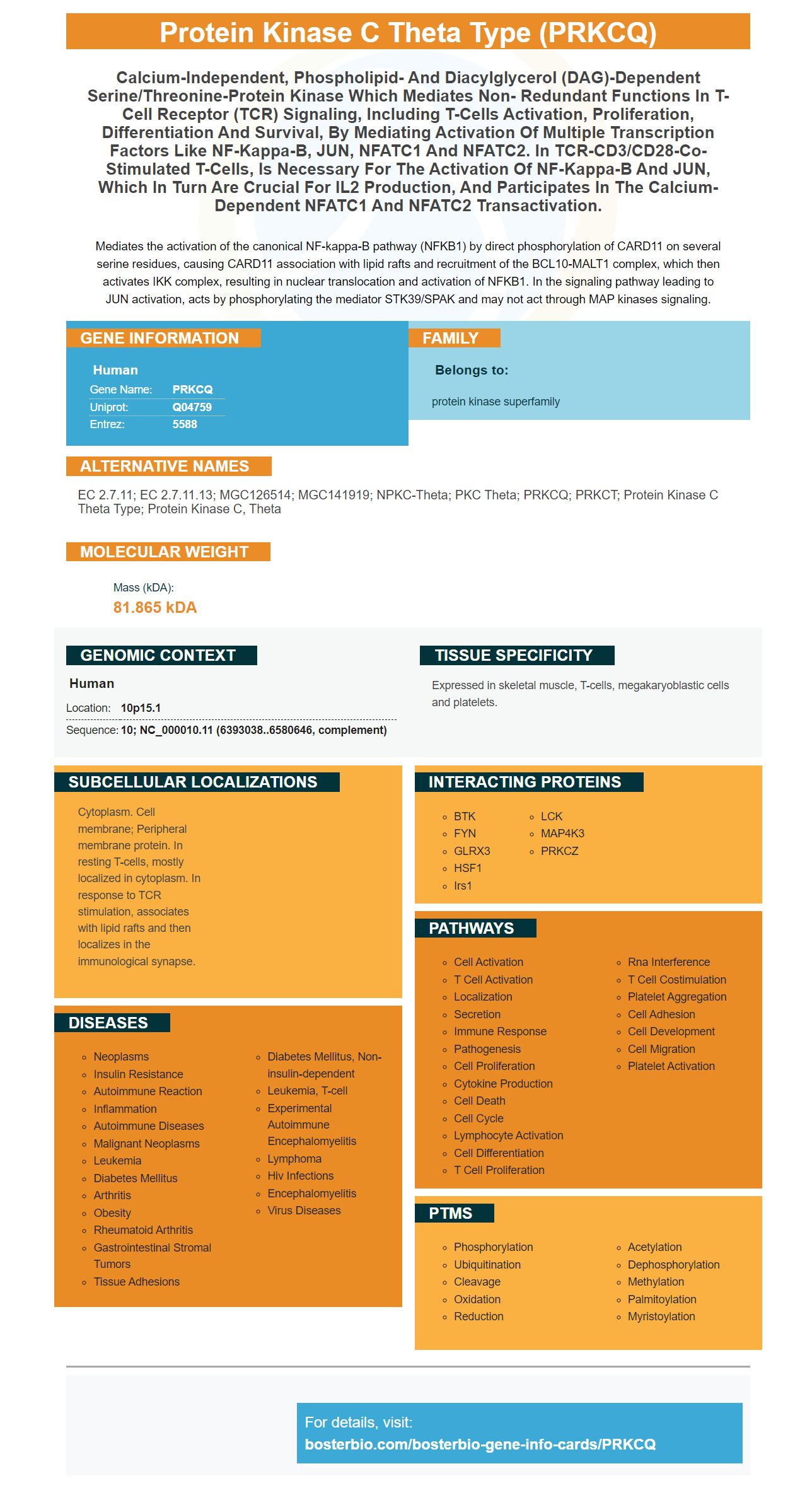
Facts about Protein kinase C theta type.
Mediates the activation of the canonical NF-kappa-B pathway (NFKB1) by direct phosphorylation of CARD11 on several serine residues, causing CARD11 association with lipid rafts and recruitment of the BCL10-MALT1 complex, which then activates IKK complex, resulting in nuclear translocation and activation of NFKB1. In the signaling pathway leading to JUN activation, acts by phosphorylating the mediator STK39/SPAK and may not act through MAP kinases signaling.
Human | |
---|---|
Gene Name: | PRKCQ |
Uniprot: | Q04759 |
Entrez: | 5588 |
Belongs to: |
---|
protein kinase superfamily |
EC 2.7.11; EC 2.7.11.13; MGC126514; MGC141919; nPKC-theta; PKC theta; PRKCQ; PRKCT; protein kinase C theta type; protein kinase C, theta
Mass (kDA):
81.865 kDA
Human | |
---|---|
Location: | 10p15.1 |
Sequence: | 10; NC_000010.11 (6393038..6580646, complement) |
Expressed in skeletal muscle, T-cells, megakaryoblastic cells and platelets.
Cytoplasm. Cell membrane; Peripheral membrane protein. In resting T-cells, mostly localized in cytoplasm. In response to TCR stimulation, associates with lipid rafts and then localizes in the immunological synapse.
Understanding Protein transfer basics will help you understand the PRKCQMarker. This article will discuss the PRKCQ Marker's ability to improve protein transfer efficiency using autoradiography film and membrane stained. It will also briefly cover the different methods for interpreting results from the protein transport experiment. Finally, we'll discuss the most important uses of PRKCQ Marker in research.
Boster Bio provides a wide assortment of high affinity primary antibodies, ELISA reagents, and reagents suitable for a wide array research applications. Boster Bio has its own labs that produce high-quality antibodies. They also make ELISA kits and reagents that are approved for use in a variety if techniques, such as IHC, WB or FC. The company offers a complimentary secondary antibody when you purchase a primary.
For flow cytometry to be successful, you need high-affinity prima antibodies. These antibodies are designed for binding to both cells as well as particles. Boster's antibodies have consistently received high citations and have been used for more than 25 years. The company also provides monoclonal and multiclonal antibodies. Numerous publications use Boster prima antibodies. To ensure the highest quality, the company is ISO-9001-certified.
Secondary antibodies are also available but cannot be used in isolation. They can be conjugated with other molecules to provide greater information and more robust answers. Researchers can use high-affinity secondary antibodies in the same experiment. These antibodies can be used for immunohistochemistry as well as histopathology. Flow cytometry is one example. It measures the amount and types of certain cells.
To measure the protein transfer efficiency by membrane staining, you must first determine the concentration of proteins in a tissue sample. Boster Bio offers protein concentration measurement and membrane stining services. Boster Bio can create Western blots premade using total proteins extracted from tissue samples. Additionally, they offer immunostaining blots for your desired protein. Upon completion, Boster will send you high-quality digital images of your results.
Next, you will need to examine the gel for proteins before and after membrane staining. This can be done using membranes made from either nitrocellulose or polyvinylidene trifluoride (PVDF). These membranes, which have lower non-specific staining than other membranes, are cheap and easy to apply, but can be fragile. They can also erase small molecular protiens. PVDF membranes have been used for this purpose because they allow for the combination and charge of negatively charged proteins.
Different fixation protocols could affect the retention and stability of proteins in the membrane. A pooled serum was subjected at 10% SDS–PAGE, before membranes were treated using either a traditional fixation method or an optimised one. In three independent experiments, band intensities were compared. Exposure times were identical. GraphPad Prism version6 and Image Lab were used to determine band intensities.
The target proteins were then detected after the gel was stained. The secondary antibody was then added to the sample, and the membrane was then washed four times with blocking buffer. The protein transfer was then completed. PVDF membranes were more sensitive, more precise, and more receptive to proteins than other membranes. The membrane staining process is fast, accurate, and simple. And, unlike most other methods, Boster Bio protein transfer efficiency by membrane staining can be optimized to match the protein concentration.
The BosterBio protein transfer efficiency by Membrane staing kit is simple. After mixing the sample in a reagent, it is incubated at 37degC for thirty to sixty minutes. When the gel is fully polymerized, it becomes solid. To avoid damage, take the gel out carefully. Boster will then mix the protein sample with the 4X Dual Color Protein Loading buffer. The protein sample must be added at a ratio of 3:1.
Wet transfer requires a high quality membrane. The sandwich is sandwiched with filters containing transfer buffer. It is fast and efficient, but can lead to bubbles between the sandwich layers. It is safe for sensitive testing. However, the process requires a lot of effort, which is why BosterBio offers its protein transfer efficiency by membrane staining kits.
SDS/PAGE can be assessed for its protein transfer effectiveness by using CHEMIDOC, or autoradiography films. Films are developed with WB Developing fixing kit and allow for a 10-minute exposure. The results can be recorded with a digital imager or luminometer. Alternatively, X-ray film may be placed on the membrane and developed for 10 minutes. For optimal results, multiple exposures may be needed.
The molecular-weight of the target proteins determines how efficient protein transfer is. For example, a low-molecular-weight protein will transfer efficiently. A protein with higher molecular masses will not transfer effectively. The optimal time and voltage should be determined for each transfer method. You must follow the instructions as different amounts may not transfer to your target membrane.
Wet-tank methods are ideal for transferring proteins of different molecularweights. Although it takes longer to complete electrotransfer it is flexible enough to optimize the transfer protocol. Towbin Transfer buffer is the best choice for this procedure. Because of its high buffering ability and pro-protein binding to membranes, it is highly recommended. The Towbin transfer buffer contains the highest concentration of sodium and potassium ions.
Image-based analysis of the protein-blotting workflow is important for a number of reasons. The blocking membrane prevents non-specific antibodies from binding to the transfer membrane, and the transferred protein is probed with an enzyme, fluorophore, or isotope. Finally, appropriate detection methods document the position of the target protein. However, a variety factors can affect protein transfer efficiency such as the type of gel and the molecular mass of the proteins.
Wet transfer is preferable to dry protein transfer. It has a lower chance of membrane failure than dry transfers. It is especially useful in large proteins. The gel is sandwiched in between two electrodes that are positively charged. The gel binds proteins and prevents them traveling to the positive electrode. However, it is important to note that the gel and filter paper are usually equilibrated in the transfer buffer.
PMID: 7686153 by Chang J.D., et al. Molecular cloning and expression of a cDNA encoding a novel isoenzyme of protein kinase C (nPKC). A new member of the nPKC family expressed in skeletal muscle, megakaryoblastic cells, and platelets.
PMID: 8444877 by Baier G., et al. Molecular cloning and characterization of PKC theta, a novel member of the protein kinase C (PKC) gene family expressed predominantly in hematopoietic cells.