This website uses cookies to ensure you get the best experience on our website.
- Table of Contents
2 Citations 1 Q&As
1 Citations 4 Q&As
3 Citations
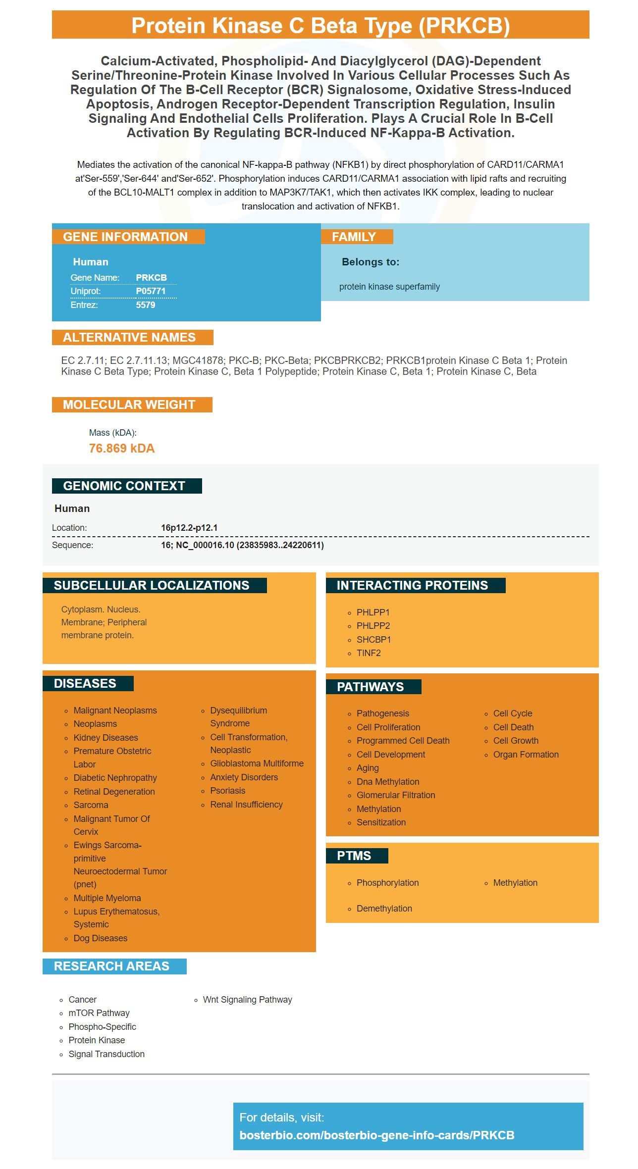
Facts about Protein kinase C beta type.
Mediates the activation of the canonical NF-kappa-B pathway (NFKB1) by direct phosphorylation of CARD11/CARMA1 at'Ser-559','Ser-644' and'Ser-652'. Phosphorylation induces CARD11/CARMA1 association with lipid rafts and recruiting of the BCL10-MALT1 complex in addition to MAP3K7/TAK1, which then activates IKK complex, leading to nuclear translocation and activation of NFKB1.
Human | |
---|---|
Gene Name: | PRKCB |
Uniprot: | P05771 |
Entrez: | 5579 |
Belongs to: |
---|
protein kinase superfamily |
EC 2.7.11; EC 2.7.11.13; MGC41878; PKC-B; PKC-beta; PKCBPRKCB2; PRKCB1protein kinase C beta 1; protein kinase C beta type; protein kinase C, beta 1 polypeptide; protein kinase C, beta 1; protein kinase C, beta
Mass (kDA):
76.869 kDA
Human | |
---|---|
Location: | 16p12.2-p12.1 |
Sequence: | 16; NC_000016.10 (23835983..24220611) |
Cytoplasm. Nucleus. Membrane; Peripheral membrane protein.
Boster Bio's AntiPKC beta 1/PRKCB marker is a sensitive and specific antiPKC gamma globulin antibody. It has been validated with positive and non-negative samples to establish its specificity. Among other benefits of this product, it rewards scientists who are the first to review it with a free product credit. It is available for commercial and research applications.
This anti-PKC beta 1/PRKCB marker is a part of the Picoband(tm) catalog and has been thoroughly tested for use in WB applications. This antibody is stable and can last up to one year at temperatures between -20 and 4 degrees Celsius. The antibody is composed of 5 mg BSA. It targets the PKC beta1 proteins from rats and mice. The product is 100% amino acid identical. You can also buy a blocking peptide to be used in place the recombinant prot.
Protein Kinase B beta is a member in the PKC gene familia. The beta subunit (of the PKC family) plays many different roles in cellular signals pathways. It is also a major receptor of phorbolesters, which are hormones-like substances that act as tumor promoters. This subunit is involved in diabetes and cancer due to its involvement in many physiological processes. It is phosphorylated at three sites: threonine 500 in the activation loop, tyrosine 642 in the turn loop, and serine 661 in the hydrophobic loop.
Both the Boster ECL chemiluminescence detection system and DAB chromogenic detection system are sensitive and specific. The detection systems can detect the presence of fluorescent and non-fluorescent compounds in samples. Each of the two types of systems has advantages and disadvantages. ECL systems employ the chromatin-dialysis technique, while DAB chromogenic detect uses the aqueous phase.
Membrane staining allows you to measure the efficiency in protein transfer. The polyacrylamide jelly transfers proteins to membranes. Membrane staining reveals whether the proteins have transferred. If there are irregular bands, uneven loading or no transfer, then the proteins did not transfer in an even fashion. This may result in an unspecific transfer of protein. You may need to wash the staining again.
Different molecular weight proteins were incubated on NC and PVDF membranes. To determine their binding ability, the membranes had to be stained with direct blu 71 after the proteins were bound. The intensity of staining were measured using scanning and density. The PVDF membrane had the same efficiency, but the NC membrane performed better in this case. The PVDF membrane is less sensitive in the second instance.
The efficiency of protein transfer largely depends on the type of protein and the type of membrane used. For example proteins with smaller molecular and/or higher molecular-weights require a membrane of lower porosity. PVDF membranes, on the other hand, are more efficient. The sensitivity, resolution and the affinity of a protein will all influence the choice of a membrane. These three factors are crucial in determining the protein transfer efficiency.
For this test, the pooled serum proteins were separated by 8% SDS-PAGE, and transferred to PVDF or NC membranes. The desired results were achieved by incubating the sample for between 30-60 min. The sample was then washed with TBS Wash buffer three times. The membrane was stained using the secondary antibody. This test takes place after three washes to remove unbound antibody.
The number of proteins that have been transferred affects the sensitivity of membrane staining to protein transfer. Inefficient protein transfer can lead to proteins being lost. A second problem is the degradation of proteins. This can lead to lower reproducibility. Fortunately, western blotting is now improved with better electrode materials and fixative reagents. Boster Bio's product line makes it easy to transfer protein easily. Western blotting can help you achieve high-quality results with more precise data.
The Autoradiography Films are sensitive to chemiluminescently-labeled samples. They offer excellent signal-to–noise ratios for increased detection efficiency. The films are light-tight and can be exposed at temperatures as low to -70°C. The advanced emulsion technology allows for excellent image resolution and background clarity. Multiple cellular structures can be captured by a single film, including the nucleus and cytoplasm.
PMID: 3755548 by Coussens L., et al. Multiple, distinct forms of bovine and human protein kinase C suggest diversity in cellular signaling pathways.
PMID: 3666134 by Kubo K., et al. Primary structures of human protein kinase C beta I and beta II differ only in their C-terminal sequences.
*More publications can be found for each product on its corresponding product page