This website uses cookies to ensure you get the best experience on our website.
- Table of Contents
1 Citations
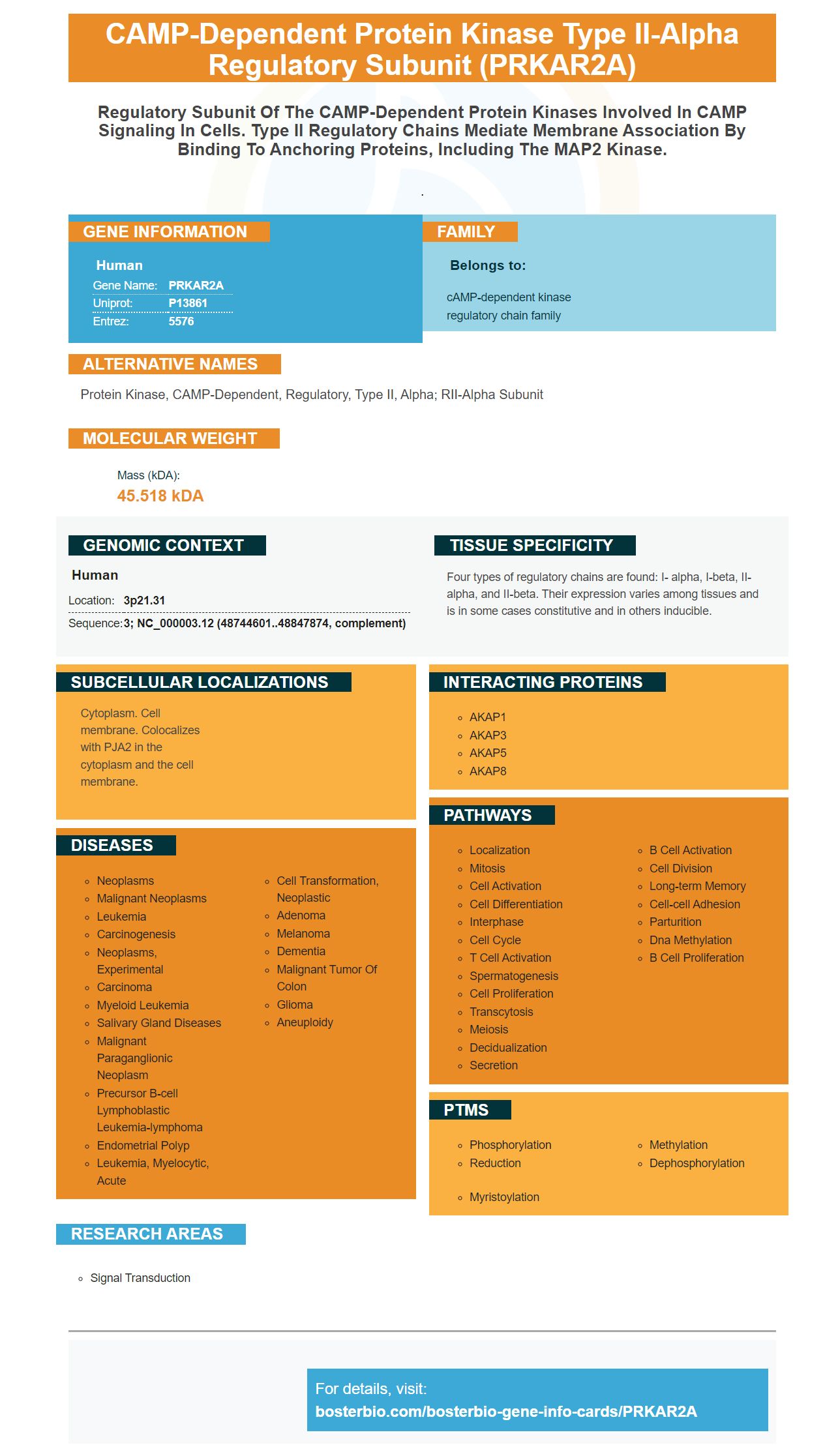
Facts about cAMP-dependent protein kinase type II-alpha regulatory subunit.
.
Human | |
---|---|
Gene Name: | PRKAR2A |
Uniprot: | P13861 |
Entrez: | 5576 |
Belongs to: |
---|
cAMP-dependent kinase regulatory chain family |
protein kinase, cAMP-dependent, regulatory, type II, alpha; RII-alpha subunit
Mass (kDA):
45.518 kDA
Human | |
---|---|
Location: | 3p21.31 |
Sequence: | 3; NC_000003.12 (48744601..48847874, complement) |
Four types of regulatory chains are found: I- alpha, I-beta, II-alpha, and II-beta. Their expression varies among tissues and is in some cases constitutive and in others inducible.
Cytoplasm. Cell membrane. Colocalizes with PJA2 in the cytoplasm and the cell membrane.
If you are searching for antibodies specific to PRKAR2A, then you've reached the right place. Boster Bio offers primary antibodies that have been widely validated on multiple platforms. They are also available in many concentrations. This allows you to use one antigen to cover many applications in a wide variety of species.
Boster Bio is the company to trust when it concerns the validation of IHC and WB applications. These products are validated using a variety of samples, which ensures high-quality and reliable results. The validation process relies on the Boster Bio laboratory for the manufacturing of the antibodies and reagents. Monitoring and optimization are key to the quality of the antibodies that are produced.
Boster Bio is an anti-body manufacturer specializing in WB/IHC-optimized monoclonal antibody, high-sensitivity ELISA kits, and picogram sensitivity ELISA kits. The company's antibodies have been validated on a panel of over 250 tissues, ensuring high quality and affinity. These antibodies are available only on individual request and covered by a quality guarantee.
WB is widely utilized to validate the specificity antibodies. WB can be used to validate an antibody's specificity if it recognizes a denatured protein. If multiple bands are observed, this may reflect multiple targets at different post-translational modification status, splice variants, and/or breakdown products. This result should cause concern and prompt further research.
Tissue microarrays are used to validate antibodies. The antigens taken from patient tissue or FFPE pellets are used in IHC/QIF. Only the most effective antibodies will stain cells. The staining intensity decreases as the dilution is increased. These validation results will be consistent to the published literature. The final result will show whether the boster's antigen-specific antibody is specific or not.
The lowest score was given to the Company 1 antibody when it was tested on IF and WB. The validation criteria were not listed in the datasheet. The datasheet for this antibody was minimal. It provided a brief background about the target as well as recommended dilutions of WB and IF. The antibody was unable to recognize proteins expressed by wild-type HEK cell lines.
The validation procedures for Picokine ELISA kits are described in the Company 6. These products are validated against 46 standard tissue samples by using proprietary blocking and coating technologies. The Company also uses a strict validation protocol to ensure consistent results from lot to lot. The Company also makes its products available for purchase online. These antibodies are manufactured in the United States and are tested under cGMP regulations.
Validation of antibodies in WB and IHC requires positive and negative cell line. The lysate from the treated and untreated cell lines shows a weak band, while the corresponding strong band is observed in the wild-type cell line. Using the same technique with a different cell line provides further validation. If Stathmin expression is restricted in BT-20 cells, antibodies may not be able to detect it.
Another method for validating antibodies is immunohistochemistry. This involves comparing the staining pattern of two antibodies to determine which one performs best. For consistent results, antibodies must not have overlapped epitopes. This Western blot method requires two samples of cell lines that express the target protein at different levels. This test is performed for recombinant expression of the target protein in the cell line and the staining signals of the two antibodies are compared with each other.
For immunohistochemistry, Prkar2a is an excellent candidate. It is found in MHb cells with substance P and Acetylcholine. It is composed of four subunits each with a distinct localization and cAMP binding affinity. All four subunits of the brain are expressed, and all four are highly expressed. The RIIa subunit, which is highly expressed, is located at the junction between two subnuclei. Prkar2a expression is absent from other brain regions as demonstrated by whole-brain 3DISH.
The RIIaKO mice reveals a clearer linkage between Prkar2a deficiency & enhanced voluntary activity. Because Prkar2a is involved in locomotor sensitization, Prkar2a deficiency is associated with enhanced motivation to exercise. PRKAR2A encodes an innovative protein that regulates and controls the habenular complex. This is part of dorsal denencephalic conduction.
PMID: 2540040 by Oyen O., et al. Human testis cDNA for the regulatory subunit RII alpha of cAMP- dependent protein kinase encodes an alternate amino-terminal region.
PMID: 9003463 by Foss K.B., et al. Molecular cloning, upstream sequence and promoter studies of the human gene for the regulatory subunit RII alpha of cAMP-dependent protein kinase.
*More publications can be found for each product on its corresponding product page