This website uses cookies to ensure you get the best experience on our website.
- Table of Contents
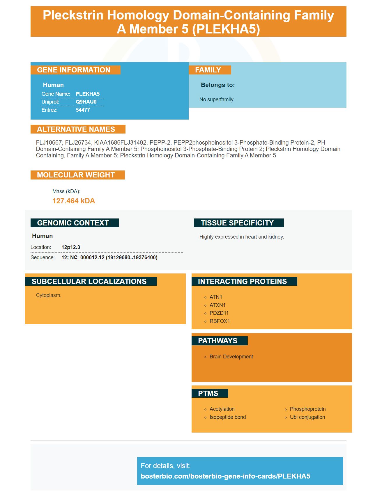
Facts about Pleckstrin homology domain-containing family A member 5.
Human | |
---|---|
Gene Name: | PLEKHA5 |
Uniprot: | Q9HAU0 |
Entrez: | 54477 |
Belongs to: |
---|
No superfamily |
FLJ10667; FLJ26734; KIAA1686FLJ31492; PEPP-2; PEPP2phosphoinositol 3-phosphate-binding protein-2; PH domain-containing family A member 5; Phosphoinositol 3-phosphate-binding protein 2; pleckstrin homology domain containing, family A member 5; pleckstrin homology domain-containing family A member 5
Mass (kDA):
127.464 kDA
Human | |
---|---|
Location: | 12p12.3 |
Sequence: | 12; NC_000012.12 (19129680..19376400) |
Highly expressed in heart and kidney.
Cytoplasm.
If you are looking for a high quality primary antibody, you may be wondering where it can be found. There are many sources that can provide information. These include Steven Boster Bio's Steven Boster Bio, Histology (high-affinity primary antibodies), and Steven Boster Bio. Read on to learn more about the PLEKHA5 Marker. You can also visit the BosterBio blog to find out more about high-affinity antibodies.
Steven Boster is known for his unique approach to solving problems throughout his career. This marker, PLEKHA5, is the ideal tool. He could do nearly anything he desired. This marker was also used by other scientists to develop new technologies, such as the PLEKHA5 fusion device. It is one of most effective methods to identify unknown substances.
PLEKHA5 is a member of the PLEKHA family of proteins. However, we don't know much about the function of this protein in the cell. p53 is the most likely candidate, but more research is needed to determine its exact function. In the meantime, the research community has a few tips to help it become a better marker for clinical research. Here are some of them.
PLEKHA5 is expressed in the gastric epithelium and other tissues. PLEKHA5 knockdown mice show a log2 fold decrease in gene expression. These cells also show a decrease in BMFS. A study of murine sarcomas found that the PLEKHA5 gene was significantly associated with the condition. Moreover, siRNA treatment of YUMUL and A375Br cells reduced their viability.
PLEKHA5 knockdown cells had more apoptotic Ki67 positive cells, two characteristics that are characteristic of apoptotic growing cells. PLEKHA5 is a key regulator of the development and acquisition of malignant phenotypes. These cell types did not show any significant effects of PLEKHA5 knockdown. In this area, more research is needed to understand the impact of PLEKHA5 on malignancies.
The promising candidate for therapeutic intervention is inhibiting PLEKHA5. It may help to reduce the spread of melanoma cells into brain metastases. Inhibiting PLEKHA5 could also inhibit the survival of melanoma cells in extracerebral sites. If the drug is approved, it could be used to improve the quality of patient's lives.
PLEKHA5, a novel protein, has been identified to be an ATP7A Trafficking Protein. ATP7A used to be thought to be found primarily in microtubules. However a new study suggests that it may also be present in actin rich cytoskeleton. Using this marker, we can develop antibodies that target both ATP7A and its ligands.
The sequences of the PLEKHA5 & PLEKHA6 protein share a high degree omlogy between their N-terminal areas. Both proteins possess tandem WW domains. PLEKHAs also include a PH-domain. The amino acid sequence of these proteins is aligned using WebLogo 3.7.4 to visualize the multiple sequence alignment.
PLEKHA5 has been found to be expressed across a range of tissues, with a large distribution within the choroid plexus. The protein also shows a better overlap with ATP7A expression in various tissues. PLEKHA5 was found to be immunolocalized within primary hippocampal neurons, blood vessels, and PLEKHA6 in blood vessels. ATP7A (and PDZD11) were immunolocalized within blood vessels.
The Swiss National Science Foundation, Swiss University of Geneva, the Service Egalite of University of Geneva and National Institutes of Health are all acknowledged by the authors. The authors would also like to thank Arielle Flinois (protocols for neuronal culture and culture of cysts) and Betty Eipper (for their work). Both contributed to antiATP7A antibodies.
ATP7A is associated with cell periphery in MDCK cells exposed to elevated levels of copper. However, the KO cells showed redistribution ATP7A from cytoplasm to the peripheral. This pattern was similar between the KO and WT cell lines. This suggests that WWPLEKHAs don't modulate ATP7A trafficking.
The quality and quantity of staining depend on the concentrations of primary antibodies and the temperature. High-affinity antibody can be used at higher concentrations with shorter incubation times. Longer incubation times help ensure that the antibodies penetrate and yield the desired signal. This does however mean that the price is higher.
The PLEKHA5 gene has an ortholog to the human protein PLEKHA5. Its full length sequence is similar to PLCKHA7 and PLEKHA6. The PH domain is different. Both proteins are located in the cytoplasm and are associated with lateral plasma membranes. This study examined plekhHa5 cell expression and subcellular plekhHA7 cell location.
PLEKHA5 is found in fibrillar cytoplasmic fibers, where it colocalizes to microtubules. PDZD11KO cells however, accumulate PLEKHA5 throughout the cytoplasm. This association is thought result of the interaction between PLEKHA5 (a protein that functions in cytoplasmic macrotubule localization) and PDZD11.
Although the PLEKHA5 genome is not known in melanoma cells but is highly expressed within cancer cell lines, it is still very prevalent. Thus, PLEKHA5 may have other functions besides its cellular role in cancer. Further research is needed in order to identify the exact cellular functions of PLEKHA5.
Ectopic localization is linked to the PH domains PLEKHA5 & PLKHA6. Met and PLEKHA5 both were phosphorylated within cancerous cells. The presence of a WW-PLKHA5 gene in cancer cells suggests that the plekha5 protein may be involved in the development of cardiac and neural tissues. It is also implicated with various systemic diseases such as hypertension, cancer, and hypertension.
PLEKHA5 not only plays a major role in tumor growth, but it is also crucial for the peritoneal spread of DGC-cells. In the mouse model, lentiviruses expressing shRNA versus PLEKHA5 was injected intraperitoneally to nude mice. PLEKHA5 shRNA was found to significantly reduce tumor formation in mice compared to controls. It also reduced the spread of tumors to the diaphragm, liver, and lungs.
DGC cells express PLEKHA5 high levels and exhibit rapid growth. They also show peritoneal dispersion and migration. It is possible to detect DGC tumors by knocking down PLEKHA5. It is not yet known if DGC cell lines express PLEKHA5 high levels in tumors. It is crucial to inhibit the PLEKHA5 gene from tumor cells in order to diagnose these malignant tumors.
PMID: 11001876 by Dowler S.J., et al. Identification of pleckstrin-homology-domain-containing proteins with novel phosphoinositide-binding specificities.
PMID: 22037487 by Yamada K., et al. Identification and characterization of splicing variants of PLEKHA5 (Plekha5) during brain development.