This website uses cookies to ensure you get the best experience on our website.
- Table of Contents
17 Q&As
16 Q&As
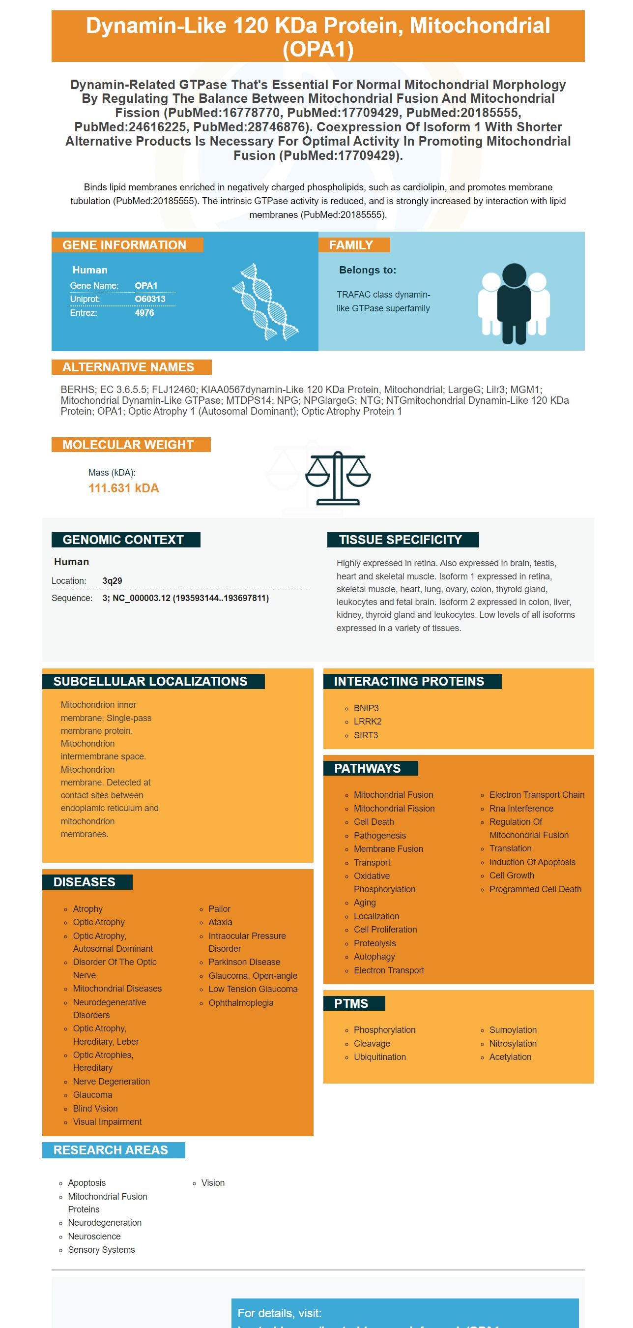
Facts about Dynamin-like 120 kDa protein, mitochondrial.
Binds lipid membranes enriched in negatively charged phospholipids, such as cardiolipin, and promotes membrane tubulation (PubMed:20185555). The intrinsic GTPase activity is reduced, and is strongly increased by interaction with lipid membranes (PubMed:20185555).
Human | |
---|---|
Gene Name: | OPA1 |
Uniprot: | O60313 |
Entrez: | 4976 |
Belongs to: |
---|
TRAFAC class dynamin-like GTPase superfamily |
BERHS; EC 3.6.5.5; FLJ12460; KIAA0567dynamin-like 120 kDa protein, mitochondrial; LargeG; lilr3; MGM1; mitochondrial dynamin-like GTPase; MTDPS14; NPG; NPGlargeG; NTG; NTGmitochondrial dynamin-like 120 kDa protein; OPA1; optic atrophy 1 (autosomal dominant); Optic atrophy protein 1
Mass (kDA):
111.631 kDA
Human | |
---|---|
Location: | 3q29 |
Sequence: | 3; NC_000003.12 (193593144..193697811) |
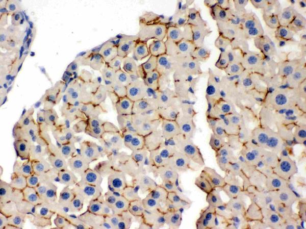
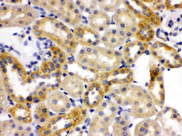
Highly expressed in retina. Also expressed in brain, testis, heart and skeletal muscle. Isoform 1 expressed in retina, skeletal muscle, heart, lung, ovary, colon, thyroid gland, leukocytes and fetal brain. Isoform 2 expressed in colon, liver, kidney, thyroid gland and leukocytes. Low levels of all isoforms expressed in a variety of tissues.
Mitochondrion inner membrane; Single-pass membrane protein. Mitochondrion intermembrane space. Mitochondrion membrane. Detected at contact sites between endoplamic reticulum and mitochondrion membranes.
OPA1 can be used to identify the presence or absence OPA1. OPA1 is an dynamin-related GTPase which is essential for mitochondrial fusion and regulation of apoptosis. The antigen is also subject to proteolytic processing. This can result in the disassembly of and release of OPA1 Oligomers as well as the caspase activator Cytochrome C. Boster has performed extensive tests on both positive and negative samples. Scientists can also apply for product credits.
In a recent study researchers discovered that the OPA1 marker is a reliable biomarker in the detection of neurodegenerative disorders. This gene encodes an atypical protein that has been involved in the development of Parkinson's disease. It is also important for the endosomal retrieval system, which regulates GARP's function and the EARP. These results suggest that OPA1 has multiple roles in the body.
It is encoded by the 11p15.5 region of the tumor suppressor gene. It is regulated by the Runx2 transcription factor. The predicted molecular mass of this gene is 43.6 kDa. A Phyre2 server estimates that this gene possesses only one WD40/bpropeller domain. These two genes are necessary for endosomal retrieval, which involves the integration of membranes.
OPA1 has been demonstrated to interact with a variety of proteins. Additionally it interacts with SNAP29, one of the proteins that participates in a variety of trafficking pathways. It also plays a role in the recycling process of the arginine transporter Can1.
There are many biological implications to high levels of LC3I/II that are found in boster biophagic cells. Inducing autophagy may increase levels of LC3B, and decrease the amount of protein aggregates. In a mouse model of sepsis, LC3B overexpression reduces lung injury. This supports the therapeutic application of LC3B-overexpression in different diseases.
Autophagy could be improved by inhibiting UBA6 and BIRC6. Both UBA6 and BIRC6 negatively regulate LC3B and LC3B, therefore inhibitory compounds that target one of these enzymes could be beneficial in treating various neurodegenerative conditions. KO of UBA6 and BIRC6 can increase autophagic clearance for pathogenic proteins.
In addition, LC3II turnover may not be necessary for all substrates. Thus, measuring the LC3-II level in autophagic cells might indicate how efficiently autophagic flow is carried out. It could also be used to study the relationship between LC3I/II levels and autophagic flux. The autophagosome activity of LC3I has been reduced by ATG7 knockdown.
Researchers need to validate the GFPLC3 assay to determine if inhibitors of autophagy are able to inhibit the production of autophagy proteins. These inhibitors should impede or reverse the formation of GFP-LC3 puncta cells that are treated with rapamycin or starvation, respectively. This assay is difficult to interpret due to its variation.
The research community has numerous applications for OPA1 markers. Recent research has shown that OPA1 is a key player in the methylation of DNA during neuronal developmental stages. According to Caglayan et al., the gene encodes two transmembrane domains. We will review some of the most efficient uses of this gene. OPA1's primary function in neuronal development is the way you can accomplish with it.
OPA1 is found in many tissues, including the brain as well as the heart. The brain is a major organ that expresses variants 3/4 4, 4b, and 6, more often than other tissues. The kidney, liver and thymus comprise other tissues that contain this gene. Exon 4 can be found in all vertebrate species. It has been preserved throughout the history of evolution. Exons 4b and 5b are ascribed to vertebrate species.
OPA1 has been examined in cells and has revealed its importance for fusion. The Chan lab published data on the processing and union of the Opa1 protein. The GTPase activity of the short isoform can be stimulated by the long isoform. However, the actions of both forms remain unclear. The OPA1 gene is important for the process of fusion. However, it's not known how it affects the cell's inner membrane.
Purified Opa1 protein was used for immunoblotting studies. Purified Opa1 was then diluted with 50-100 milliliters of ice-cold lysis buffer containing 0.5 percent Nonidet P-40, 50 mM Tris-HCl (pH 8.0), 150 mM NaCl a complete protease inhibitor mix. Whole-cell lysates were then centrifuged at 150000xg for 15 min. The supernatants were gathered for analysis.
ATP bioluminescence assay kit manufactured by Beyotime Biotechnology, was used to quantify intracellular ATP production. After preparing the membranes according to the manufacturer's instructions and then incubating them with Boster Bio's OPA1 marker. A chemiluminescence enhancement method was used to measure the intensity of DCFDA fluorescence.
The previous research has revealed that OPA1 can be used to identify neuronal cells. It enhances chemiluminescence in brain tissue. The protein level of L-OPA1 was significantly decreased after tMCAO, while the levels of S-OPA1 and total OPA1 decreased. This study suggests that OPA1 overexpression may decrease oxidative damage following cerebral ischemia.
Researchers found that OPA1 overexpression reduced the size of brain infarcts as well as preserved mitochondrial bioenergetics in animal models. These findings suggest that OPA1 could be a potential therapeutic target in stroke. OPA1-v1DS1 prevents neuronal death and maintains mitochondrial bioenergetics. This could be a key factor in stroke prevention.
TSINGKE, a biotech company based in Beijing, has developed the OPA1 marker. The company designed PCR primers to identify the marker which include GAPDH and the IL-1b. The OPA1 marker is a highly expressed gene that plays an important role in the process of cellular signaling. It is tightly controlled in human cells. It is also essential in the process of developing drugs and is essential for many studies in human and animal cell lines.
In animal studies in animal studies, the OPA1 gene is implicated in regulating basic cellular processes that include cell proliferation and differentiation. OPA3 was previously linked to cancer cell proliferation and growth. Eliminating the OPA3 gene from Panc-1 cells resulted the reduction of 30% in proliferation. The direct count of cells in shRNA-OPA3 cells showed a significant reduction in the rate of proliferation, however this was not accompanied by a significant increase in the number of cells dying and Annexin V-positive cells.
The upregulation of the OPA1 gene in cells can help maintain mitochondrial energy metabolism. OPA1 could be maintained in cells by upregulation. K-ras, which is an important transcription factor in the proliferation of oncogenic cells and regulates OPA3. OPA1 upregulation increases the activity antioxidant enzyme ATPase.
OPA1 encodes a protein that serves multiple functions in the human body. It regulates mitochondrial fusion as well as the process of apoptosis. During proteolytic processing, OPA1 oligomers break down and are released as caspase activator cytochrome C. Boster Bio validates its antibodies using ELISA and immunofluorescence. The company offers product credits for research conducted by qualified researchers.
The OPA1 gene is crucial for the proper functioning of mitochondria. Its expression is controlled by other genes, including cyclin D. One study injected OPA1-v1DS1 into adult male SD rats and gave them a 90-min transient mid cerebral artery occlusion. In vitro assays detected OPA1 expression.
PMID: 20843780 by Wang W., et al. Identification of rare DNA variants in mitochondrial disorders with improved array-based sequencing.
PMID: 11810270 by Delettre C., et al. Mutation spectrum and splicing variants in the OPA1 gene.