This website uses cookies to ensure you get the best experience on our website.
- Table of Contents

1 Citations 8 Q&As

1 Citations 7 Q&As


Facts about Oxidized low-density lipoprotein receptor 1.

Its association with oxLDL induces the activation of NF-kappa-B through an increased production of intracellular reactive oxygen and a variety of pro- atherogenic cellular responses including a decrease of nitric oxide (NO) release, monocyte adhesion and apoptosis. In addition to binding oxLDL, it serves as a receptor for the HSP70 protein involved in antigen cross-presentation to naive T-cells in dendritic cells, thereby participating in cell-mediated antigen cross-presentation.
| Human | |
|---|---|
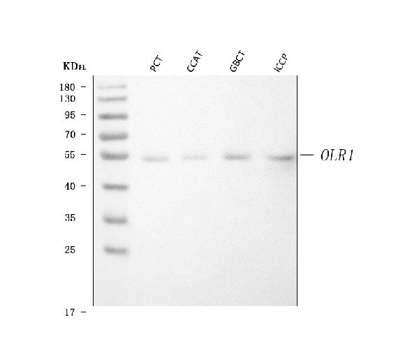
| Gene Name: | OLR1 |
| Uniprot: | P78380 |
| Entrez: | 4973 |

| Belongs to: |
|---|
| No superfamily |

CLEC8A; CLEC8ASLOX1; C-type lectin domain family 8 member A; hLOX-1; Lectin-like oxidized LDL receptor 1; Lectin-like oxLDL receptor 1; Lectin-type oxidized LDL receptor 1; LOX1; LOX-1; LOX1ox LDL receptor 1; LOXIN; OLR1; oxidised low density lipoprotein (lectin-like) receptor 1; oxidized low density lipoprotein (lectin-like) receptor 1; oxidized low-density lipoprotein receptor 1; oxidized low-density lipoprotein receptor 1, soluble form; Ox-LDL receptor 1; SCARE1; scavenger receptor class E, member 1; SR-E1
Mass (kDA):
30.959 kDA

| Human | |
|---|---|
| Location: | 12p13.2 |
| Sequence: | 12; NC_000012.12 (10158300..10172191, complement) |
Expressed at high level in endothelial cells and vascular-rich organs such as placenta, lung, liver and brain, aortic intima, bone marrow, spinal cord and substantia nigra. Also expressed at the surface of dendritic cells. Widely expressed at intermediate and low level.
Cell membrane; Lipid-anchor. Cell membrane; Single-pass type II membrane protein. Membrane raft. Secreted. A secreted form also exists. Localization to membrane rafts requires palmitoylation.


PMID: 9052782 by Sawamura T., et al. An endothelial receptor for oxidized low-density lipoprotein.
PMID: 9763655 by Li X., et al. Assignment of the human oxidized low-density lipoprotein receptor gene (OLR1) to chromosome 12p13.1-->p12.3, and identification of a polymorphic CA-repeat marker in the OLR1 gene.
*More publications can be found for each product on its corresponding product page