This website uses cookies to ensure you get the best experience on our website.
- Table of Contents
16 Q&As
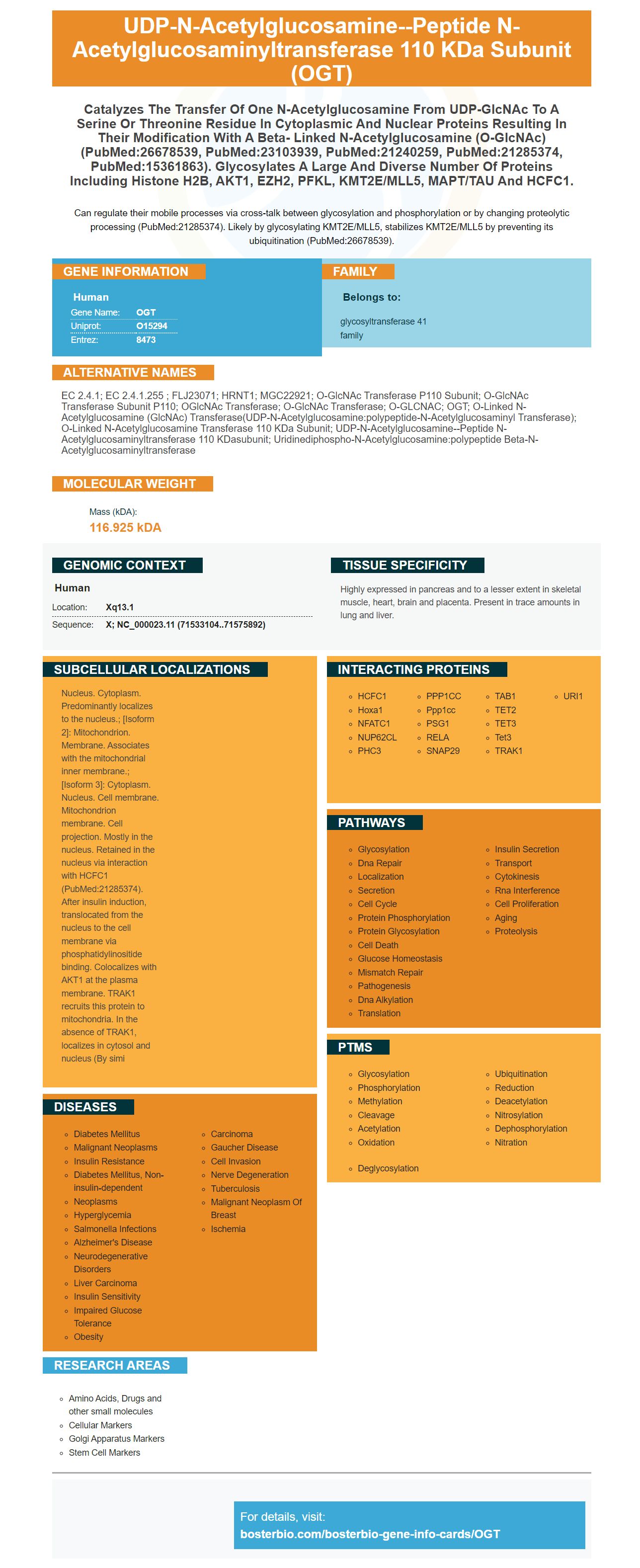
Facts about UDP-N-acetylglucosamine--peptide N-acetylglucosaminyltransferase 110 kDa subunit.
Can regulate their mobile processes via cross-talk between glycosylation and phosphorylation or by changing proteolytic processing (PubMed:21285374). Likely by glycosylating KMT2E/MLL5, stabilizes KMT2E/MLL5 by preventing its ubiquitination (PubMed:26678539).
Human | |
---|---|
Gene Name: | OGT |
Uniprot: | O15294 |
Entrez: | 8473 |
Belongs to: |
---|
glycosyltransferase 41 family |
EC 2.4.1; EC 2.4.1.255 ; FLJ23071; HRNT1; MGC22921; O-GlcNAc transferase p110 subunit; O-GlcNAc transferase subunit p110; OGlcNAc Transferase; O-GlcNAc Transferase; O-GLCNAC; OGT; O-linked N-acetylglucosamine (GlcNAc) transferase(UDP-N-acetylglucosamine:polypeptide-N-acetylglucosaminyl transferase); O-linked N-acetylglucosamine transferase 110 kDa subunit; UDP-N-acetylglucosamine--peptide N-acetylglucosaminyltransferase 110 kDasubunit; uridinediphospho-N-acetylglucosamine:polypeptide beta-N-acetylglucosaminyltransferase
Mass (kDA):
116.925 kDA
Human | |
---|---|
Location: | Xq13.1 |
Sequence: | X; NC_000023.11 (71533104..71575892) |
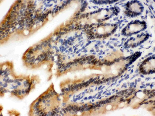
Highly expressed in pancreas and to a lesser extent in skeletal muscle, heart, brain and placenta. Present in trace amounts in lung and liver.
Nucleus. Cytoplasm. Predominantly localizes to the nucleus.; [Isoform 2]: Mitochondrion. Membrane. Associates with the mitochondrial inner membrane.; [Isoform 3]: Cytoplasm. Nucleus. Cell membrane. Mitochondrion membrane. Cell projection. Mostly in the nucleus. Retained in the nucleus via interaction with HCFC1 (PubMed:21285374). After insulin induction, translocated from the nucleus to the cell membrane via phosphatidylinositide binding. Colocalizes with AKT1 at the plasma membrane. TRAK1 recruits this protein to mitochondria. In the absence of TRAK1, localizes in cytosol and nucleus (By simi
The OGT marker has many uses, including prenatal stress, neurodevelopmental problems, and ovarian cancer. In this article, you'll discover its uses and discover how it promotes exosome release. This protein may help detect the presence of ovarian cancer cells. It also plays a key role in the immune response to cancer. If you'd like to learn more, continue reading.
A new study suggests that the OGT gene may serve as a potential biomarker for prenatal stress, which may help to identify babies who are at a greater risk for developing neurodevelopmental disorders. The researchers studied the placentas of pregnant mice and found that the female OGT gene, known as Ogt, is less expressed in males than in females. The OGT gene is an important mediator of the effects of EPS, and its decreased expression in male placentas may lead to abnormal neurodevelopment in male offspring.
The research found that prenatal stress changed the expression of several key genes, including VEsicular vGluT-1 and GLT1 messenger RNA. In the frontal cortex, VEsicular vGluT-1 levels were higher, while messenger RNA levels of GLT1 were higher. However, the effects of prenatal stress on these gene expressions were more pronounced in the hippocampus than in the frontal cortex.
Prenatal stress is also associated with increased levels of the HPA system. The HPA system in pregnancy is similar to that in controls, but at an increased level. The study also showed that the levels of OGT in rats exposed to high levels of prenatal stress increased in the fetus, compared to controls. In addition, the increased levels of this hormone could also lead to depression in the mother.
The results of this study suggest that the Boster Bio OGT is a biospot for prenatal stress. Prenatal stress has been associated with altered gene expression in the offspring, and it is associated with increased vulnerability to aversive events in later life. A biomarker for prenatal stress may help in monitoring these changes. However, there are still several questions remaining.
The development of a biomarker for neurodevelopmental disorders is a critical step towards addressing this pressing health problem. These conditions require the objective quantification of biological processes to identify subgroups whose patients are likely to benefit from particular treatments and to identify risk factors for autism and related disorders. Biomarkers for neurodevelopmental disorders also allow for the rapid assessment of change. Boster Bio is one such biomarker.
The Boster Bio anti-GFAP antibody was tested for sensitivity on different platforms including IHC and Western blot. It reacts with Human, Mouse, and Rat cells. It is also available in ELISA kits for research purposes. Boster Bio also provides immunological reagents through tebu-bio. The company believes that the biomarker has great potential for long-term autism diagnosis.
The Boster Bio OGT is a new in vitro diagnostic test that detects the expression of Wilms tumor protein, also known as CA125. It is an essential transcription factor that is involved in organogenesis, morphogenesis, cell growth, and differentiation. This marker is highly expressed in ovarian cancer. Because of its sensitivity and specificity, it is a useful diagnostic biomarker for ovarian cancer. It is also useful in assessing the prognosis of patients.
In the current era of precision medicine, ovarian cancer is the most common gynecologic cancer death in women. It is a global health and life-threatening disease, affecting women all over the world. Treatment options are limited by the late-stage of the disease, which results in a high mortality rate. Current treatment options include platinum-plus-paclitaxel-based chemotherapy and radical surgical tumor debulking. However, these therapies only have a 40% survival rate. Early detection and prediction can change this.
Ovarian cancer is one of the most common types of uterine cancer in women, with a 5-year survival rate of about 47%. Chemotherapy and aggressive debulking are standard treatments for ovarian cancer, but resistance to these treatments has increased the incidence of recurrence in these women. One possible reason for this is the role of exosomes in chemoresistance. Exosomes transport key regulatory proteins that reduce the damage to cells in tumors during chemotherapy.
A novel study suggests that the OGT marker promotes exosome release from ovarian cancer cells. Ovarian cancer cells express this protein, and its release leads to tumor growth, invasion, and spheroid formation. Exosomes also convert local fibroblasts into cancer-associated fibroblasts (CAFs), supporting tumorigenesis. Specifically, tumor cells upregulate the expression of TGFb1 and enhance the permeability of the basement membrane, facilitating the tumor cells' migration and invasion.
OGTR, an oncogenic gene, promotes exosome release from ovarian tumors. Exosomes are small vesicles that carry unique signatures of proteins, lipids, DNA, RNA, and DNA. These molecules have important roles in ovarian cancer, and identifying exosomes in early stages of disease may help clinicians better identify the tumor's potential for metastatic spread.
It is unclear exactly how these tumor cells function and which molecules are involved in exosome release. Ovarian cancer cells have been known to release exosomes as a means of promoting metastasis. Exosomes released by ovarian cancer cells also promote immune system responses, which in turn protect metastatic cells from apoptosis. Exosomes from ovarian cancer have been shown to induce T-cell arrest, a process called premetastatic niche. Moreover, these tumor cells can activate monocytes, thereby inducing IL-6 secretion.
Despite its low prevalence in ovarian cancer, exosomes have important roles in tumor regulation. Exosomes contain ncRNAs that promote tumorigenesis. Exosomes are an integral part of the immune system and have been shown to be essential in ovarian cancer cells. However, more research is needed. To further understand the mechanisms underlying the OGT-induced exosome release in ovarian cancer, additional research is needed.
Exosomes in ovarian cancer cells contain an extracellular molecule called MMP-1. This protein helps tumor cells in establishing a pre-metastatic niche before metastasis. MMP1 is also a bad indicator for ovarian cancer patients. Exosomes transfer MMP1 mRNA to the peritoneal mesothelium, which induces apoptosis through self-destruction.
PMID: 9083068 by Lubas W.A., et al. O-linked GlcNAc transferase is a conserved nucleocytoplasmic protein containing tetratricopeptide repeats.
PMID: 11773972 by Nolte D., et al. Human O-GlcNAc transferase (OGT): genomic structure, analysis of splice variants, fine mapping in Xq13.1.