This website uses cookies to ensure you get the best experience on our website.
- Table of Contents
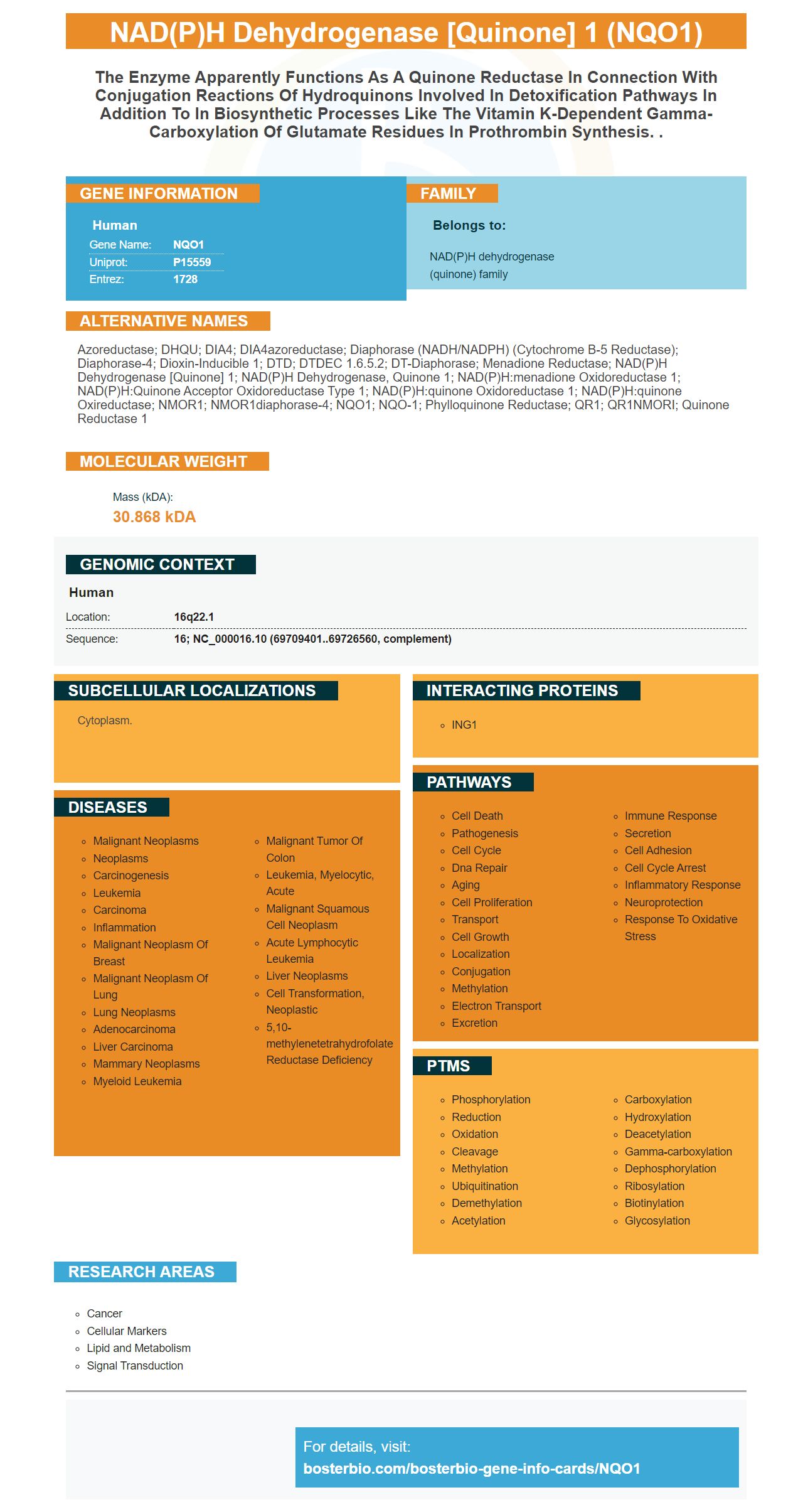
Facts about NAD(P)H dehydrogenase [quinone] 1.
Human | |
---|---|
Gene Name: | NQO1 |
Uniprot: | P15559 |
Entrez: | 1728 |
Belongs to: |
---|
NAD(P)H dehydrogenase (quinone) family |
Azoreductase; DHQU; DIA4; DIA4azoreductase; diaphorase (NADH/NADPH) (cytochrome b-5 reductase); Diaphorase-4; Dioxin-inducible 1; DTD; DTDEC 1.6.5.2; DT-diaphorase; Menadione Reductase; NAD(P)H dehydrogenase [quinone] 1; NAD(P)H dehydrogenase, quinone 1; NAD(P)H:menadione oxidoreductase 1; NAD(P)H:Quinone acceptor oxidoreductase type 1; NAD(P)H:quinone oxidoreductase 1; NAD(P)H:quinone oxireductase; NMOR1; NMOR1diaphorase-4; NQO1; NQO-1; Phylloquinone reductase; QR1; QR1NMORI; Quinone Reductase 1
Mass (kDA):
30.868 kDA
Human | |
---|---|
Location: | 16q22.1 |
Sequence: | 16; NC_000016.10 (69709401..69726560, complement) |
Cytoplasm.
You've found the right place to find an antibody specifically for the NQO1 marker. Boster Bio makes antibodies for all of the most crucial biomedical applications. These antibodies are tested on an extensive variety of platforms and have positive and negative samples to ensure high specificity and affinity. Boster also rewards its initial reviewers with product credits. Boster rewards scientists around the globe for their contributions to science.
If you're in search of an NQO1 ELISA kit you've arrived at the right place. Boster Bio offers several ELISA kits for NQO1. These kits are suitable for measuring the concentration of NQO1 within a variety of samples, including serum, plasma and urine. Boster Bio NQO1 ELISA Kits come with all the instructions on how to use.
The ELISA kit MBS705270 microwell stripplate ELISA, is to detect and quantify the presence of NAD (P.H) dehydrogenase, quinone-1 within biological samples. This kit is designed around an antibody-NQO1 interaction. The detection limit is set at 100 pg/ml based on the mean of 50 samples that were not dilute.
NQO1 overexpression significantly increases acylcarnitines and confers improved metabolism in mice fed HFD. These changes are driven by signaling pathways at the plasma membrane. These processes may be important in regulating inflammatory and immune processes. Incredibly, the overexpression of NQO1 is associated with improved lipid management and a diminished inhibitory effect of mitochondrial hyperacetylation of proteins.
In addition, overexpression of NQO1 provides protection against HFD-mediated changes to the whole-body metabolism. These effects were confirmed by metabolomics analysis that was not targeted using mass-spectrometry in the serum and liver. These experiments showed that NQO1 was a protective factor against the effects of HFD on the whole body metabolic changes. Additionally, mice that were overexpressed with NQO1 had lower levels of the insulin receptor B-sub and AS160 both of which are important for glucose uptake via insulin.
The mechanisms that link NQO1 overexpression to improved serum lipid profile are not fully understood. It is not clear what function NQO1 plays in regulating mRNA transcription. It is believed to enhance metabolism by regulating muscles and eWAT as well as fatty acid bi-oxidation. NQO1 also plays a part in sirtuin-mediated epigenetic regulation of fatty acid metabolism in adipocytes.
To assess the effects of NQO1 on liver function Small liver samples were collected from Keap1 fl/fl, mice with KO and WD. Small liver specimens were fixed in 10% neutral formalin and embedded in 4% paraformaldehyde for H&E staining. Tissue samples were harvested surgically and stored at -80°C. All animal experiments were conducted under the supervision and guidance of the Johns Hopkins University Animal Care and Use Committee.
Two compounds that cause IFN-g-induced in mice were utilized to test for NQO1 activity in RAW264.7 cells. Combining these two compounds allowed us test the effectiveness of these compounds in triggering the NQO1 activity. These experiments were conducted with compounds that inhibited iNOS in murine hepatoma cell lines and activating NQO1 activity.
These results suggest that NQO1 expression confers a protective effect on liver function in diets that are induced by HFD. The NQO1 transgenic mice were significantly smaller than WT mice and had higher glycogen deposition. Furthermore, they showed lower levels of fat infiltration and inflammation. The lipid-related changes that occur in NQO1 expression are in line with a central role for adipose tissues in the regulation of insulin sensitivity and liver Steatosis.
Tissue samples were fixed in the presence of 4% paraformaldehyde. They were then permeable with 0.5 percent TritonX 100 for 10 minutes. The cells were then blocked using 3% bovine serum albumin fraction V or Solarbio A8020 for 15 minutes at RT. The cells were incubated with the NQO1 antibody (1:200 Santa Cruz Biotechnology).
Transgenesis of NQO1 causes improvements in glucose homeostasis, insulin sensitivity, and lipid excess management. The improvement that NQO1 induces in the whole body glucose metabolism is thought to be mediated by increased constitutive NRF2 transcriptional activity. Additionally, NQO1 overexpression inhibits mTORC1 protein expression, suggesting improved liver function.
Different fields are studying the roles of NQO1 markers. This molecule is an important component in the PMRS which decreases the amount of ubiquinone. It also plays a role in the progression and growth of cancer. It is well-known that certain human NQO1 polymorphisms are associated with higher risk of cancer. In addition, polymorphic NQO1 variants are less active and less affinity to FAD than the wild-type. This could be due to the inadvertent mobility of the NQO1 protein's key parts.
Although it is not regulated by the 20S proteasome is important, NQO1 blocks degradation of proteins. In addition, NQO1 interacts with other proteins involved in this process. NQO1-interacting proteins also have intrinsically disordered regions which prevent their degradation by the 20S proteinasome. The proteins then become damaged, which can cause dysfunction and disease. Therefore the NQO1 marker can aid in determining what the function of these proteins.
It is also possible that NQO1 is directly involved in superoxide scavenging capability. Cells have numerous mechanisms to regulate the levels of superoxide. The superoxide dismutase system (SOD) enzyme system removes and produces superoxide. The superoxide reaction rate of NQO1 is four orders of magnitude lower than that of SOD's enzyme dismutation.
In addition to its role as a redox switching device, NQO1 undergoes structural changes in response to reduced pyridine nucleotides. The protein's oxidized state is able to bind reduced Pyridine nucleotides which alters the structure of NQO1. In addition, the altered conformation of NQO1 influences its interactions with mRNA and proteins.
Research has proven that NQO1 has multiple roles in the metabolism quinones. It is a cell source for NAD+ and could play a role as mediator of metabolic syndrome. The enzyme also shields proteins from degradation by proteasomes. The polymorphism found in the NQO1 gene has been implicated in various human diseases such as respiratory disorders and cancer. This gene is also associated in the metabolism of benzene.
The connection between NQO1 null and the amount of BCC cases has been proven. The risk of developing the disease is significantly increased if one has an NQO1 null gene. The polymorphism results in lower BCC count than that of the polymorphism. However the polymorphism in SQO1 causes a modest decrease in the levels of protein.
PMID: 2843525 by Jaiswal A.K., et al. Human dioxin-inducible cytosolic NAD(P)H:menadione oxidoreductase. cDNA sequence and localization of gene to chromosome 16.
PMID: 1657151 by Jaiswal A.K.; Human NAD(P)H:quinone oxidoreductase (NQO1) gene structure and induction by dioxin.